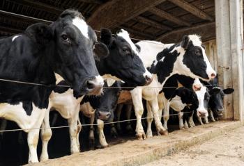
The state is the 22nd to report the virus in cows, and the outbreak next door has been linked to 3 human infections, including 1 death.
The textedbook coedited by veterinarians from Brookfield Zoo Chicago, The Ohio State University, and San Diego Zoo Safari Park, is the first comprehensive imaging reference dedicated to nondomestic mammals.
Zoo and wildlife veterinarians now have a dedicated resource for interpreting radiographs, CT scans, MRIs, and ultrasonographic images of nondomestic mammals. Published by Wiley, Diagnostic Imaging of Zoo and Wild Mammals is the first comprehensive reference devoted to diagnostic imaging in these species, according to a news release from Brookfield Zoo Chicago.1
The 1,008-page textbook includes contributions from 58 veterinarians and radiologists worldwide.1,2 It was coedited by Michael J. Adkesson, DVM, MBA, Dipl. ACZM, Dipl. ECZM (ZHM), president and CEO of Brookfield Zoo Chicago; Eric T. Hostnik, DVM, MS, Dipl. ACVR-DI, Dipl. ACVR-EDI, associate professor at The Ohio State University College of Veterinary Medicine in Columbus and primary radiologist for the Brookfield Zoo Chicago Radiology Consulting Service; and Matthew E. Kinney, DVM, Dipl. ACZM, director of veterinary services at San Diego Zoo Safari Park in California.2
Anatomic differences among nondomestic mammals, which range from rhinoceroses to pangolins, can make diagnostic images especially difficult to interpret. Reference materials for many of these species have historically been limited.1
“When a veterinarian is learning to interpret a CT scan of a rhinoceros or an X-ray of a pangolin, they need to know what normal anatomy looks like before being able to recognize what might be wrong,” said Adkesson. “For many of the species we care for, those references have been limited and scattered across different sources. This textbook is the resource I wish I’d had when I was training to become a zoo veterinarian.”1
The book was developed over several years and contains hundreds of annotated diagnostic images and clinical photographs. It also gives practical guidance on acquiring images with digital radiography, ultrasonography, and CT and MR imaging.1,2 Topics include anesthesia, training animals to participate voluntarily in imaging procedures, and radiation exposure and safety for animals and people. Each species group is discussed in depth, with coverage of normal and abnormal anatomy and common diseases.1,2
Coverage spans the following groups:2
A companion website provides high-resolution images from the book.1 The text is designed for veterinary students, residents, and interns, as well as practicing zoo and wildlife veterinarians, exotic animal practitioners, and veterinary radiologists.1,2
Chapters are coauthored by board-certified specialists in zoological medicine and veterinary radiology. The pairing connects the practical experience of veterinarians caring for zoo and wildlife species with the expertise of diagnostic imaging specialists.1
“Zoo veterinarians and radiologists bring different perspectives to the same image,” said Hostnik. “By bringing those areas of expertise together, this book gives veterinarians a new resource to learn not only how to interpret an image, but what those findings mean for an animal’s care.”¹
Kinney noted the value for clinicians facing uncertain cases. “This textbook gives veterinarians a stronger foundation for making clinical decisions while helping those entering the profession build their skills,” he said. “It brings years of collective experience into a resource they can use throughout their careers.”1
The book’s cover features a CT scan of Layla, an eastern black rhinoceros previously cared for at Brookfield Zoo Chicago. Safely imaging an anesthetized, 2,000-lb rhinoceros posed significant logistical challenges. Interpreting the scans was also difficult because few comparable studies were available.1
Layla’s case helped inspire wider efforts to expand access to imaging references. These include the Zoo & Aquarium Radiology Database, which a cohort of zoos and aquariums launched in 2024. The group includes Brookfield Zoo Chicago, The Ohio State University, and San Diego Zoo Safari Park.1
References