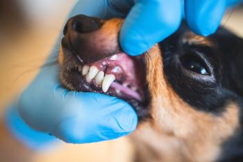
Mandibular fracture repair using acrylic external fixators (Proceedings)
Mandibular fracture is a relatively common injury. Surgical repair of the mandible is complicated by the presence of tooth roots and a mandibular canal with its neurovascular contents.
Mandibular fracture is a relatively common injury. Surgical repair of the mandible is complicated by the presence of tooth roots and a mandibular canal with its neurovascular contents. These occupy the body of the fractured bone leaving no appreciable linear marrow space. Consequently, most fractures in the horizontal ramus cannot be repaired using plates or pins without causing significant iatrogenic damage to the teeth. Any roots that are penetrated with pins or screws will most likely require root canal treatment of extraction. Additionally, regaining normal occlusion in this narrow curved bone is just as important as successful bone healing.
Fractures that extend caudoventrally, as shown in the radiograph, are considered unfavorable because the action of the masticatory muscles distracts the fragments. In contrast, in a rostroventral fracture the fragments tend to be apposed by masseter contraction. However, as far as fixation is concerned, one might consider a favorable fracture to be one that is mid-body with healthy teeth on each side. When a mandibular fracture is located rostral to the mandibular first molar tooth in an area that has healthy teeth both rostral and caudal to it, the teeth can be used as part of a solid intraoral external fixator. The tooth roots act functionally similar to transcutaneous pins in a Kirschner apparatus. An acrylic or composite resin splint is then applied directly to the crowns of the teeth to function as the side bar. This provides extremely solid fixation along the tension side of the fractured bone. Since it is on the tension-band side of the mandible, when in function, even with a caudoventral fracture, muscle action will tend to appose the fragments.
To apply this type of splint, preserve any teeth that are firmly attached regardless of their proximity to the fracture. They can contribute to the stability of the repair, and future root canal treatment or extraction can be done as needed after the jaw has healed. The first step is to place interdental wires to stabilize the mandible, ideally including a minimum of two teeth (four roots) on each side of the fracture. The wiring serves to hold the fragments in alignment and the teeth in correct occlusion while making the rigid splint. There are multiple wiring techniques that can be used. A simple and very adequate technique is Stout's multiple loop wiring. The easiest way to apply the wires is by placing a hypodermic needle through which the wire can be passed. Usually 25 ga wire works well for this, although on small patients 28 ga is sufficient. The wire does not accomplish the fixation, but acts as a type of rebar to reinforce the stiffer polymer that will be added later. The wire is first placed behind the most caudal tooth to be included in the splint. The buccal wire should be long enough to extend past the most rostral tooth to be included, adding enough to allow twisting with the other side. The lingual wire needs to be a few cm longer since it will be used interproximally to engage the buccal wire (see diagram). With each interproximal loop around the buccal wire, one twist is applied to hold it in place. The fracture is manually reduced, the buccal wire is pulled tight, and the two ends (buccal and lingual) are twisted together rostral to the most rostral tooth. Then each loop is tightened, beginning with the most caudal, to snug them tightly around each tooth at the cervical area and underneath the wider crown. After twisting, the loops of the wires should be bent dorsally into the interproximal areas to avoid interference with occlusion against the palatal surfaces of the upper teeth when the mouth is closed.
The next step is to close soft tissue defects using an absorbable suture material. Then place either acrylic (e.g. Jet Acrylic) or composite (e.g. Protemp Garant) over wires and on the lingual and buccal sides of the tooth crowns. The buccal splint should be thin to avoid interference with the opposing dentition. The lingual side can be thicker for strength, and the two sides should meet in each interproximal space to fully engage the teeth for retention. Removal of the splint is easier if the tip of each tooth is left uncovered to help identify their exact locations within the splint. The final step is to use an acrylic bur to remove any material that would interfere with occlusion, and to smooth the entire surface of the splint to prevent any areas that could be sharp or uncomfortable to the tongue, lips, or cheeks.
For the rigid material, either acrylic or composite resin can be used. Each of the materials has advantages and disadvantages and may be better suited to specific cases and patients. Acrylic is inexpensive, and is resistant to cracks or fractures. The best placement method is to layer it onto the teeth, alternately placing powder and a drop of liquid as the splint is built up. This method avoids the significant heat generation that occurs from a large mass polymerizing at once when the liquid is mixed together with the powder. It also allows the fluid plastic to conform perfectly to the teeth and wire, creating ideal mechanical retention. If the materials are mixed together then placed around the teeth, early polymerization decreases the flowability and compromises the adaptation to the fine dental anatomy and undercuts. The disadvantage of the acrylic is the time it takes to apply, set, adjust and smooth the splint. Composite resin, in comparison, is very fast and easy to apply when using an auto-mix product. These are designed for human dental work to quickly make a temporary crown for a patient to use while waiting for the lab to craft the final crown. The material is extruded from an application gun with a mixing tip that completely mixes the two paste phases together, beginning the automatic self-cure. It is extruded directly onto the teeth and wires. The set is fast enough that the animal can be extubated during application, allowing the mouth to be closed and the teeth occluded as it hardens, thereby preventing any interference of the final splint. The disadvantages are the cost of the materials, and the brittle nature of the material that can be more easily cracked and fractured by external forces. On the positive side, this brittle nature makes the splint much easier to remove than the acrylic splints after the bone has healed.
After healing in 6-8 weeks the splints are removed using a combination of high speed and low speed burs and very careful chipping.
If there are no teeth adjacent to the fracture, bone screws can be placed in the edentulous fragment to provide a purchase on the bone. It is also sometimes combined with circlage around the mandible, particularly for long oblique fractures.
This method of intraoral splinting provides solid fixation with good occlusion, and is non-invasive to minimize further trauma to the surrounding tissues.
Newsletter
From exam room tips to practice management insights, get trusted veterinary news delivered straight to your inbox—subscribe to dvm360.


