

|Articles|August 1, 2009
Managing fleas, ticks and vector-borne diseases (Proceedings)
Author(s)Byron L. Blagburn, MS, PhD
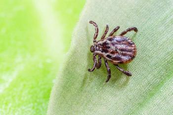
Ticks are second only to mosquitoes in the number of diseases that they transmit.
Advertisement
Importance of ticks
- Ticks are second only to mosquitoes in the number of diseases that they transmit.
- Ticks feeding may cause irritation, anemia, hypersensitivity reactions, and toxicosis.
- Ticks may transmit numerous pathogens including helminths, protozoa, viruses, bacteria (including rickettsiae) and fungi.
- Several pathogens transmitted by ticks are potential zoonotic agents.
- Two families of ticks are important in veterinary medicine
Ixodidae (hard ticks)
- Environmental stages found in open areas on vegetation.
- Mouthparts visible from dorsal surface.
- Body capable of limited expansion (dorsal surface covered by leathery scutum [see below]).
- Balance water using salivary secretions.
- Secrete cement from salivary glands for attachment.
- Female hard ticks feed and engorge slowly over long periods.
- Imbibe many times their body weight in host blood.
- Females lay thousands of eggs per oviposition in environment.
Argasidae (soft ticks)
- Environmental stages live in nests and burrows of hosts.
- Mouthparts not visible from dorsal surface.
- Body is soft and easily expandable.
- Life cycle consists of many nymphal stages.
- Females feed intermittently and lay eggs in masses.
- Soft ticks do not produce cement for attachment.
Important ixodid (hard) tick species found on companion animals
- Rhipicephalus sanguineus (Brown Dog Tick)
- Dermacentor variabilis (American Dog Tick); Dermacentor andersoni (Rocky Mountain Wood Tick)
- Ixodes scapularis (Black-legged Tick; Deer Tick); Ixodes pacificus (Western Black-legged Tick)
- Amblyomma americanum (Lone Star Tick); Amblyomma maculatum (Gulf Coast Tick)
Ixodid (hard) tick structure and function
- Body is divided into head (capitulum [contains mouthparts]), and body (idiosoma).
- Mouthparts consist of structures that function in tactile sensing (palps), lacerating host skin (chelicerae), and anchoring the tick to the host (hypostome).
- Body is covered by a complex waxy somewhat inflexible exoskeleton (tegument) that protects the tick from water loss and predators.
- A scutum (dorsal shield) covers the entire surface of adult male tick, but only a portion of the dorsal surface of female ticks.
- The scutum is sometimes colored with iridescent white or yellow patches (ornate tick) or may be devoid of such ornamentation (inornate ticks).
- Partial coverage of the dorsal surface of female ticks allow for enormous expansion during feeding and engorgement.
- Ticks have an excretory system (malpighian tubules), "liver" (fat body), an open circulatory system with a blood-like hemolymph, a respiratory system (net-like tracheae with openings called spiracles), a rather complex nervous system and a number of sensory structures that detect chemical, thermal, light and mechanical stimuli. A multifunctional organ located on the legs (Haller's organ) receives stimuli used during questing (crawling on vegetation to gain entry onto a host) and host detection. Some ticks have eyes.
Ixodid tick developmental cycles
• Ticks develop through four distinct life cycle stages.
o Egg, larva, nymph, adult
o Larvae, nymphs, and adults are similar in appearance, but differ in size and numbers of legs (larvae possess 6 legs; nymph and adults possess 8 legs).
o Stages increase in size from larva to adult.
o Larvae and nymphs are without features of sexual dimorphism.
• Ticks that infest companion animals are 3-host ticks.
Advertisement
• Most species utilize a different species of host at each stage; R. sanguineus utilizes the dog during each developmental phase.
• Ixodes scapularis (Black-legged Tick; Deer Tick) may parasitize in excess of 100 host species representing three vertebrate classes (mammals, birds, reptiles).
• Certain ticks (i.e. R. sanguineus) can complete their life cycles in weeks, while others require two years or more.
• Eggs are laid by replete females in sheltered environments off the host.
• Larvae and each subsequent stage seek a host, feeds to repletion, drop from the host and either molts to next stage or deposit eggs (replete females).
Behavior of ticks in the environment
- Free-living ixodid ticks reside in numerous environments including forests, savannahs, fields, shrubs and brush.
- They are capable of developmental arrest (diapause) to allow them to survive periods of environmental stress
- Diapause is triggered by environmental changes such as day length, temperature changes, and seasonal changes.
- Ticks seek hosts by questing on vegetation; types and height of vegetation is determined by the host they seek (lower vegetation for smaller hosts; higher vegetation for larger hosts)
- Ticks utilize vibrations, shadows produced by changing light patterns, body heat and odors and chemicals as host-seeking cues.
- Ticks literally "grab" hosts as they pass in close proximity to questing sites.
Feeding habits, pathogen transmission, vector competence
- After gaining access to the host, ticks search for a desired attachment site.
- Attachment occurs by cleaving the host skin with the chelicerae, insertion of the hypostome (anchor) and production of adhesive cement and salivary secretions.
- Tick saliva contains numerous moieties that inhibit immune responses, coagulation of blood, and exert anti-inflammatory and early analgesic effects.
- The tick body undergoes enlargement and expansion (greatest in female) during imbibition of blood.
- As ticks feed, they ingest pathogens that may be circulating in the host's blood.
- Pathogens undergo replication in tick gut cells and are disseminated to numerous tick organs (i.e. salivary glands).
- Pathogens gain entry into a new host when the next stage (after molting) feeds on susceptible hosts (transstadial transmission) or by larvae that hatch from eggs through which the pathogen was passed (transovarian transmission).
- Successful transmission can occur via salivary secretions, coxal fluid, regurgitated gut contents, contaminated mouthparts, or a combination of these mechanisms.
- Whether a tick will serve as a successful vector depends on several factors
o Size of bloodmeal ingested, virulence of pathogen, susceptibility of host.
o Capability of pathogen to infect and replicate in the tick.
o Successful dissemination of the pathogen throughout the tick's body.
o Transstadial or transovarial habits of the pathogen.
o Interactions between multiple microbes infecting the same tick.
Tick control
• Treatment of the Animal (numerous available agents; the following are marketed by the veterinary profession) >
o Amitraz (Preventic®, Promeris™ for Dogs)
o Fipronil (Frontline®, Frontline® Plus)'
o Permethrin/Imidacloprid (K9 Advantix™, Vectra 3D™)
o Selamectin (Revolution®)
• Treatment of the environment
o Habitat modification
o Host elimination
o Targeted acaricides
• Integrated Tick Control
o Combination of animal treatment and environment treatment/management.
Vector-borne disease control
• Prevent tick infestation by using appropriate tick avoidance behavior (difficult).
o Avoid tick infested areas
o Wear light-colored clothing (allows you to see tick crawling on clothing)
o Use a chemical repellent such as DEET, (Picaridin??)
• Prevent tick feeding (ticks may gain access to pet host, but do not attach or feed)
o Amitraz (Preventic [collar]), Fipronil (Frontline [spray]), and Permethrin/Imidacloprid (K9 Advantix [spot-on]) can prevent vector transmission.
• Treat host with antimicrobial medications (therapeutic or prophylactic regimens).
Canine ehrlichiosis/anaplasmosis
Canine ehrlichiosis and anaplasmosis are multisystemic diseases that may be caused by different genera and species of gram-negative obligate intracellular bacteria. The taxonomy of this group of organisms has undergone significant changes due to new information derived from molecular biological characterizations. Most ehrlichial species of importance to companion animal veterinarians use ixodid ticks as vectors. Some are transmitted by a single tick species; others may utilize multiple tick vectors. Ehrlichia canis, the principle cause of canine ehrlichiosis, is transmitted in salivary secretions by the ubiquitous brown dog tick (R. sanguineus). Infections are acquired by larvae and nymphs and are transmitted by the next life cycle stage after the molt and successful attachment to a new canine host (transstadial transmission). Transmission from adult female ticks to her ova (transovarial transmission) does not occur. Clinical canine ehrlichiosis occurs as three phases. The acute phase of infection persists for 1- 3 weeks. The hallmark of the acute phase is the development of vascular inflammation and subsequent thrombocytopenia. Thrombocytopathia and hyperglobulinemia also are characteristics of the acute phase of disease. Dogs either eliminate the organism or enter the subclinical phase. During this phase, the parasite is restricted to somewhat dormant occupation of visceral (predominantly splenic) macrophages. The subclinical phase may last only for weeks, but can persist for years. During this phase many dogs remain antibody positive and some retain abnormal hemograms. Mechanisms of disease during the final or chronic phase of ehrlichiosis is poorly understood, but probably results from immune events induced by the parasite. Features of this phase include bleeding tendencies (thrombocytopenia), non-regenerative anemia, pronounced hyper-globulinemia and thrombocytopathy. Clinical finding in dogs represent a wide array of abnormalities and presenting signs. These include lethargy, anorexia, weight loss, epistaxis, melena, petechial and echymotic hemorrhage, retinal hemorrhage and hematuria. Additional findings might include enlarged lymph nodes, pale mucus membranes (anemia), and splenomegaly. Occasionally ocular disease (anterior uveitis, retinal changes) and CNS signs such as ataxia, proprioceptive defects, head tilt, nystagmus and seizures have been reported. Diseases caused by Anaplasma phagocytophilum (AKA Ehrlichia equi, Erhlichia phagocytophilus, HGE Agent), Ehrlichia chaffeensis and Ehrlichia ewingi are similar to E. canis except that polyarthritis appears more common with E. ewingi. Diagnosis of canine ehrlichiosis/ anaplasmosis is based on history, clinical signs, hemogram and chemistries, and the results of immunologic and/or molecular biologic testing. Keep in mind that dogs can be infected with multiple vector-borne agents, given the wide geographic ranges of ixodid ticks and their capability to feed on multiple hosts. The availability of commercially available serological tests (some capable of detecting multiple pathogens) can greatly assist veterinarians in diagnosing ehrlichiosis. However, cross-reaction between the different species occurs only within genotypic groups. This can be a positive aspect of diagnosis if response to treatment is successful for all members of the group (not always the case). It can be a hindrance if infection is caused by a species not detected by the particular test employed. Specimens must be referred to specialty laboratories for molecular based tests. Some reference laboratories may also perform serologic tests for Ehrlichia species other than those caused by E. canis. Dogs are also host to E. platys, E. ewingi, E. chaffeensis, Anaplasma phagocytophilum, and potentially with A. risticii. The treatment of choice for Ehrlichiosis/Anaplasmosis is doxycycline (10 mg/kg for 3-4 weeks). Other effective drugs include tetracycline, oxytetracycline, minocycline, and chloramphenicol. Only marginal success was achieved with imidocarb diproprionate. Ehrlichial infections in cats are uncommon (only 30-40 cases worldwide). Cats are host to E. canis, A. phagocytophilum, and A. risticiii. Humans can be infected with E. chaffeensis, E. canis, E. ewingi, and A. phagocytophilum.
Canine borreliossis (Lyme disease)
Canine Lyme borreliosis (CLB) is an infectious vector borne (potentially zoonotic) arthropathy that is enzootic in the northeastern US. Other smaller foci of infection occur in the upper midwest and the west. CLB is transmitted in the US by two species of ixodid tick. Ixodes scapularis is the primary vector in the northeastern, southern and midwestern US. Ixodes pacificus serves that same purpose in the west. The causative agent of CLB is Borrelia burgdorferi, one of more than 20 such small bacteria (spirochetes). Other species of Borrelia are responsible for the disease in other parts of the world. Differences in strains or species of the organisms can account for differences in the presentation and/or severity of resulting disease. Spirochetes gain entry into hosts (whether dog or human) in the saliva of the offending tick species. Severity of resulting disease is likely the result of the numbers of ticks that infest the host, as well as the number of organisms present and introduced by the ticks. As was mentioned for other agents, the disease syndrome is generally the result of the infected host's immune response. CLB is transmitted transtadially by nymphs and adults. Nymphs likely account for most human infections. The primary reservoir host in nature is the white-footed mouse. Clinical disease in dogs generally occurs 2-6 months after exposure to infected ticks. Signs include fever, shifting leg lameness, regional lymphadenopathy, and malaise. Polyarthritis is reported as the most common sign accompanying clinically significant canine infections. Glomerulopathy (azotemia, uremia, proteinuria, peripheral edema) has been observed occasionally, as has central nervous system disease. Diagnosis of CLB based on history, clinical signs, laboratory findings, results of serologic tests (IFA, ELISA), and rarely, detection of organisms by culture. An available point of care ELISA test that detects the so-called C6 protein is helpful in differentiating true infection titers from those that are vaccine induced. This test also detects antibodies to Ehrlichia canis and Ehrlichia chaffeensis, Anaplasma phagocytophilum, and antigens produced by the canine heartworm, Dirofilaria immitis. Idexx Corporation can quantify antibodies to C6 protein. This quantitative test can be helpful in identifying a treatment strategy in an asymptomatic dog, monitoring responses to treatment, and monitoring antibody responses in an asymptomatic dog over time. Tetracyclines and Clavamox are the treatments of choice for Lyme borreliosis.
Rocky mountain spotted fever (RMSP)
RMSP is a disease of man, dogs and small mammals caused by Rickettsia rickettsii, an organism related to Ehrlichia sp. Dogs are infected following infestation by and subsequent feeding of ixodid ticks of the genus Dermacentor (D. variabilis, D. andersoni). Recent reports also suggest that Rhipicephalus sanguineus also can transmit RMSP. Ticks serve as both vectors and reservoirs for the disease, since the agent can persist and develop in successive generations of ticks without the necessity of infecting a vertebrate host. RMSP occurs throughout the United States but is seen with more frequency in the southeast in an area encompassing the Carolinas westward through Tennessee, Arkansas and Oklahoma. RMSF results from infection and replication of R. rickettsii in microvessels (arterioles and venules), causing disseminated vasculitis. Following an incubation period of 2 days to 2 weeks, the following clinical syndromes may result: lymphadenopathy, myalgia, cough, dyspnea, edema of face and extremities, congestion of vessels in the conjunctiva and sclera, abdominal pain, and petechiae/rash. Interestingly, rash, which is a principal presenting sign in humans, occurs only rarely in dogs. Diagnosis is based on history (including presence of ticks or exposure to ticks), clinical signs, laboratory data (thrombocytopenia), and serology. Tetracycline, (22 mg/kg, orally, 3 times daily for 14-21 days) is very effective against RMSP. Doxycycline and chloramphenicol have also been used.
Feline cytauxzoonosis
Cytauxzoonosis is an often fatal tick-borne disease of rural outdoor cats caused by a dimorphic, biphasic protozoal parasite called Cytauxzoon felis. Cytauxzoonosis has been described in cats from many states in the southeastern US including Texas, Arkansas, Louisiana, Oklahoma, Missouri, Tennessee, Alabama, Georgia and Florida. Wild felids (e.g. bobcats) are probably natural hosts. Stages of the parasite occur in erythrocytes (piroplasms) and in leukocytes (schizonts). Both intraerythrocytic and leukocytic stages are structurally similar to parasites within the genus Theileria. Intraerythrocytic piroplasms are ingested by American Dog Ticks (Dermacentor variabilis) or perhaps other species. It is likely that a complex developmental cycle occurs in the tick which culminates in the production of infectious zoites. These zoites are injected when infected ticks feed on a susceptible feline host. Infection of mononuclear cells (macrophages) leads to the development of intracellular schizonts. These so-called "foamy" macrophages are found in a variety of organs including lung, spleen, liver and lymph nodes. It is this tissue phase of parasite development that is responsible for the clinical syndrome. Affected cats often develop a rapid course of disease which includes anorexia, dyspnea, lethargy, dehydration, depression, icterus, pallor, and high fever. Regenerative anemia and thrombocytopenia also have been observed. Hemoglobinuria and bilirubinuria are rare sequelae. The period of clinical illness is extremely rapid. Death usually occurs less than a week after development of initial clinical signs. The pathogenesis of the disease is not known but probably results from a combination of the obstructive effects of the proliferative tissue schizonts, and perhaps their production of toxic, vasoactive, and pyrogenic byproducts. The intraerythrocytic piroplasms may destroy erythrocytes or enhance their destruction and phagocytosis. Diagnosis of cytauxzoonosis is based on recognition of the clinical syndrome in outdoor cats with a history of exposure to ticks. Intraerythrocytic piroplasms are described as either "signet ring"-shaped bodies or bipolar "safety pin" forms that usually occur singly within infected erythrocytes. Necropsy usually reveals dehydration, pallor, icterus, and enlarged, edematous, and petechiated lymph nodes. The spleen and liver may be enlarged. The lungs are often congested and edematous. Hydropericardium is often observed. The lungs, liver, lymph nodes and spleen usually contain numerous schizont-infected ("foamy") macrophages. Feline cytauxzoonosis has been treated successfully with imidocarb diproprionate as per babesiosis in dogs, however, the second treatment should be administered within one week. The combination of atovaquone and azithromycin mentioned for treatment of babesiosis has also been used successfully to treat feline cytauxzoonosis. Tick control remains the best means of preventing the disease.
Canine babesiosis
Babesiosis is a tick borne disease of dogs caused by several different species or subspecies of Babesia. Taxonomy of organisms in this group are currently under intense study. Babesia spp. that infect dogs in the Unites States are all thought to be transmitted by Rhipicephalus sanguineus. The possibility also exists that certain species are transmitted horizontally between infected dogs. Babesia spp. produce large (B. canis vogeli) or small (B. gibsoni, B. conradae) intraerythrocytic piroplasms. Those of B. c. vogeli are usually 4-5 μm, often occur paired within erythrocytes, and usually are bilobed. Those of B. gibsoni are 1-2 um, occur individually in erythrocytes, and are usually round to oval, or ring-shaped. Piroplasms of B. conradae are also smaller than B. c. vogeli. These intraerythrocytic stages are the only stages that occur in dogs. Ticks ingest piroplasms during feeding. A sexual cycle of development in the tick vector culminates in the production of infective zoites. Feeding on susceptible hosts by infected tick vectors leads to injection of infectious zoites and subsequent parasitemia. Successful transmission usually requires lengthy (at least 2-3 day) feeding by ticks. The piroplasms reproduce by binary fission within infected vertebrate erythrocytes. Other means of transmission may include transplacental transmission, transfusion, use of contaminated vaccination needles or instruments used during wound repair or surgery, and bite wounds resulting from dog fights. The latter appear to be causes for enzootics of B. gibsoni in certain breeds in the United States. Clinical signs of babesiosis vary depending on the species or subspecies of Babesia involved. However, common features of the disease include hemolytic anemia, thrombocytopenia, lethargy, fever, anorexia and hemoglobinuria. Many infected dogs are either subclinical carriers or become carriers after brief bouts of disease. Diagnosis of canine babesiosis is based on recognition of intraerythrocytic piroplasms, positive serologic tests, or response to treatment. Molecular-based diagnostic tests are available in laboratories conducting specialized research on these agents. Babesiosis can be treated with imidocarb diproprionate (6.6 mg/kg IM; repeat in 2 weeks). Imidocarb appears to be effective against B. c. vogeli. Atovaquone (13.5 mg/kg PO TID) and azithromycin (10 mg/kg PO Q 24 hr) in combination for 10 days are more effective against the smaller piroplasms. Supportive care may be necessary in severe or complicated cases.
American hepatozoonosis
Hepatozoonosis is a canine disease caused by Hepatozoon americanum that is emerging in prevalence, particularly in the southeastern United States. It is a tick-transmitted hemosporazoon, distantly related to Babesia and the animal malaria-like organisms. The tick vector has been identified as Amblyomma maculatum. Other species also may serve as vectors in other areas of the US. Dogs suffering from hepatozoonosis present with a stiff gait, inactivity, weight loss, and myalgia. Other signs include bilateral ocular discharge and periosteal proliferations of the femur, pelvis and vertebrae. The latter sign appears to occur most commonly in young dogs. Other aids in diagnosis include profound leucocytosis (mature neutrophilia; 70,000-200,000 cells/•l), and elevated serum glucose and alkaline phosphatase. The disease is confirmed by demonstration of foci of pyogranulomatous inflammation in affected muscles. Also present are large cystic structures resembling onions, with a compact, often darkly staining center surrounded by concentric lamellar-like rings. These stages represent schizont-like structures present in the coccidia and coccidia-like organisms. Acute American hepatozoonosis is best treated with a combination of sulfadiazine/clindamycin/pyrimethamine. Long term (24 month) administration of decoquinate has been helpful in preventing relapses.
Canine and feline bartonellosis
Principle Bartonella species infecting dogs and cats include Bartonella vinsonii subsp berkhoffi and B. henselae. Canine bartonellosis (Bv) may present as endocarditis and/or granulomatous hepatitis. Some reports indicate that polyarthritis and lameness are also common findings. Anemia and thrombocytopenia have been reported in approximately 50% of the canine cases. The pathogenetic potential of B. henselae is questioned by some. Some reports implicate B. henselae as a cause of lymphadenopathy and stomatitis in cats. However, cats in these reports were co-infected with feline immunodeficiency virus. Serologic positivity is indicative of exposure or active infection. PCR tests confirm active infection. Improved culture techniques will likely increase our capability to recover B. henselae from infected cats. Bartonellosis has been treated with azithromycin (5-10 mg/kg PO q24 hours for 5 days). Humans with particular immunodeficiencies have been infected with Bartonella spp.
Biology and behavior of fleas
Fleas are wingless insects that belong to the order Siphonaptera (named because of their siphon-like mouthparts). Of the approximately 2000 species of fleas, 95% are found on mammals; fewer species are found on birds and other hosts. The species that most commonly infests dogs and cats is Ctenocephalides felis; less common species include Ctenocephalides canis, Echidnophaga gallinacea and Pulex simulans. Occasionally, the odd rodent or rabbit flea will be found on pets that prey on or scavenge rodents or rabbits.
Fleas, like many insects, undergo a complex cycle of development. Adult fleas feed immediately after infesting dogs and cats. In fact, data indicates that host blood is demonstrable in both male and female fleas generally in less than 3 minutes after gaining access to a host. Egg production begins within 24 hours of the first acquisition of a blood meal. During feeding, the body weight of the adult female flea can increase by 140%. Female fleas are prolific egg layers and can produce an average of 27 eggs per day, totaling 1,350 eggs or more in 50 days. Eggs hatch in 1 to 10 days, depending on temperature and relative humidity (RH). At 95 F and 70% RH, 50% of eggs hatch in 1.5 days. At 70% RH and 55 F, 50% of eggs hatch in 6 days. Maggot-like larvae that hatch from eggs develop through 3 larval stages. Larvae are susceptible to heat and desiccation and cannot survive below 50% RH or above 90 F. At 75 F and 78% RH the larval stage develops to the cocoon (pupa) in about 11 days. Adults emerge from eggs in about 5 days when the temperature is 80F and RH is 80%. However, the pre-emerged adult can remain in the pupa for up to 174 days. Emergence of adults from cocoons in stimulated by heat, pressure, movement, and C02 produced by the host. During the warmer summer months, the entire flea life cycle can be completed in 2-3 weeks.
Control of fleas and flea induced diseases requires a thorough understanding of the life cycle of fleas and the attributes and efficacies of flea control agents. It is important to remember that most flea life stages (eggs, larvae and pupae) remain off the host and generally inaccessible to on-animal agents. Pre-emerged adult fleas in the cocoon can be especially difficult to control. As mentioned above, unstimulated adults can emerge over extended developmental periods, convincing the unknowing pet owner that control efforts are having little effect. This is particularly true when these stages are in locations where the pet spends time, unbeknownst to the pet owner (e.g. under the bed in the carpeted bedroom).
Abiotic factors, such as temperature and relative humidity, drive the flea's life cycle. Cooler, drier climates result in longer life cycles, causing adults to emerge over a longer period of time. Warmer, humid climates support shorter flea life cycles.
A third flea biology characteristic that should guide our control efforts is the lack of C. felis host specificity. Cat fleas can infest various mammalian species, including some urban-adapted wildlife, creating infestation sources in the pet's environment that aren't apparent to the pet owner.
Finally, recall the female flea's prolific egg-laying potential. A small residual population of surviving fleas can produce many eggs in a short time. We now know that fleas survive even the most effective control agents, and these fleas can serve as a continuing environmental infestation source. If we communicate these points to pet owners, we can select and use products more effectively and eliminate many misperceptions about lack of efficacy of flea control agents.
Flea allergy dermatitis
Flea allergy dermatitis (FAD) is the most common allergic skin disease of dogs and cats in certain regions of the US. It is caused by an atypical and exaggerated immune response to antigens present in flea saliva. At present, at least 15 potentially immunogenic (allergenic) components have been described. It is important to note that these are complete antigens and not haptens. Dogs with FAD can present with several types of hypersensitivity: Type 1 (immediate) hypersensitivity; Type IV delayed hypersensitivity; and cutaneous basophil hypersensitivity (CBH). Type I hypersensitivity is a humoral response which occurs in as little as a few minutes. It is triggered by immunoglobulin E (reaginic antibody) binding to mast cells resulting in the release of inflammatory mediators such as histamine, serotonin, and leucotrienes. Type IV reactions are cell-mediated and involve interactions of T lymphocytes. Release of numerous lymphokines results in the release of pruritogenic inflammatory mediators. CBH is a transient delayed type reaction in which basophils comprise the principal cell population. Type 1 and Type IV reactions (particularly Type I) are the reaction basics sought in intradermal skin tests for flea allergy.
It is important to note that flea bite dermatitis (FBD; the typical reactions to irritation caused by flea bites) and FAD are two distinctly different conditions. Some believe that all cases of flea bite dermatitis involve some degree of allergy. In my experience, non-allergic dogs usually present with fleas and demonstrate few signs of typical FAD. Normally, they have mild skin irritation, acute moist dermatitis ("hot spots") or acral lick granulomas. Allergic dogs (many times between the ages of 3 and 5 years) usually present with one or more of the following: crusted papules with erythema, alopecia, and/or lichenification or hyperpigmentation - usually on the dorsal lumbosacral area, caudomedial thighs, or ventral abdomen. In many cases, atopic dogs also suffer concurrently from FAD. In true cases of FAD, the ears, feet and face are usually devoid of lesions. Dogs with FBD and FAD many times are infected with Dipylidium tapeworms. FAD in dogs must be differentiated from food and atopic allergies, other parasitic dermatoses such as lice and Cheyletiella, dermatophytosis, demodicosis, and superficial pyoderma. Dogs intermittently exposed to fleas are more likely to develop FAD than dogs that are exposed chronically to fleas. Consequently, if a dog is treated irregularly or flea control is discontinued until fleas reappear, this intermittent nature of flea challenge is more likely to result in the development of FAD. We have observed this is our colony of cats used as propagation subjects for fleas. We infest them weekly with fleas. Since cats tend to groom many fleas from the haircoat we are, in essence, pulsing them with fleas at weekly intervals. This has lead to the frequent development of FAD in our colony.
Flea control
Successful control of pet flea infestations usually involves a combination of strategies. These include host-targeted and environmental insecticides, and mechanical means of reducing or eliminating environmental flea stages. Mechanical means of environmental control include washing of pet bedding or bed cloths in areas frequented by pets. Vacuuming of carpets, furniture cushions, rugs or other substrata with a vacuum machine containing a "beater bar" will reach deeper in carpet or furniture fabric, and the associated vibration will stimulate adult fleas to emerge from puparia. Assure that vacuum bags are disposed of properly to prevent recolonization of the home with fleas stages previously removed by vacuuming. Treatment of indoor and outdoor environments with insecticides requires both a knowledge of what to use and where to use it. For this reason, I suggest that pet owners consult with a licensed pest control specialist for such applications.
Numerous safe and effective host-targeted flea control agents are available. Available agents include topical (imidacloprid, dinotefuran, fipronil, metaflumizone, selamectin) and oral (spinosad, nitenpyram) adulticides. Topical products can be combined with available tick control actives to yield combination flea/tick control products. However, because they reside in the superficial layers of the skin, their residual efficacy can be can be affected by water immersion and shampoos. Certain de-scaling and follicle-flushing shampoos are more likely to affect residual flea control than are simple detergent (grooming) shampoos. Some flea control formulations also combine adulticides with insect growth regulators (IGRs: methoprene, pyriproxyfen) or insect development inhibitors (IDIs: lufenuron). Still others such as spinosad and nitenpyram exert their effects so quickly that they do not allow fleas to survive long enough to lay viable eggs.
Fleas as vectors for disease
Fleas can be vectors for parasites and other disease agents. Among them are Bartonella spp. (see above also), hemoplasmas (previously Hemobartonella spp.), the plague bacillus, tularemia, cat flea typhus-like disease and murine typhus
Bartonella spp. are gram negative, hemotropic, aerobic bacteria that may cause infection in dogs, cats, humans and other animals. Bartonella hensalae, the causative agent of "cat scratch disease" is transmitted horizontally between cats and dogs via fleas or by blood transfusion, and to humans via cat bite or scratch and subsequent percutaneous inoculation of bacteria. Canine infection may result in granulomatous hepatitis and peliosis hepatitis. Cats are usually asymptomatic. However, infection has been associated with fever, endocarditis, uveitis, urinary tract disease and stomatitis. Cats that were infected experimentally developed lymphadenopathy and myalgia. Disease in humans may present in several ways: bacillary angiomatosis, peliosis, fever, optic neuritis, granulomatous disease, and osteomyelitis. Very severe or fatal disease can occur in humans that are immunocompromised. Diagnosis is based on clinical signs and specific serologic or PCR results. All dogs and cats should be on effective flea control regimens to decrease the likelihood of transmission. Treatment of dogs or cats consists of azithromycin (10 mg/kg PO q24 hr for 7days and every other day for 5 weeks) and doxycycline (10 mg/kg PO q12 hr for 2-4 weeks).
Bartonella vinsonii subsp berkhoffii is a cause of canine bartonellosis. Ticks are probably the principle vectors, although fleas may play a role in transmission. Wild canids (particularly coyotes) are likely reservoir hosts. Canine infections have been associated with granulomatous rhinitis, lymphadenopathy, endocarditis, myocarditis, cutaneous vasculitis, anterior uveitis, and polyarthritis. Bartonella vinsonii was recovered from a human with endocarditis. Diagnosis is via serology and/or PCR. Treatment includes either azithromycin or doxycycline.
Rickettsia felis is a small gram-negative, coccobacillus that may cause flea-born spotted fever, or cat flea typhus-like disease in humans. Infected cats are usually asymptomatic. Signs in humans may include: rash, headache, nausea, vomiting, diarrhea, myalgia, abdominal pain, conjunctivitis or CNS involvement. R. felis is transmitted via infected flea feces. Opossums are the usual reservoir hosts. Increasing numbers of reports of human infections suggest the R. felis-infected fleas are common. Certainly, use of effective flea control products can help reduce the prevalence of R. felis in the feline population.
Rickettsia typhi is the causative agent of murine or endemic typhus. Humans develop signs similar to those described above for R. felis. The pathogenicity of R. typhi in dogs and cats is not known. The route of infection is similar to R. felis. Rodents appear to be the usual reservoir hosts.
Feline infectious anemia, or more accurately feline hemotropic mycoplasmosis, is caused by Mycoplasma spp. (formerly Hemobartonella spp.). The causative agents are Mycoplasma haemofelis and/or Mycoplasma hemominutum (also called Candidatus Mycoplasma hemominutum). A third species, Candidatus Mycoplasma turicensis was recently described from Swiss cats. Infected cats may be asymptomatic, they may have mild disease, or they may be severely infected in which case they are weak, anorexic, anemic, dehydrated and jaundiced. Transmission can occur via fleas, ticks, blood transfusion, and from queen to kittens. Signs are usually more severe in FeLV-infected cats. Diagnosis is achieved by observing intra-erythrocytic mycoplasmas in stained blood smears. Presence and numbers of organisms in circulating blood can vary. PCR has also been used successfully for diagnosis. Tetracycline, oxytetracycline and doxycycline are effective treatments for clinically ill cats. In severely affected cats, concurrent administration of corticosteroids may reduce host-mediated destruction of red blood cells. In the most severe of cases, blood transfusion may be necessary. Effective flea and tick control are the foundations of prevention.
Fleas may also transmit parasites such as Dipylidium caninum, Hymenolepis nana, and Dipetalonema (Acanthocheilonema) reconditum. Our interest in Ctenocephalides felis, the common flea of dogs and cats, is increasing due its role in the transmission of several of the above agents.
References available on request
Advertisement
Related Content
Advertisement


Latest CME


Advertisement
Advertisement
Trending on dvm360
1
Q&A: Managing anemia, appetite loss, and phosphorus in feline chronic kidney disease
2
Preventing lost dogs on July 4: Stress cues, safe spaces, and recall training
3
Veterinary Scene Down Under: Digital platform for business administration and avian flu news
4
Flex Forecast: July/August 2026
5