
|Articles|March 1, 2006
Hypertensive encephalopathy in feline patients
Hypertensive encephalopathy (HyE) is an acute neurological syndrome, which can be characterized by any of the following clinical signs: nausea, vomiting, abnormal vocalization, ataxia, tremors, photophobia, blindness, frequent blinking, head pressing, stupor and/or seizure activity.
Advertisement
Hypertensive encephalopathy (HyE) is an acute neurological syndrome, which can be characterized by any of the following clinical signs: nausea, vomiting, abnormal vocalization, ataxia, tremors, photophobia, blindness, frequent blinking, head pressing, stupor and/or seizure activity.
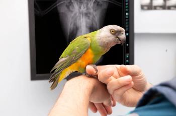
Photo 1: Indirect Doppler blood pressure technique.
It could occur as a consequence of acute or chronic hypertension both in humans and animals. In cats, the most common reason for systemic hypertension is acute or chronic renal disease. Authors [MP Littman: Spontaneous systemic hypertension, J Vet Intern Med. 8(2):79-86, 1994] have described the clinical signs in naturally occurring hypertension. Also, HyE is described as a potentially fatal complication of renal transplantations (AE. Kyles et al.: Management of Hypertension Controls Post-operative Neurologic Disorders After Renal Transplantation in Cats, Vet Surg 28:436-441, 1999).
A recent article (C.A. Brown et al.: Hypertensive encephalopathy in cats with reduced renal function, Vet Pathol 42:642-649, 2005) describes the pathophysiology and histopathological findings of HyE in cats with hypertension that followed marked surgical reductions in renal mass with subsequent azotemia. Blood pressures in this study were measured by indwelling arterial transducers.
Advertisement
Macroscopically, the symptomatic cats had signs of global brain edema, such as flattening and widening the cerebral gyri and herniation of the cerebellar vermis through the foramen magnum in one cat. Half the cats had similar changes. There were no macroscopic changes on the brain noted in the other (asymptomatic) cats.
Microscopically, the edema was more pronounced in the white matter, with vacuolization of the neutrophil and myelin separation. Changes in the small blood vessels were characterized further: There was arteriolar hyalinosis (mural hyaline deposits) observed with the presence of neutrophils and macrophages around the affected vessels. According to Dr. Brown's article, this hyaline deposition might precede the destruction of the smooth muscle of the arteriole (fibrinoid vascular necrosis) that is commonly observed in human patients with HyE. Fibrinoid vascular necrosis of the small arteries is a sequella of deposition of plasma proteins — including fibrin — in the vessel wall at a more advanced stage of persistent high blood pressure in humans (D.I Graham, P.L. Lantos: Greenfield's Neuropathology, Vol. 1, ppg. 292-296). Hyperplastic arteriosclerosis was another vascular change that was observed in the symptomatic cats. This abnormality was characterized by concentric hyperplasia of the smooth muscle of the vessel wall, causing thickening of the arteriola, due to the increased intraluminal pressure.
According to the Greenfield's neuropathology, these previously mentioned degenerative arteriolar changes are probably consecutive. The asymptomatic cats did not have arteriolar changes.
It has been suggested that the cerebral arterial blood flow is regulated in a constant level by vasoconstriction of the cerebral arteriole during systemic hypertension and by vasodilation during systemic hypotension. In case of an acute or chronic severe systemic hypertensive event (renal disease, catecholamine release from phaeochromocytom, etc.) there is a forced dilation of the resistance vessels, due to excessive hyperperfusion. The high pressure causes disruption of the blood-brain barrier resulting in multifocal leakage to the extravascular space, progressing to cerebral edema. The constant leakage of the plasma leads to the previously mentioned fibrin deposits and eventually arteriolar degeneration. Veterinarians have long been aware of the ocular lesions leading to blindness, which result from systemic hypertension in cats [F. Maggio et al.: Ocular Lesions Associated with Systemic Hypertension in Cats: 69 cases (1985-1998), JAVMA, Vol 217, No 5: 695-702, 2000].
It is important for the clinician to be aware of the HyE syndrome. In humans, early re-establishment of the normal arterial blood pressure rapidly closes the blood-brain barrier before major structural injury of the blood vessels occurs. Early and appropriate treatment of the hypertension can reverse the neurological symptoms. The veterinary clinician can employ a Doppler technique (Photo 1) to measure blood pressure accurately in feline patients. It would be prudent to routinely monitor this parameter in elderly, azotemic, hyperthyroid or other cats whose clinical signs are nonspecific and vague.
Treatment and control of hypertension before severe neurologic or ocular lesions occur is preferable to the alternative of emergency therapeutic measures in a time of clinical crisis.
Dr. Nanai is a resident of the European College of Veterinary Neurology/Neurosurgery at the Animal Emergency and Referral Center in Fort Pierce, Fla.
Dr. Lyman is a graduate of The Ohio State University College of Veterinary Medicine. He completed a formal internship at the Animal Medical Center in New York City. Lyman is a co-author of chapters in the 2000 editions of Kirk's Current Veterinary Therapy XIII and Quick Reference to Veterinary Medicine.
Newsletter
From exam room tips to practice management insights, get trusted veterinary news delivered straight to your inbox—subscribe to dvm360.
Advertisement
Related Content
Advertisement


Advertisement
Advertisement
Trending on dvm360
1
USDA approves new Anti-IL31 monoclonal antibody injection
2
Q&A: Set nutrition before surgery to improve outcomes
3
Products+Services360: AI-powered predictive pet health tool and other products
4
Wildlife linked to H5N1 spread in Wisconsin dairy farm
5