
|Articles|December 1, 2003
Early foal losses in mares
Author(s)Ed Kane, PhD
A mare is bred. She conceives. A foal is planned for. Tragically in a substantial number of cases, within 40 days the embryo has succumbed to death.
Advertisement
A mare is bred. She conceives. A foal is planned for. Tragically in a substantial number of cases, within 40 days the embryo has succumbed to death.
For every mare bred the hope is for a healthy, thriving foal. When unsuccessful, there are several questions to ask. Discounting the sire effects, early embryonic loss involves the deficiency of the reproductive physiology of the mare. Has the mare been repeatedly infertile or barren? Did the embryo not successfully pass the oviduct to the uterus? Was it a failure of the uterus to accept and maintain the embryo? Was it the failure of the mare's hormonal physiology to maintain her pregnancy?
Thirty-two day pregnancy. This image shows a 32 day pregnancy in an Arab mare.
Though not as acute or seemingly devastating as viral abortion or mare reproductive loss syndrome, "early embryonic death is the biggest cause of reproductive inefficiency in the mare," says Northwest Equine Reproductive Laboratory (NERL) Director Gordon Woods, DVM, Ph.D. According to the NERL Web site, 'although their mothers may have conceived them without trouble, as many as one-third of embryos die in the oviduct by the time they are 4 days old.'
Defining early embryonic loss (EEL)
Early embryonic loss, or pregnancy loss before day 40 of gestation, accounts for most pregnancy losses in mares.
"This loss represents a considerable economic loss to the equine industry in the form of increased costs associated with additional breeding of mares and/or decreased foal production," says Dirk Vanderwall, DVM, University of Idaho, Northwest Equine Reproductive Laboratory.
Data from several studies, both experimental and from the field, show that between day 10 and 40 of gestation, there is a mean incidence of embryonic loss, as detected by ultrasonography, of 7 percent, with a range of 2.5 to 25 percent. In a study of 2,562 pregnancies in Thoroughbred mares in New Zealand, 76.5 percent of all detected pregnancy losses occurred before day 49.
In recent years, it is ultrasound technology, as early as 10 days post ovulation, that enables veterinarians to more critically examine the status of a mare's pregnancy. It allows earlier, more accurate (98 percent) detection of the embryonic vesicle than previously possible with rectal palpation, and has increased the information available on embryonic development and subsequent embryonic loss.
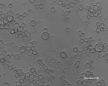
Twins: This ultrasound image shows day 15 twins that are in direct contact with each other.
Embryos lost within 40 days post-ovulation, may be due to the failure of the endocrine system, the embryo, or the uterine environment.
Factors affecting early pregnancy loss
Potential causes include intrinsic, extrinsic and embryonic factors. Intrinsic factors include oviduct, uterus (endometrial disease), endocrine (progesterone, estrogen), maternal age, lactation, and foal-heat breeding. Extrinsic factors include stress, nutrition, season, climate, sire effects, effects of rectal palpation or ultrasonography, and iatrogenic factors.
Embryonic factors include chromosomal anomalies and immunogenetic influences.
The environment and function of the oviduct may play an important role in embryonic loss.
Intrinsic factors-oviductal environment
In the mare, transport of the embryo through the oviduct (five to six days) to the uterus is delayed compared to other species. The embryo is in the late morula or early blastocyst stage by the time it reaches the uterus. The mare is also unique because of the retention of unfertilized oocytes within the oviduct. Early embryos are often transported past unfertilized oocytes that are retained at the ampullar-isthmic junction of the oviduct in mares.
Failure of this mechanism responsible for the selective passage of embryos through the oviduct is a possible factor for embryonic loss. In a study looking at oviduct transport though, only one case of complete oviduct blockage was noted in 2,622 mares.
Uterine environment-periglandular fibrosis and endometritis
Advertisement
Uterine secretions are critical to the survival and the development of the embryo. Proteins and phosphatase, responsible for acidity of the uterus, are dependent on progesterone secretion to help maintain pregnancy.
More important to early embryonic loss are histopathologic changes of the endometrium. Studies have indicated reduced fertility and higher incidence of embryonic and early fetal loss in mares with periglandular fibrosis or endometritis.
Periglandular fibrosis or scarring of the endometrium is a common cause of early fetal loss in mares between 40 and 90 days of gestation, with a relationship to early foal losses prior to 40 days. Fibrosis, essentially chokes-off the endometrial glands, preventing adequate nutritional support of the embryo and decreases the likelihood that a particular mare can carry a foal to term.
The severity of fibrosis is a function of the mare's age, the number foals she has produced over her lifetime, and the level of uterine infection she has been exposed to. Older mares are more likely to have scarring and other uterine changes due to previous pregnancies, though the uterine scarring, per se, may not be a primary underlying cause of early embryonic death, though severe fibrosis has been shown to increase embryonic mortality.
Endometritis may result in the loss of the embryo in three possible ways.
First, an increased number of inflammatory cells in the uterus, initiated by infection, may cause destruction of the embryo.
Second, the organism causing the infection (usually a bacteria) may have a direct effect on the embryo.
Third, uterine irritation-caused infection may cause the mare to return to heat, through the release of prostaglandin, which affects progesterone secretion and therefore maintenance of pregnancy.
Endometritis or uterine inflammation often follows breeding. Mares are somewhat unique among domestic species in that semen is deposited directly into the uterus during natural or artificial insemination. Breeding therefore introduces contamination into the uterus. Mares undergo a natural period (36-48 hours) that allows the uterus to clear itself of excess sperm and contaminants, restoring sterility prior to the arrival of the embryo from the oviduct, four to five days later.
The sperm and contaminants trigger the immune system, which initiates a cascade of events, drawing white blood cells into the uterus, beginning the clearing process. Therefore, in the normal, healthy mare the clearing process will negate the effects of endometritis, and allow for a healthy uterus prior to the arrival of the embryo.
"We know a lot more today about the cascade response of the endometrium and its contamination in terms of the underlying causes of residual or post-breeding endometritis," says Barry Ball, DVM, Ph.D., University of California, Davis. "Older mares that have degenerative changes in the uterus seem to have reduced ability to clear, which otherwise is the normal inflammatory response that we see post-breeding. If that lack of clearing persists for several days, then there becomes a concern of its effect on the conceptus or the embryo when it arrives in the uterus."
The question is the relative importance, suggests Ball.
"My feeling is that there is not much data from the standpoint of causation regarding endometritis. Defects in the oocyte may have a bigger impact, particularly in older mares, though older mares also have a higher incidence in post-breeding endometritis as well. Sometimes it is hard to separate those factors."
Progesterone is required for survival of the equine embryo. Progesterone released by the healthy corpus luteum of the ovary maintains the calm state of the uterus during pregnancy.
Progesterone deficiency
Low levels of progesterone have been associated with embryonic loss.
"Although reduced progesterone concentrations have been implicated in spontaneous embryonic loss in mares, the cause-and-effect relationship between reduced progesterone and embryonic loss is not well understood," says Ball.
Before the maternal recognition of pregnancy, which occurs from days 14 to 16 in mares, three mechanisms that may lead to reduced progesterone levels and corresponding embryonic loss have been proposed: (1) uterine-induced, premature luteolysis; (2) primary luteal insufficiency; and (3) failure of the conceptus to block luteolysis. Premature luteolysis, the early dissolution of the corpus luteum, can be induced by endometrial irritants, such as endometritis. Primary luteal insufficiency, which results from decreased progesterone production by the mid-cycle corpus luteum, has been associated with short interovulatory intervals and decreased progesterone levels at day 12. The presence of the conceptus in the uterus prevents luteolysis and maintains progesterone secretion from the corpus luteum by blocking or reducing prostaglandin F2a secretion by the endometrium.
The early equine conceptus is mobile within the uterus from the time of first detection with ultrasonography until day 15 or 16. This mobility has been proposed to be necessary for luteal maintenance and the recognition of pregnancy. As the embryo moves within the uterus it touches the walls of the endometrium, likely sending a receptor trigger blocking prostaglandin release. Failure of the conceptus to block luteolysis because of poor embryo mobility or underdevelopment has been proposed as a cause of embryonic loss.
There is a decline in fertility with increased mare age.
Maternal age
Some of this decrease is due to a proportion of aged mares not detected pregnant after breeding, although much of the increase is due to a higher incidence of pregnancy loss with increased maternal age. Though no critical age for increased pregnancy loss has been identified, increased loss has been noted in mares greater than 13 years of age.
"Increased pregnancy loss is also paradoxically seen in very young mares (11-16 months of age)," says Ball. "Mares appear to be most susceptible to pregnancy loss when they are very young or very old."
"Embryonic loss rates are much higher in aged mares," adds Vanderwall.
Ball also notes, "Clearly, as the mare gets older, the rate of embryo losses goes up."
Early embryonic loss needs to be differentiated from fertilization failure, which is essentially the same for young and older mares. 'The cumulative incidence of embryonic loss between fertilization and day 40 may be as low as 20 percent in young mares to more than 70 percent in aged mares. Under field conditions, the detected incidence of embryonic loss between days 14 and 40 is about 10-15 percent in young mares, and 25-30 percent for aged mares, greater than 18 years of age.
In aged, subfertile mares embryonic losses may occur very early prior to detection with ultrasonography.
When monitored day two to day 14, embryonic loss was 62-73 percent in subfertile mares compared to 0-9 percent in normal mares. In cases where embryos were recovered early (from day seven to day 10) significantly fewer embryos were recovered from subfertile (commonly barren) mares than from normal mares.
From studies at the Northwest Equine Reproduction Laboratory, it was determined that early embryonic loss in aged mares was the failure of the embryo.
Woods transferred embryos from older to younger mares. They did not survive. The conclusion was that of potentially 'bad eggs', not the health of the older uterus, or the endocrine system, but the embryos themselves.
"The age-related component to increased embryonic loss in mares, which is very similar to what is seen in women, seems to be associated with decline in oocyte quality or defective oocytes," Ball explains.
"There is not a lot of information on exactly what the defects are at the cellular level, and how they occur," Vanderwall says. "Carnevale did one study using microscopy. She did see some differences morphologically between oocytes from older mares compared to oocytes from young ones. She was not able to determine where the structural changes were in the oocytes from the older mares, or whether the structural changes she saw were directly related to the lower quality of those eggs from those older mares. She wasn't able to draw a line between those two. That remains to be determined. She saw differences between the eggs of young and old mares, but whether or not those differences are really the cause for lower quality of the eggs from old mares has not been determined.
"Another theory of why aged mare eggs are less viable is at the chromosomal level. Factors considered are either gross chromosomal abnormalities, missing chromosomes, extra chromosomes, or potentially defects at the DNA molecular level. There is no confirming data in the horse to document that there are chromosomal problems that are the underlying problem in those old mare eggs."
There is increasing data that in these older mares as those embryos are developing, they may have a delay in their embryonic development.
"That has direct implications for things like embryo transfer," Vanderwall suggests. "For years we would repeatedly flush the embryos from the mare's uterus on day seven after ovulation. The day seven embryo of the older mare was possibly defective and delayed in development. It's possible that although according to the calendar she was seven days post ovulation, her embryo biologically was a day or two delayed in its development, and therefore did not result in a viable pregnancy due to its defective condition.
"From the standpoint of overcoming fertility problems in these older mares, although the oocyte quality is a big issue in these aged mares, it is also possible that the oviduct plays a role in the embryo's demise. For some mares, and often it's the older mares, they've got abnormalities of the oviduct (or the uterus), that precludes the mare either herself from getting pregnant and maintaining the pregnancy, or even precludes the ability to flush an embryo out for transfer."
Dr. Kane is a freelance writer for equine topics and senior nutritionist with Stuart Products, Inc., Bedford, Texas. He holds a Ph.D in equine physiology and nutrition from the University of Kentucky.
Newsletter
From exam room tips to practice management insights, get trusted veterinary news delivered straight to your inbox—subscribe to dvm360.
Advertisement
Related Content
Advertisement


Advertisement
Advertisement
Trending on dvm360
1
Veterinary relief organization earns NOMV certification for mental wellness
2
Extracurriculars at VMX focus on wellness, giving back and entertainment
3
Researchers create canine cognitive decline guidelines
4
Researchers are working to learn more about equine herpesvirus
5