

|Articles|April 1, 2009
Cuterebriasis in cats (Proceedings)
Author(s)Dwight Bowman, MS, PhD
There are 34 species of Cuterebra in North America.
Advertisement
There are 34 species of Cuterebra in North America. There are related genera that parasitize rodents in South America. One genus, Alouttamyia, is found in the necks of howler monkeys and has been reported on several occasions from humans. None of these genera from South America have yet been reported from cats.
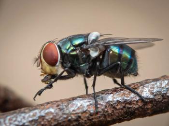
The adults flies are about the size and shape of bumble bees, and the fully formed bots have to be large enough to contain the fully developed fly. Thus, the bots are also very large. Adult Cuterebra spp. live only a few weeks, just long enough to mate and lay eggs. Male botflies find and defend lek territories in areas of congregation. When virgin females fly into the site, they are they chased by a male who will grab her in midair and copulate with her after they land. The mated female then goes off to lay her eggs; the male function is complete – they never feed nor bother a rodent. The female lays egg, some species on grass stems, wood chips, and bark along narrow trails or rodent runs near the opening to the rodent burrow, other enter the burrow and lay their eggs inside. The larvae develop in the egg and hatch in response to the temperature increase and motion associated with a passing host. They then jump onto the host.
Newly hatched larvae are moist and stick to the fur of passing animal; the larvae then crawl into a body opening. The larvae cannot enter unbroken skin. In the host, the larvae remain as first stage maggots within the nasopharyngeal region for 6 to 8 days, then they migrate to the area of the trachea, through the tracheal wall into the thoracic cavity, through the diaphragm into the abdominal cavity, and then to their subcutaneous locations. Once within the subcutaneous site, the larvae molt and continue to grow within the developing warble. Larval maturation from infection to when the larva leaves the warble, ranges from slightly less than 3 to over 8 weeks; rodent bots mature in three to four weeks and rabbit bots in 4 to 6 weeks. The third-stage larva leaves the host, burrows into the soil, where it stays for months and years. In areas with winters, the flies all emerge in the spring, in other parts of North America, they are active all year.
Cats (and sometimes dogs) are infected with these maggots in a manner that causes significant and sometimes fatal disease. The typical presentation in the cat without signs is a maggot developing within a warble within the skin on the cheek, back, or some other location on the body. Cats most likely are infected as they prowl in areas frequented by rodents and rabbits. It is possible for very young kittens to be infected with larvae crawling about on the queens fur. It is not known how the larvae enter the cat, but it would be assumed that they enter through the mouth, nose, or anus as they do in the rodent and lagomorph hosts. The maggots migrate around in the viscera of the cat and then make their way to the warble in the skin where they undergo the majority of their growth. It is important to realize that the maggots do not enter the host at the site of warble development, because unfortunately, in some cats, the migrating larvae get lost on their way to the skin, and it is these larvae that cause severe disease in the affected cat.
Signs in infected cats depend on where the larvae migrate within the body. Their migration through the lungs or upper respiratory system causes respiratory distress. Maggots have been reported from the globe and the anterior chamber of the eye. The maggots sometimes enter the nervous system causing severe acute neurological disease with signs consisting of anything from changes in behavior to blindness, paralysis, or sudden death. Typically, neurological signs develop in 1 to 2 weeks, but sometimes up to 4 to 10 weeks, after respiratory signs. At necropsy of cats dying with neurologic signs, maggots may be found intracranially or within the spinal cord.
Cuterebra maggots also occur on occasion in humans. Baird et al reviewed 55 cases of human Cuterebra myiasis. Most cases were subcutaneous abscesses or opthalmomyiasis, and all were infected with only a single larva. Three cases were tracheopulmonary myiasis with only one showing complete development of the maggot. Moreover, four patients were described with Cuterebra cutaneous myiasis in New Hampshire in 2004; another case occurred in 2008.
Treatment of cuterebriasis usually requires the surgical extraction of the bot or bots. When mature bots are present within a warble, they will extract themselves very shortly, but they can be assisted in their leaving of the warble by the expansion of the pore and extraction with forceps, using care not to crush the bot. Squashing the larva may result in a severe tissue reaction resulting from a Type I hypersensitivity-like reaction. Smaller larvae in the skin require removal by careful dissection. Larvae within the soft tissues of the mouth, nasal sinus, larynx, or eye also require surgical extraction. For intracranial larvae or larvae in the spinal cord, treatment is likely the extraction of the larva.
Ivermectin has been shown to be effective against Cuterebra species at 0.1 mg/kg, and is well tolerated when given at 0.3 mg/kg. Ivermectin (0.3 mg/kg subcutaneously on alternate days for 3 treatments) in association with corticosteroids has been reported in 2 cats with neurologic signs; in both cats the eventual outcome of the infection was not improved, although the larvae in 1 case at necropsy was decomposing. At Cornell, a limited number of cats with presumptive cuterebriasis associated with an upper respiratory syndrome during late summer and fall have responded to the above regimen of ivermectin treatment (but given orally) combined with prednisone (1 mg/kg, PO, q 12 h for 3 weeks, then 1 mg/kg, PO, q 24 h for 3 weeks).
Advertisement
Related Content
Advertisement


Latest CME


Advertisement
Advertisement
Trending on dvm360
1
PerioVive: Regenerative Support Through Hyaluronic Acid for Every Dental Procedure
2
Weekly Vet Report: First clinical use of TMJ arthroscopy in dogs, Lyme-lepto combo vaccine approval, & more
3
New World screwworm: The flesh-eating parasite returning to the spotlight
4
Veterinary hospice offers cats and clients familiar comforts (Part 1): Familiarize clients with veterinary hospice
5