|Articles|January 1, 2006
Correcting polyuric disorder could put incontinence on hold
Author(s)Johnny D. Hoskins, DVM, PhD, DACVIM
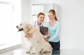
Q. Please review urinary incontinence in dogs.
Advertisement
Q. Please review urinary incontinence in dogs.
A. Dr. Jodi L. Westropp and Dennis J. Chew at the 2005 American College of Veterinary Internal Medicine Forum in Baltimore gave a lecture on urinary incontinence in dogs. Some relevant points in this lecture are provided below.
A thorough history from the owner that presents his/her dog with a micturition problem should be obtained. Differentiations should be made between pollakiuria, polyuria and urinary incontinence because each complaint is handled quite differently. A dog may also present with multiple problems such as urinary incontinence and polyuria. Depending on the underlying cause for the urinary incontinence, correcting the polyuric disorder may lead to significant improvement in urinary incontinence.
Ectopic ureters are the most common underlying cause of urinary incontinence in young dogs and cats. The location of ectopic ureters typically occurs in the urinary bladder-urethra junction, various locations along the urethra, and rarely in the vestibule. Urinary incontinence is the most common clinical sign in dogs with ectopic ureters and is usually diagnosed in dogs before 1 year of age; however, ectopic ureters should be considered in any dog with urinary incontinence. Breeds reported to be at risk include the Golden Retriever, Labrador Retriever, Siberian Husky, Newfoundland and English Bulldog. Although unilateral ectopic ureters have been reported to be more common, bilateral ectopic ureters are significantly more common, which suggests that careful imaging of the urinary tract should be performed before surgery to obtain the best clinical outcome. Ectopic ureters are uncommon in male dogs.
A diagnosis of ectopic ureters can be made by excretory urography, fluoroscopic urethrography, abdominal ultrasound, cystoscopy, helical computed tomography (CT) or a combination of these diagnostic procedures. CT has the advantage of evaluating the entire urinary tract and has 91 percent sensitivity and 100 percent specificity, although this procedure is usually significantly more costly and requires referral. Urine cultures should always be performed in dogs with suspected ectopic ureters because urinary tract infections appear to be quite common with this disorder. Other congenital abnormalities can also occur in dogs with ectopic ureters such as a hypoplastic bladder or urethra, pelvic bladder, ureterocele, renal agenesis or hydroureter. For these reasons, it is essential to evaluate the entire urinary system prior to surgery.
The preferred treatment for dogs with ectopic ureters is surgical correction (success rates vary between 50-75 percent after surgery). The poor success rate could be due to a variety of causes including incorrectly identifying the terminal portion of the ectopic ureter, the presence of multiple ureteral openings, concurrent urethral sphincter mechanism incompetence or a combination of these. Urodynamic testing is recommended prior to surgery because abnormal urethral closure pressures in dogs with ectopic ureters are noted.
Decreased urethral closure pressure can occur due to malformations of the spinal vertebrae (Manx cats), dysautonomia and lumbosacral disorders such as intervertebral disc disease, degenerative myelopathy, diskospondylitis and trauma. A thorough neurologic examination should be performed on all dogs that present for urinary incontinence.
Urethral sphincter mechanism incompetence (USMI) (acquired urinary incontinence occurs mostly after spay) is a diagnosis of exclusion once all other disorders have been ruled out. Urinary incontinence can occur after spay of the female dog and the onset of signs can vary from immediately to 10 years after surgery. Leaking urine while asleep appears to be the most common complaint. Urinary incontinence can be daily or episodic and range from mild to very severe. There appears to be higher risk for larger breed dogs after spay compared to small breeds and certain breeds appear over-represented (German Shepherds, Boxers, Spaniels and Doberman Pinchers). The underlying cause of USMI is unclear and estrogen deficiency is probably not the sole cause of the urinary incontinence because estrogen concentrations are similar between continent anestrous dogs and incontinent spayed dogs.
Studies regarding the timing of neutering are contradictory, but according to the most recent data, early spaying (prior to the first heat cycle) had a reduced incidence of urinary incontinence (9.7 percent vs. 18 percent) in large-breed dogs. However the severity of the urinary incontinence was more pronounced in the early spay group. This relative disadvantage of early spaying is inconsequential when compared with the benefits of reduced incidence of urinary incontinence and protection against mammary tumors. The causes of USMI are probably multifactorial including hormone imbalances, genetics, obesity, age-related changes in urethral musculature and perhaps position of the neck of the urinary bladder.
Advertisement
The diagnosis of USMI can be made based on signalment, history and lack of any other cause of urinary incontinence found on diagnostics. Because many of these dogs are older, it is crucial to evaluate these dogs for polyuria. If another disorder causing polyuria is documented (e.g., hyperadrenocorticism), one should treat the disorder first before suggesting more invasive treatments for the urinary incontinence. A urethral pressure profile is the gold standard and further urodynamics should be considered in refractory dogs. Other neurologic abnormalities pertaining to the urinary bladder such as detrusor instability may occur simultaneously and contribute to urinary incontinence.
Medical intervention
Medical management of USMI includes the use of drugs aimed at improving urethral tone via the alpha-1 adrenoceptors. Phenylpropanolamine (PPA) is currently the drug that results in continence in the most dogs, but this drug needs to be given at least two to three times daily. Clinically, some have had more successful outcomes using the extended release formulation but there are no published results comparing extended release versus standard formulation. The dose can be titrated from 1-2 mg/kg SID-TID. Side effects in dogs include restlessness, anxiety, hypertension and tachycardia. Phenylpropanolamine is not recommended in dogs with cardiac disease or hypertension.
Estrogens may also be used for USMI and these hormones are thought to sensitize the alpha-1 adrenoceptors and indirectly result in an improvement in the closure pressure. Due to the possibility of bone-marrow suppression, the lowest possible dose of estrogen should be used. Diethylstilbestrol (DES) is still commonly used; however, premarin (conjugated estrogen) has been administered with successful resolution of urinary incontinence in many dogs. Bone-marrow suppression has been described in dogs receiving older generation depot estrogens and in those receiving much higher doses of DES. If the dog is still experiencing urinary incontinence while receiving phenylpropanolamine, estrogen can be given concurrently. DES can be prescribed at 0.5-1.0 mg per dog once daily for five to seven days and then decrease the dose to the lowest effective dose, often once or twice per week.
Submucosal urethral collagen injections are now available for animals that are refractory to medications or for owners who do not wish to continually medicate their pets. Dogs are placed under general anesthesia and three to four collagen deposits (Contingen(r), Bard collagen implant) are injected in a circular fashion approximately 1.5 cm distal to the trigone via the cystoscope. Some dogs still require medications after this procedure, but greater continence is usually gained following the implants when drugs were previously ineffective. A second series of implants may be needed to improve continence in some dogs. Twenty-seven of 40 (68 percent) dogs in a recent study were continent for a mean 17 months (one-64 months range). In 10 of 40 dogs, continence was improved following collagen treatments, and in six of these 10 dogs full continence was gained with medication. Some dogs with initial full continence deteriorated after one year. Retreatment with collagen is usually easier and often successful in gaining continence in these dogs. In another recent study, mean continence scores following collagen implant treatment increased considerably over scores while on medication.
Novel treatment
Novel treatment of USMI in ovariectomized dogs has been reported with use of GnRH (gonadotropin releasing hormone) analogs. Luteinizing hormone (LH) and follicle stimulating hormone (FSH) increase dramatically in spayed dogs probably due to a lack of feedback function on the hypothalamic-pituitary system. Activation of FSH and LH receptors in the urethra and urinary bladder may have some effects that increase incontinence in some dogs by mechanisms that are not yet clear. It is theorized that urinary incontinence might be controlled by suppressing FSH and LH. This can be accomplished by administration of GnRH-analogues that will down-regulate the GnRH-receptors in the pituitary gland. In one study, complete continence was gained in seven of 11 dogs following treatment with GnRH analogs for a mean of 247 days (50 to 738 days). The addition of PPA treatment to the remaining four dogs resulted in complete continence for three. All dogs had previously failed conventional medical therapy for USMI. FSH and LH concentrations were increased in these bitches before treatment and were decreased following treatment often to undetectable levels. Estrogen treatments that are effective in regaining continence in about two-thirds of dogs with USMI may exert their effects in a similar way by decreasing FSH and LH.
Urinary retention
Urinary retention can present as urinary incontinence. Mechanical causes for urinary retention include urethroliths, urinary bladder and/or urethral neoplasia, proliferative urethritis, urethral strictures, urethral foreign bodies, urethral plugs in cats, prostatic diseases (abscess, paraprostatic cyst, benign prostatic hypertrophy) and extraluminal compressions. Functional obstructions can be seen with suprasacral or brainstem disease (upper motor neuron bladders), urethral spasms that usually occur secondary to urethritis or a mechanical obstruction, idiopathic detrusor-urethral dyssynergia, and administration of phenylpropanolamine. Urinary bladder dysfunction can lead to detrusor atony and secondary overflow incontinence. Urinary bladder dysfunction can occur secondary to a prolonged obstruction (functional or mechanical), neurogenic causes (sacral cord lesions, pelvic nerve injuries, peripheral neuropathy), pharmacologic agents (anticholinergics, tricyclic antidepressants, opioids) or be idiopathic.
Mechanical obstructions
The diagnosis of overflow incontinence is made based on history, thorough physical and neurologic examination and thorough imaging of the urinary tract. Survey radiographs, cystourethrograms and cystoscopy can all be beneficial to evaluate the dog for mechanical obstructions. An enema should be performed prior to radiographic studies in order to fully evaluate the distal urethra. Cystoscopy is useful to evaluate the urethral mucosa and obtain samples for biopsy and culture. If no mechanical obstructions are present, urodynamic studies can help provide insight for functional obstructions and may even be able to help localize the area of injury.
Treatment for mechanical obstructions is to remove the obstruction if possible or treat the lesion pharmacologically (e.g., piroxicam for neoplastic disorders or proliferative urethritis). Secondary medications may still be warranted if urethral spasticity occurs. Alpha-1 adrenoceptor antagonists (phenoxybenzamine, prazosin) can be used to help relax the internal urethral sphincter. In some cases, such as reflex dyssynergia, a skeletal muscle relaxant such as valium is also beneficial. Once these drugs have taken effect or a urinary catheter is in place, parasympathomimetics can be started. Bethanechol, a muscarinic agent, helps to restore urinary bladder tone and facilitate urinary bladder emptying.
An overactive urinary bladder occasionally results in urinary incontinence, although more commonly it causes pollakiuria. Most often dogs with detrusor hyperreflexia have an underlying cystitis caused by bacteria, cystic calculi, neoplasia, polyps or drugs (cyclophosphamide). Occasionally, idiopathic detrusor hyperreflexia can occur and medical management can be beneficial in controlling signs. A cystometrogram is the gold standard for evaluating urinary bladder function in these dogs when previous diagnostics have not delineated a cause. Spontaneous contractions of the detrusor do occur in some normal dogs and sometimes in dogs with USMI.
In dogs with low urethral tone, spontaneous detrusor muscle contractions result in further incontinence; spontaneous contractions of the detrusor in normal dogs does not result in incontinence due to normal urethral tone. Oxybutynin and flavoxate have resulted in continence in dogs that have failed to improve with PPA alone. Oxybutynin, tolterodine and some newer anticholinergics as well as tricyclic antidepressants (imipramine, clomipramine) have anticholinergic properties that can be considered for treatment of dogs with refractory idiopathic urinary incontinence.
Dr. Hoskins is owner of DocuTech Services. He is a diplomate of the American College of Veterinary Internal Medicine with specialities in small animal pediatrics. He can be reached at (225) 955-3252, fax: (214) 242-2200, or e-mail:
What's your question? Send your pediatric/geriatric related questions to: Pediatric/Geriatric Protocol, DVM Newsmagazine, 7500 Old Oak Blvd., Cleveland, OH 44130. Your questions will be answered by Dr. Hoskins in upcoming columns.
Newsletter
From exam room tips to practice management insights, get trusted veterinary news delivered straight to your inbox—subscribe to dvm360.
Advertisement
Related Content
Advertisement


Advertisement
Advertisement
Trending on dvm360
1
Conference Insider: VMX will celebrate “champions of care” in 2026
2
The 2025 year in review
3
Paws and profits: Small Door Veterinary appoints new chief financial officer, and other updates
4
Q&A: What the 2023 ACVIM update means for leptospirosis vaccination
5