

|Articles|September 1, 2004
Computed tomography angiography
Author(s)Ed Kane, PhD
Eventually it will be possible to do high-quality MRI on standing horses.
Advertisement
In a recent paper discussing magnetic resonance imaging (MRI) in live horses, Susan Dyson, DVM, Center for Equine Studies, Animal Health Trust in Newmarket, United Kingdom, notes, "Foot pain is a common cause of equine lameness, and there have been significant limitations of the methods available for the diagnosis of the causes of foot pain (radiography, nuclear scintigraphy and ultrasonography)."
Radiography is limited to assessment of mineralized tissues, and a 40-percent change in bone density is necessary to identify variations, so subtle abnormalities might be missed. Nuclear scintigraphy is highly sensitive, but false positives are fairly common. Ultrasound is limited by the anatomy of the foot, and interpretation might be confounded by the inability of its application.
In her comparison to these other modalities, Dyson concludes that MRI has several advantages over these other procedures: It is adaptable; it provides three-dimensional images; the entire foot is accessible, and it provides physiological information that is anatomically specific. Additionally MRI provides cross-sectional imaging and high sensitivity for alterations of the actual composition of the soft-tissue structures.
CTA scans are done in about 30 minutes, while most facilities doing MRI are taking between one and two hours. The CTA technique combines the use of X-rays with computerized analysis of the images.
According to Tim Mair, DVM, Bell Equine Veterinary Clinic in Kent, United Kingdom, the high soft-tissue contrast afforded by MRI makes it ideal for assessment of articular cartilage, ligaments, tendons, joint capsules, synovium and bone marrow. Their system was the first one in the world performing MRI on standing horses. In addition to the equipment expense, the limitations of MRI for non-standing, recumbent horses, are lengthy scan times and the concerns of general anesthesia. With MRI, positioning the horse for imaging is labor-intensive, though the image acquisition is not. Also, MRI image analysis is time consuming and requires considerable experience.
Optimization of the equine limb MRI scanner is still in progress, but to date, it has proved capable of producing diagnostic images of the foot and fetlock of standing horses.
"We believe this technique could revolutionize the assessment of certain musculoskeletal lesions of the distal limb," Mair suggests.
The ability to perform MRI in the standing equine patient using the open U-shaped magnet is a considerable advantage over conventional closed magnets that require general anesthesia and fairly lengthy scanning times, up to one to two hours. There are several of these standing-horse units recently available and being used in Europe and the United States. However, the open magnet is, by necessity, of lower field strength, which results in a lower signal-to-noise ratio, which in turn results in lower resolution or longer imaging times for the same resolution as a higher-strength magnet. In addition to permitting use in standing horses, this equipment has advantages to the veterinary profession in terms of lower initial cost and lower operating costs.
Mair concludes, "This system will allow the application of MRI technology in general equine practice, which would be unlikely with higher-field closed magnets because of their inherently higher cost."
Eventually it will be possible to do high-quality MRI on standing horses, but the technology has yet to mature. The diagnostic quality of these systems is not as good as the higher-filtering systems, such as the one at Washington State University.
"Without degrading that system, its just not yet mature enough that it comes close to what we're talking about when we anesthetize a horse and use a high-filtering system," notes Dr. Russell Tucker, DVM, Washington State University (WSU) Teaching Hospital. "The diagnostic quality and resolution are not the same."
At WSU, Tucker has worked extensively with MRI, as well as conventional imaging via ultrasound and radiography. MRI gives a totally different look at the tissues than does conventional radiography or computed tomography (CT).
"When you look at an MRI compared with CT, it's night and day as far as all the structures you get to look at," Tucker says.
They are looking at soft tissue and the osseous damage as indicated by fluid accumulation and by changes of the magnetic properties of the tissue. WSU's MRI, the first of its kind in the world, is designed for human use. Therefore, they have adapted the device to allow horses to go into it while the horses are under general anesthesia.
"It is quite possible to do what we call magnetic resonance angiography, which gives you the same detail of vascularity that CT angiography does," Tucker says.
Advertisement
For most of the vascular studies that WSU does, they don't have to inject contrast material, but solely use the fact that under the magnetic influence flowing blood will have its own magnetic inherent contrast.
"I think it is important to note that with the anesthetized laterally recumbent horse, there is probably a lot of disruption of the normal flow, some introduced flow deficits that may not be truly representative of true lesions," Tucker stresses. "In time, with high-quality MR units, perhaps we will have a more-valid, physiologically true mapping of the vascularity as opposed to horses lying down on their side."
Contrast enhanced MR or CT is not routinely used in the equine. The major advantage of contrast is that it changes a purely morphologic assessment into a functional test.
The Basics of Computed Tomography Angiography
For human patients CT angiography (CTA) is a recently developed technique that uses X-rays to visualize blood flow in arterial vessels throughout the body, from arteries serving the brain to those bringing blood to the lungs, kidneys and the arms and legs. CTA combines the use of X-rays with computerized analysis of the images. Beams of X-rays are passed from a rotating device through the area of interest in the patient's body from several different angles to create cross-sectional images, which then are assembled by computer into a three-dimensional picture of the area being studied.
Dr. Sarah Puchalski, DVM, University of California-Davis, Veterinary Medical Teaching Hospital, and colleagues have developed the CTA technique that allows equine veterinary practitioners to assess the subtleties of foot injury and lameness, to diagnose with accuracy and to then be able to treat these injuries with greater proficiency. The major benefit is that it is a better diagnostic tool for bony abnormalities, though previously no one was using it for soft-tissue injuries.
"We began using CT angiography to evaluate the vasculature of the distal limb after traumatic injuries and realized that contrast enhanced CT could be an alternative to magnetic resonance," Puchalski states.
With contrast enhancement, inflammatory lesions tend to show up very well.
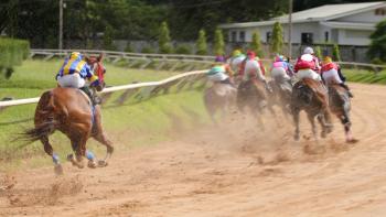
Lameness arising from the heel, previously termed navicular syndrome, is one of the most commonly diagnosed lameness conditions in performance and pleasure horses. The diagnosis is usually non-specific, and treatment programs often are prolonged. Historically, this pain-causing lameness was thought to arise from the navicular bone itself, but it has been shown that a number of different soft-tissue injuries—including deep digital flexor tendonitis, collateral desmitis, navicular suspensory ligament injuries and digital cushion injuries—also possibly are occurring. Current diagnostic techniques, including radiography, ultrasound and nuclear scintigraphy, frequently fail to identify the injured structure. Contrast enhanced CT allows for evaluation of both the soft tissue and bony structures in a cross-sectional manner, eliminating the problems of superimposition of bones and the hoof capsules.
Initially, CTA in the horse was performed to assess the distal limb vasculature. The technique for ultrasound-guided catheterization of the median artery was developed for this purpose. Conversely, the contrast agent in the enhanced CT is delivered directly to the distal limb via the arterial supply. Contrast agents are used in cross-sectional imaging of both MRI and CT to define areas of increased perfusion and altered permeability. This generally allows better margin definition in both neoplastic and inflammatory lesions.
In the equine in general, contrast-enhanced studies are cost and time prohibitive because the dose to an adult horse is between 1 liter and 2 liters. This technique of median-artery catheterization has allowed for selective contrast-enhanced CT studies of the soft tissues of the distal limb without having to administer large quantities of contrast material in a non-specific fashion, such as the jugular vein.
Interestingly, the purpose of the CTA procedure for the median artery catheterization was developed for orthopedic breakdown injuries. After evaluating the images for the vascularity, the hypothesis was raised that horses with soft-tissue injuries to the foot would have altered vascular supply and possibly contrast enhancing lesions in the soft tissues. The technique for guiding the catheter into the median artery with ultrasound was refined further. It is a bit tricky approach, but is doable. You can get it with ultrasound guidance. There is a research group in the College of Veterinary Medicine at the University of California-Davis that does perfusion and permeability studies in conjunction with the biomedical engineering department. They have been developing software to do time density curves and the mathematical computation for measurement of contrast permeability and perfusion.
"We were discussing that and just thought that it would interesting to see what a horse's foot might look like," Puchalski says. "Our initial hypothesis was that soft-tissue structures of the foot will have a repeatable pattern of contrast enhancement and that the anatomy of the foot would be readily recognized on CT. Secondarily, we hypothesized that contrast-enhanced CT will identify specific soft-tissue injuries within the foot by the accumulation of contrast material in inflamed tissues," she explains.
The work to describe the patterns of enhancement and the anatomy of the foot of the horse has been done and they are currently looking at clinical cases.
CT is recognized in diagnostic imaging as the modality of choice for lesions of bone, and an MRI generally is accepted as less optimal for bone lesions. Currently, the protocol for imaging the equine distal limb includes evaluation of the bone structures and the soft-tissue structure, as well as their contrast-enhancing patterns. This procedure is proving to be useful for identifying both lesions of bone and soft tissue.
The CTA procedure
Each horse is anesthetized and positioned in lateral recumbency within the gantry of the CT scanner. The dependent forelimb is the limb that generally is scanned, although they recently have scanned both forelimbs of several clinical cases.
The medial aspect of the carpus is clipped and aseptically prepared for the catheter insertion. A high-resolution linear ultrasound probe is used to identify the median artery, and ultrasonographic guidance is used to place a regular over the needle catheter into the artery. Once the catheter is placed, it is connected via pressure tubing to a remotely controlled pressure injector. Prior to any contrast administration two scout views—amounting to lateral and DP radiographs—are made. The scout views are used as guidance to determine the location for the CT scanner. Cross sectional images of 5-millimeter thickness are then obtained from the mid portion of the first phalanx to the distal extent of P3. This initial study is used to evaluate the bone column and to compare the cross-sectional post-contrast images. The next study is a repeat of the first run of cross sectional images except that the pressure injector is connected to the median artery catheter, and diluted iodinated contrast material is injected for the duration of the second scan. The contrast material will extravasate and accumulate in regions of altered permeability and perfusion secondary to inflammation. A third study is performed, and this study documents the wash-in and wash-out of contrast material in a given area, chosen after an abnormality is identified. Using custom-imaging software, a time-density graph can be developed and compared to the time-density graph of a normal horse for any given area. The graph allows for quantitative comparison of the amount of contrast in the tissues not in the vasculature.
With CTA, an iodinated contrast material shows up as "white" upon X-ray. It attenuates the X-ray beam more than the surrounding soft tissues. The contrast material is infused directly into the arteries. It gives you a localized specific delivery of the contrast agent. If the vasculature is normal, the contrast agent should stay within the vascular space. If there is inflammation, neoplasia or re-vascularization, then an increased number of blood vessels, will cause an increased amount of contrast material to go into the area. The beauty of CT is that it can be measured quantitatively. The amount of X-ray attenuation increases linearly with the amount of contrast material that is present. If it's whiter, then it means there is more contrast material. With each insult or injury-damaged tissue, it is going to be a range from glaringly obvious to a subtle lesion. There is a set of values for how much normal tissue should enhance compared to injured tissue. Normal tissue should not show very much enhancement if at all, while injured tissue would enhance significantly. With CT angiography, there is a quantitative measure called Hounsfield units.
"What we look for is the number of Hounsfield units that the tissue increases," Puchalski explains.
For example, the DDFT, should not increase more than 10 Hounsfield units. With contrast, with injuries, it enhances more than that, from 20-40 units. The nice thing about it is that you can actually measure it, which is very helpful when trying to identify more subtle lesions, i.e. areas of inflammation, tissue edema, and neovascularization, increased blood-vessel recruitment. With injury, not only is there measurably increased vascularization but also an increase in the amount the blood vessels leak, or the vascular permeability. The contrast agent leaks out of the blood vessels into the interstitial space and just sits there. Instead of just washing out with the normal blood flow, it is retained, i.e. leaked into the interstitial space, shown as enhancement.
These procedures would not be possible without helical CT scanning capabilities. The CT gantry is where image acquisition occurs. The gantry is a circular structure where X-ray generator is placed across from a row of X-ray detectors. During the helical scanning procedure, the CT gantry continues to spin and acquires images without resetting itself due to "slip-ring" technology, whereby an electrical connection is maintained without electrical cords. The older machines stopped after a 360-degree rotation and then would have to back-rotate and repeat its movement. With helical scanning, the machine continues through 360 degrees and continues spinning in the same direction. It is useful in two ways. You can advance the table that the patient is resting on as the gantry continues to spin creating a helical image path around the patient. The other thing that you can do is to continue to acquire images within a prescribed amount of time indefinitely. They've taken an image every other second giving a contrast bolus, stopping the infusion of contrast material and watching how it washes out of the tissue. The idea is that in the normal non-injured horse with normal tissue, the contrast material will come in and wash out with the normal vascular flow. As the blood leaves the area, the material will leave the area. In injured tissue, the contrast material just sits there. In a time density curve, you actually can plot Hounsfield units vs. time on a graph and compare your clinical case to graphs from normal horses. The horse that has inflammation or contrast retention in the interstitial space will not wash out in a normal fashion.
Cases of CTA Use
Cases where this technique has shown major benefits usually have a relatively lengthy history of lameness with a consistent blocking pattern of alleviating the lameness with a palmar digital (PD) nerve block. Specifically, they have identified contrast-enhancing lesions of the deep digital flexor tendon (DDFT) and CL of the distal interphalangeal (DIP) joints.
"In my opinion, the use of CTA is really critical for people who are dealing with these foot-sore horses," Puchalski says. There are a few different causes of heel pain in horses. It depends on whose literature you read, but there are definitely some horses that have pain arising from the navicular bone itself, and another subset of horses that have pain arising from soft-tissue injury, true tendonitis or desmitis. It is the DDFT that can exhibit true tendonitis or tendon tearing. Those injuries, as they're below the coronary band and the bulb of the heels, cannot be diagnosed on ultrasound, which would be the typical modality for diagnosing tendon injuries anywhere else. The hoof capsule is in the way, and the probe has to be perpendicular to the tendon fibers in order to get a true image. As the tendon wraps down and around the navicular bone, it changes direction or curves. So it is very difficult to get perpendicular to it while penetrating the hoof capsule. Its anatomy presents a significant problem to properly assess it with ultrasound. The other place that injuries can occur is the collateral ligaments of the DIP, which could tear. Desmitis or actual tearing can occur. That can be seen on ultrasound for a limited length from its origin, but as soon as it goes below the coronary band—which it still has significant length of—it can't be seen on ultrasound, either. Those are two specific areas that are difficult to diagnose by any other modality.
Advice for the Use of CTA for the Future
MRI is very sensitive to these types of foot-tissue injures, but only gives you a morphologic assessment of the tissues, CTA provides a functional test, a quantitative measure and more detailed enhancement. MRI is very useful morphologically for these types of injuries, though you can't do the dynamic studies such as with CT angiography.
CTA is a more-efficient process; scans are done in about 30 minutes, while most facilities doing MR are taking between one and two hours. The actual CT scanning procedure takes about a minute. The balance of the procedure is involved in positioning within the CT scanner and the catheterization. The actual CT scan, acquiring the image takes less than a minute. It's a real key feature of CT vs. MRI, which takes about two hours to acquire an image.
"I think that CT would be a very good investment for a private practice for many reasons," Puchalski recommends.
CT is much more versatile for the head and sinuses, as well as limbs. Helical scanners have been available for a long time in human medicine. The current push in the medical field is to move from single detectors to multiple detectors, so that a lot of helical scanners are becoming available on a secondary or "used equipment market", which is very beneficial for veterinary medicine.
"In my opinion, it is only a matter of time until veterinary clinics will take advantage of this technology," she says. "As a researcher, I would like to do a comparison of MRI and CT in the same animal, and I think someone needs to develop a technique for giving MRI contrast material."
Continuing education is important. Both CT and MR require a significant amount of continuing education. Particularly if many private practices are obtaining MR and CT, then both are very difficult in interpretation. A steep learning curve is necessary to become trained in interpretation as well as use. Veterinarians need to stay on top of continuing education to say that they are doing a good job. At this time, there is a dearth of available resources for the practitioner to become educated. Universities and those skilled with both modalities must provide advanced education to equine practitioners.
The area of the foot, lameness and the navicular bone of the horse is a sight of a very common and serious clinical problem. "The earlier we can detect these injuries, the better off we are going to be as far as treating them," Tucker explains. "We are very excited about the opportunity that we have for a much-earlier and more-reliable diagnostic imaging modality to intervene when there is still an opportunity for success."
Advertisement
Related Content
Advertisement


Latest CME


Advertisement
Advertisement
Trending on dvm360
1
FDA issues emergency use authorization for ivermectin solution to prevent screwworm in horses
2
Every 30 extra minutes under anesthesia raises complication risk in brachycephalic patients, underscoring the importance of peri-anesthetic management
3
From the CVO: I’m off the clock
4
Paws and profits: VetEvolve names first chief veterinary officer, NVA appoints two board members, and more updates
5