

|Articles|November 1, 2014
Colic: The most common equine killer
Author(s)Ed Kane, PhD
With close to a million reported cases annually, equine colic is responsible for more deaths than any other disease group-except old age. Learn how doctors are rapidly assessing and treating this condition.
Advertisement
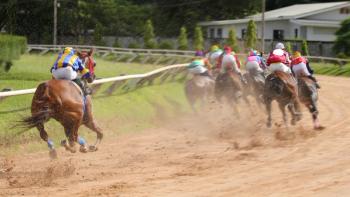
This past May, two-time Horse of the Year Wise Dan underwent emergency colic surgery after routine training at Keeneland racecourse in Lexington, Kentucky. Trainer Charlie LoPresti says the 7-year-old competitor showed signs of abdominal discomfort after his morning run. Racetrack veterinarians attempted to treat the condition medically, but when the horse showed no improvement, the staff transported him to Rood and Riddle Equine Hospital where Scott Hopper, DVM, MS, DACVS, performed surgery. “I don't care if he races again as long as you save his life,'” LoPresti told Hopper.1
Racehorse Wise Dan developed colic after a training run. After undergoing emergency surgery to repair a nephrosplenic and small intestine entrapment, he made his return to racing, winning two races (and counting). (Photo by John Kaiser.)
Nephrosplenic entrapment
Wise Dan had a nephrosplenic entrapment, a condition in which the colon becomes stuck over the band of tissue between the kidney and the spleen. “Most of time, we can resolve nephrosplenic entrapments with a drug called phenylephrine, which shrinks down the spleen. Then we try to reduce the entrapment by jogging or lunging the horse,” Hopper says. Another method of treatment is anesthetizing and “rolling” the horse. “We lunged Wise Dan for awhile, which seemed to reduce the nephrosplenic entrapment, but after he returned to his stall, his pain level escalated rapidly. He had already gone through a few doses of drugs, so we decided to go to surgery.”
It turns out that Wise Dan's colic was different from most cases. Not only did he have a nephrosplenic entrapment, but the small intestine was entrapped as well. “Between the previous process of jogging and lunging him, and the process of getting him onto his back for surgery, his colon was out,” Hopper says. “But we could see that there was still a portion of his small intestine that was entrapped and a bit bruised.” The colic surgery was straightforward and fairly quick, “putting things back where they belong.”
The process involves going through the entire organ system front to back, making sure the intestinal tract is decompressed and put back in its normal anatomical location. “Nephrosplenic entrapments are common,” Hopper says. “We often see these in yearlings, getting ready to go to sale.”
An age-old condition
Equine colic is responsible for more deaths than any other disease group except old age, according to Nathaniel A. White, DVM, MS, DACVS, at the 2005 AAEP Focus Meeting in Quebec City.
Advertisement
A 1998 National Animal Health Monitoring System study looked at the incidence of colic in U.S. horses and the condition's economic impact on more than 1,000 horse operations in 28 states. The annual incidence of colic was 4.2 events per 100 horses. And the cost that year to horse operations (including death and surgical treatment) was estimated at $115 million.2
The USDA Animal and Plant Health Inspection Service reported that thoroughbreds were more likely to develop colic (19.9 per 100 horses per year) than were stock horse breeds, such as quarter horses, paints and Appaloosas (3.5 instances of colic per 100 horses per year). Also, foals were significantly less likely to develop colic than were horses in all other age catagories.2
The UC-Davis 2008 Horse Report noted that despite the progress made in equine medicine during the past 30 years-including significant advances in abdominal surgery, postoperative treatment and intensive care for horses-colic is still considered the most common cause of death in adult horses and accounts for a large proportion of equine emergencies. The report estimates the number of cases at 920,000 annually, with 64,000 horses facing life-threatening problems because of colic.3
Reported equine colic involves gastrointestinal (GI) distension, GI obstruction or blockage, GI obstruction or blockage with partial or complete shutoff of the blood supply, and enteritis, colitis or inflammation of the bowel wall.3 These conditions may produce gas accumulation in the colon or cecum, accompanied by pain and decreased GI motility. The horse also may experience feed impactions in the large or small colon that also produce abdominal pain, with minimal fecal output. Other complications include sand impactions, especially for horses fed on the ground or grazing pastures in sandy areas; enteroliths, usually struvite (magnesium-ammonium-phosphate) or a nidus of metal, plastic or gravel that forms a mass within the gut; colon displacement, usually associated with colon tympany, colon torsion or twisting that can obstruct the GI tract and GI blood flow; and strangulations of the small intestine (often lipomas) or incarcerations of the bowel in the epiploic foramen (the small opening between the liver and pancreas).3
Ultrasound and advanced diagnostics
“Colic is certainly not going to be eliminated from the practice any time soon, so the most we can hope for as clinicians is efficient and minimally invasive diagnostics that help us diagnose and manage this condition as accurately and appropriately as possible,” says Sally Ness, DVM, DACVIM, instructor in equine and farm animal internal medicine at Cornell University's College of Veterinary Medicine. “The abdominal ultrasound has become an essential part of a thorough colic workup. With the availability of portable and affordable machines, the standard of care, even in a field situation, has made ultrasound a key component of colic diagnostics.”
Today clinicians can ultrasound colicky horses in the field, in hospitals and just about everywhere in between. The ability to visualize the location and appearance of the abdominal organs provides a wealth of diagnostic information (Figures 1 and 2).
Figure 1: An ultrasonogram obtained from the left flank of a horse with a nephrosplenic entrapment of the large colon. Note the gas-distended viscus (left side of image) located dorsal to the spleen (right side of image). The bowel is entrapped within the nephrosplenic space and is displacing the spleen off of the body wall.
Figure 2: An ultrasonogram obtained from the right 8th intercostal space of a horse with a diaphragmatic hernia. Note the thickened loop of small intestine (SI) observed adjacent to the lung and dorsal to the diaphragm within the thoracic cavity, as well as the section of large colon (LC) observed ventral to the diaphragm within the abdominal cavity.
“An ultrasound examination offers clues that we can't acquire through any other routine colic diagnostic procedure, short of exploratory laparotomy,” Ness says. “For example, a piece of bowel can herniate across the diaphragm into the thoracic cavity and cause colic. Without ultrasound, it would be difficult to determine that there was bowel in the thorax. With ultrasound, it becomes what we call the ‘five-second diagnosis.' It's not always that easy, but when it is, it results in a rapid assessment, a directed treatment plan and oftentimes a better overall prognosis for the patient. In many cases, ultrasound helps clinicians make an accurate diagnosis before the horse ever gets near the surgery table. This allows us to provide more complete and accurate information to the owners as they consider treatment options. Every colic presents a diagnostic challenge, and we're always trying to figure out the answer before the surgeons have a chance to do it for us in the operating room.”
Ultrasound is not only useful for diagnosing horses with colic, it's also a valuable tool for monitoring their response to medical management or surgical recovery. “We frequently use ultrasound in the postoperative period to monitor GI motility, assess incision integrity and look for sources of fever, such as pneumonia, peritonitis or incisional infection,” Ness says. “As in Wise Dan's case, there are well-described ultrasound findings associated with nephrosplenic entrapments. In those cases, ultrasound can be helpful in monitoring the response to medical management and ultimately making the decision to pursue surgery. Still, not all cases follow the rules. Sometimes you think you know what's going on, and at surgery a case may surprise you.”
Wise Dan back to the races
Soon after his colic surgery, Wise Dan made a steady recovery, and by mid-July he was back posting morning workouts. “It will be great when he runs and wins,” says Hopper. “It will be icing on the cake.”
This past August, Wise Dan returned to racing in the Bernard Baruch Handicap at Saratoga Race Course, a 1 1/16-mile run on the grass. He won by a nose. In October, he won the million-dollar Shadwell Turf Mile at Keeneland-from off the pace by more than a length.
References
1. Novak C. Wise Dan undergoes colic surgery. The Bloodhorse May 16, 2014.
2. USDA APHIS. Info Sheet. Incidence of colic in U.S. horses.
3. Colic: An age-old problem. CEH Horse Report 2008:26(1).
Advertisement
Related Content
Advertisement


Latest CME


Advertisement
Advertisement
Trending on dvm360
1
Veterinary Emergency Team in Texas deploys in response to NWS cases
2
Dermatology health and the microbiome: How they interact for healthy skin from within
3
Beyond the exam room: Pride month in veterinary medicine
4
USDA approves canine vaccination for Lyme disease and leptospirosis
5