

|Articles|June 8, 2016
Clinical Rounds: Canine thyroid carcinoma in 4-year-old American bulldog
Let's head back to the halls of the University of Tennessee's College of Veterinary Medicine to help save this dog with a palpable thyroid mass.
Advertisement
The Clinical Rounds team is from the Department of Small Animal Clinical Sciences, College of Veterinary Medicine, The University of Tennessee, Knoxville, Tennesee.
Clinical Rounds coordinator: Jeanne Larson, DVM, DACVIM (oncology)
Clinical pathology: Lisa Viesselmann, DVM
Radiology: Lee Emery, DVM
Surgery: Brett Darrow, DVM
Anatomic pathology: Caitlin Brown, DVM
Medical oncology: Emily Manor, DVM
Radiation oncology: Isabella Pfeiffer, DMV, DECVIM-CA (oncology), DACVR (radiation oncology)
Jeanne Larson, DVM, DACVIM (oncology)Thyroid tumors are most common in middle-aged to older dogs, and breed predilections have been seen in Golden retrievers, beagles, and Siberian huskies.1 While small, benign thyroid adenomas have been identified in necropsy studies of dogs, nearly all thyroid masses in dogs that are palpable on physical examination are malignant (carcinomas).1 While it is most common to palpate a mass in the ventral neck, ectopic thyroid tumors may also be identified.
Some thyroid carcinomas are well-encapsulated, while others are deeply invasive into underlying structures. These tumors are also associated with a moderate to high metastatic potential, most commonly to local lymph nodes (submandibular, medial retropharyngeal and prescapular) and the lungs. In dogs, most thyroid carcinomas (60%) are nonfunctional. About 30% of dogs with thyroid carcinoma are hypothyroid secondary to destruction of normal thyroid tissue, and 10% of dogs are hyperthyroid.2
CASE PRESENTATION
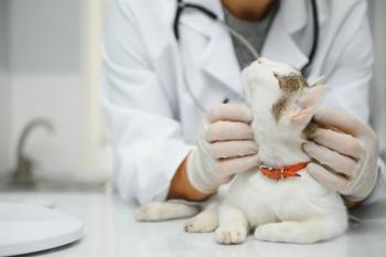
A 4-year-old castrated male American bulldog was referred to the University of Tennessee Veterinary Medical Center Oncology department after the owner palpated a mass in his ventral neck. The dog had no clinical signs and was otherwise healthy. On physical examination, the mass was located subcutaneously, to the left of midline and ventral to the trachea. The mass measured about 3 x 5 x 3 cm and was freely moveable (Figure 1). The submandibular and prescapular lymph nodes palpated small and soft, and no other abnormalities were found on physical examination. A fine-needle aspirate of the mass was performed, and cytology was consistent with neuroendocrine cells, with thyroid gland origin considered most likely based on the location.
Figure 1. A photo of the patient in this case, a 4-year-old castrated male American bulldog with an incidentally found mass in the left ventral neck.
Tumor staging and treatment
A complete blood count, serum chemistry profile and urinalysis were performed, which showed no significant abnormalities. A serum thyroxine (T4) concentration was within the normal reference range. Three-view thoracic radiographs revealed no evidence of pulmonary metastatic disease, but mild right heart enlargement was noted. An echocardiogram showed no structural heart abnormalities.
Because the mass was freely moveable, surgical removal was pursued. Histopathology confirmed thyroid carcinoma with complete excision and evidence of vascular invasion. Postoperative chemotherapy was discussed as a treatment option, and ultimately the owner declined chemotherapy and elected to monitor the dog every three months with a physical examination and three-view thoracic radiographs.
Follow-up
One year later, at a routine recheck, one 5-mm pulmonary nodule was noted on thoracic radiographs. Chemotherapy was again discussed as a potential treatment option, and continued monitoring was pursued. This pulmonary nodule progressed in size slowly, and six months later, two additional nodules were noted. At a recheck several months later (two years from original diagnosis), two masses were palpated in the cervical region, measuring approximately 3 x 4 x 3 cm and 1.5 x 1.5 x 2 cm. These masses were evaluated by ultrasonographic examination of the neck, and fine-needle aspirates were obtained via ultrasound-guidance. Cytology confirmed a neuroendocrine origin.
Additional treatment
Additional treatment options were discussed, including surgical removal of the recurrent masses or radiation therapy to the ventral neck. A computed tomography (CT) scan was performed to further evaluate the area and plan radiation therapy. Palliative radiation therapy (treatment with 8 Gy once weekly for four weeks) was pursued. Transient acute side effects of radiation were observed, including gagging and regurgitation due to suspected laryngitis and esophagitis. These side effects were managed with sucralfate and anti-inflammatories. Hair loss and mildly reddened skin was observed at the radiation site.
At a recheck evaluation two months later, the masses on the neck were no longer palpable. Recheck thoracic radiographs showed no progression of the pulmonary nodules. The patient continues to do well at home, now 2.5 years after his initial diagnosis.
References
1. Wucherer KL, Wilke V. Thyroid cancer in dogs: an update based on 638 cases (1995-2005). J Am Anim Hosp Assoc 2010;46:249-254.
2. Withrow SJ, Vail DM, Page RL. Tumors of the endocrine system. In: Withrow and MacEwan's small animal clinical oncology. 4th ed. St. Louis, Missouri: Elsevier;2007.
Click on the doors below to get six different perspectives on this case.
Advertisement
Related Content
Advertisement


Latest CME


Advertisement
Advertisement
Trending on dvm360
1
New World screwworm tracker: US case count and response update for the week of June 26, 2026
2
Weekly Vet Report: FIP's shorter treatment window, lone star ticks driving red-meat allergies & more
3
What the new AAHA Diabetes Management Guidelines for Cats say about SGLT2 inhibitors
4
What your team won't tell you: Why good intentions aren't enough to understand your practice culture
5