Chronic pain: Decreased activity starts vicious cycle of problems
Q. Please provide educational material about canine chronic pain management for the client that is not commercially prepared.
QPlease provide educational material about canine chronic pain management for the client that is not commercially prepared.
AAt the recent Southwest Veterinary Symposium of 2003 in Fort Worth, Texas, Dr. Darryl L. Millis from University of Tennessee presented an excellent talk on chronic pain management that would be appropriate for the client and is not commercially based. Selected aspects from his presentation follow.
Chronic pain is frequently associated with orthopedic conditions, such as osteoarthritis. Other conditions resulting in chronic pain include neoplasia, neurologic conditions, myopathies and chronic inflammatory conditions.
Animals with chronic painful conditions generally do not display many of the dramatic painful behaviors associated with acute pain. For example, dogs with osteoarthritis may have restricted activity, limited ability to perform, muscle atrophy, pain and discomfort, decreased range of motion and decreased quality of life. Other signs of chronic pain include reduced appetite, licking the area and laying quietly in an area free of other activity.
As animals reduce their activity level because of chronic pain, a vicious cycle of decreased flexibility, joint stiffness, loss of strength and decreased cardiovascular fitness occurs.
Management of dogs with chronic pain should include medication and physical modalities. Medication for chronic painful conditions should reduce pain and discomfort, decrease the severity of clinical signs, maintain an acceptable quality of life, improve strength and fitness, slow the progression of the underlying disease if possible and promote the repair of damaged tissue. Surgical treatment may focus on correcting the underlying condition, or performing a salvage procedure such as a total hip replacement for hip dysplasia or amputation for osteosarcoma.
Management of dogs with chronic pain due to osteoarthritis includes anti-inflammatory and analgesic medications, disease-modifying osteoarthritis agents, weight reduction, low-impact exercise programs and physical modalities, alteration of the environment. The management of chronic osteoarthritis is a lifelong commitment and requires diligent effort and regular follow-up assessments.
Managing pain
Many inflammatory mediators may be involved in chronic painful conditions, including prostaglandins, leukotrienes, interleukins and metalloproteinases. It is possible that nonsteroidal anti-inflammatory agents (NSAIDs) provide benefits to arthritic dogs in several ways. One of the primary modes of action is the reduction of inflammatory mediators, especially prostaglandins, in the peripheral tissues and in the central nervous system. The inflammatory cascade is initiated when cell membrane phospholipids are acted on by phospholipase to produce arachidonic acid. Cyclooxygenase (COX) and lipoxygenase then act on arachidonic acid to produce eicosanoids, such as prostaglandins and leukotrienes.
NSAIDs inhibit the COX enzyme, thereby decreasing the production of inflammatory mediators and reducing pain associated with osteoarthritis. Recently, two forms of the COX enzyme have been identified, COX-1 and COX-2. COX-1 is a constitutive enzyme and is normally produced in relatively constant amounts and has "house-keeping" functions, such as protection of the gastric mucosa, maintaining renal perfusion and production of platelet thromboxane A2. The COX-2 enzyme is inducible and its production increases in response to inflammation.
In addition to the induction of COX-2 in peripheral tissues with inflammation, COX-2 is also induced in the central nervous system. The inhibition of the COX-1 enzyme by NSAIDs is believed to be responsible for adverse side effects, including gastric ulceration, platelet dysfunction and decreased renal perfusion.
Non-selective COX inhibitors inhibit both COX-1 and COX-2 enzymes. The identification of the two COX isoforms has resulted in the development of products that preferentially or selectively inhibit the inducible "bad" COX-2 enzyme while sparing the COX-1 enzyme. Selective inhibition of COX-2 with preservation of COX-1 should reduce the adverse effects associated with the GI tract, kidneys and platelets. The concept of COX-1:COX-2 ratios helps in the understanding of the relative ability to inhibit the various forms of cyclooxygenase. A ratio >1 indicates that the drug inhibits more COX-1 activity than COX-2 activity, while a ratio <1 indicates that the drug inhibits more COX-2 activity than COX-1 activity. Theoretically, fewer side effects should occur with a COX-1:COX-2 ratio >1.
Unfortunately, there are no standard ways of measuring this ratio, and ratios may be significantly affected by the type of in vitro or in vivo test system, the substrates used, and the incubation times and conditions of the test system.
The best guidelines are the clinical efficacy of a particular drug, while maintaining an acceptable level and degree of side effects. NSAIDs that are frequently used include deracoxib, carprofen, etodolac, and aspirin. Phenylbutazone, meclofenamic acid, meloxicam, and piroxicam have also been used, and ketoprofen is approved for use in other countries and has been used in the USA. Acetaminophen with or without codeine is occasionally used in dogs but should not be used in cats.
Deracoxib is a COX-2 specific inhibitor, as defined by the World Health Organization, while carprofen, etodolac, and meloxicam have some preferential selectivity to inhibit COX-2. While no NSAID has been shown to clearly be more efficacious than others in the relief of chronic pain, some dogs apparently have a better response to some drugs as compared with others. Veterinarians should not be reluctant to perform therapeutic trials to evaluate the efficacy of various NSAIDs in a particular animal to determine which will provide the best clinical improvement without side effects. Two-week trials of various NSAIDs with adequate animal evaluation should be performed to determine which medication provides the best response. Before prescribing any medication, the animal's health status, especially liver and kidney function, should be assessed. It is important to educate owners regarding potential side effects.
Slow-acting disease-modifying osteoarthritic agents are substances which are thought to alter the course of osteoarthritis by improving the health of articular cartilage or synovial fluid. Nutraceuticals are nutritional supplements believed to have a positive influence on cartilage health by providing precursors necessary for repair and maintenance. Glucosamine and chondroitin sulfate are routinely combined as disease-modifying agents.
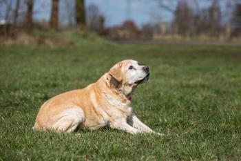
Obesity likely contributes to the progression of osteoarthritis in dogs. In a study of 48 Labrador Retrievers, a control-fed group was allowed food ad libitum, while a limit-fed group was fed 25 percent less than the control-fed group. The hip, shoulder, elbow, and stifle joints monitored over an eight-year period. Radiographic signs of osteoarthritis were significantly less common in the limit-fed group, with osteoarthritis of the hip occurring in 15 of 22 in the control group and in only three of 21 of the dogs in the restricted fed group. Similar trends were apparent in the shoulder joint. Additionally, weight loss results in less joint pain and a decreased need for medication to treat osteoarthritis. Weight reduction of 11-18 percent of the initial body weight of obese dogs resulted in significantly improved hind limb lameness associated with hip osteoarthritis. Another study investigated 16 moderately overweight to obese dogs which were clinically lame from hip dysplasia. Dogs were 113-129 percent of optimal body weight with body condition scores of 6 to 8 out of 9, had radiographic evidence of hip osteoarthritis, and resented hip manipulation. After undergoing a weight loss program with 20 to 60 minutes of daily leash walking, dogs lost 3.9 to 12 kg and body condition scores improved to 4 to 5 out of 9.
In addition to restricting intake of the normal diet and eliminating treats, prescription diets are available that can dramatically assist in achieving and maintaining ideal body weight. The animal's ribs should be easily palpable and there should be a waist when the animal is viewed from above. Exercise is a vital component of weight loss for osteoarthritis treatment. Caloric restriction alone in obese animals results in decreased resting metabolic rate. Acute exercise increases the resting metabolic rate for two to 48 hours. Frequent exercise over an extended period may prevent the reduced resting metabolic rate associated with a diet and caloric restriction. Therapeutic exercise is intended to increase muscle mass. As lean mass increases, the resting metabolic rate increases, making it easier to burn calories. While exercise is very important to weight loss, a low calorie diet is also important for weight loss because a tremendous amount of exercise is required to alter body composition in the absence of caloric restriction. Exercise does not appear to induce an increase in energy intake in obese animals, but it does in non-obese animals. Providing 20 to 60 minutes of daily leash walks will likely benefit overweight animals with diagnosed osteoarthritis.
The benefit of physical modalities for the treatment of dogs with pain secondary to osteoarthritis has been increasingly recognized. Cryotherapy, heat, therapeutic exercise, aquatic exercises, and transcutaneous electrical nerve stimulation all have the potential to help reduce pain in dogs with osteoarthritis. Heat and cold help to temporarily reduce the pain associated with osteoarthritis. Cryotherapy may reduce inflammation and slow conduction of painful stimuli during periods of exacerbation of arthritis, while heat is generally believed to soothe painful areas. In preparation for exercising, warming and stretching affected muscle groups and joints during a "warm-up" period is recommended. This promotes blood flow to the area and collagen extensibility, and decreases pain, muscle spasms, and joint stiffness. Heat is contraindicated if swelling or edema is present in the limb or joint. Heating agents such as moist or dry hot packs, circulating warm water blankets, and warm baths typically heat the skin and subcutaneous tissues to a depth of 1-2 cm. Therapeutic ultrasound results in tissue heating up to 5 cm deep. Ultrasound frequencies of 1 and 3 MHz are typically used. Any stretching should be done during the latter part of warming or immediately after. Therapeutic and aquatic exercise programs in animals with moderate to severe osteoarthritis of multiple joints help to increase function and weightbearing. It is generally believed that low impact exercises in normal dogs do not cause osteoarthritis.
The therapeutic exercise should reduce body weight, increase joint mobility, and reduce joint pain through the use of low-impact weight-bearing exercises designed to strengthen supporting muscles. Muscles act as shock absorbers and strengthening of periarticular muscles may help protect joints. Mild weight-bearing exercise also helps stimulate cartilage metabolism and increases nutrient diffusion. Heavy training programs or exercise in animals with abnormal joint biomechanics may result in changes which predispose to the development of osteoarthritis. Animals should not be forced to exercise during periods of increased lameness because inflammation may be exacerbated. Overloading joints is minimized by performing low-impact activities, such as controlled leash walking, walking on a treadmill, jogging, swimming, and going up and down stairs or ramp inclines.
Exercise should be adjusted to account for the normal variation in lameness associated with osteoarthritis so that there is no increased pain after activity. It is better in the early phases of exercise training to provide three, 10-minute sessions spread throughout the day rather than one, 30-minute session. Avoiding sudden bursts of activity helps prevent acute inflammation of arthritic joints. Swimming and walking in water are some of the best activities for dogs. Training in an underwater treadmill may increase peak weight bearing forces by 5-15 percent. Following exercise, a cool down period is beneficial. A slower paced walk may be initiated for five minutes, followed by ROM and stretching exercises. A cool down massage may help decrease pain, swelling and muscle spasms. Finally, cryotherapy (cold packs or ice wrapped in a towel) may be applied to painful areas for 15-20 minutes to control post-exercise inflammation. The use of transcutaneous electrical nerve stimulations (TENS) may also help reduce pain and lameness associated with osteoarthritis. In one study of dogs with stifle osteoarthritis, a single 30-minute application of TENS resulted in an approximately 5 percent increase in peak weight-bearing forces. The maximal effect persisted for several hours, and dogs remained above baseline weightbearing for 24 hours.
Newsletter
From exam room tips to practice management insights, get trusted veterinary news delivered straight to your inbox—subscribe to dvm360.


