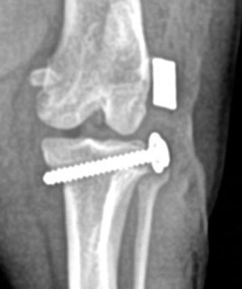
Canine arthroscopy provides better picture for diagnosing joint, orthopedic problems
Arthroscopy used for diagnosis and treatment is the standard of care in man and the horse. Canine arthroscopy has lagged in use. Reasons for this include technical difficulties, cost and perception on behalf of the veterinarian that open arthrotomies are as good. A Japanese surgeon, Dr. Takgi, is credited with early efforts in diagnostic and surgical arthroscopy.
Arthroscopy used for diagnosis and treatment is the standard of care in man and the horse. Canine arthroscopy has lagged in use. Reasons for this include technical difficulties, cost and perception on behalf of the veterinarian that open arthrotomies are as good. A Japanese surgeon, Dr. Takgi, is credited with early efforts in diagnostic and surgical arthroscopy. Today, arthroscopic surgery is the standard of care for many human orthopedic conditions. It is generally recognized that people recover more quickly with reduced morbidity. There are many veterinary orthopedic surgeons who routinely use contemporary arthroscopic equipment in treating many canine and equine orthopedic conditions.
Insertion of CCL after debridement.
Equipment
It is necessary to make a sizeable purchase to begin diagnostic arthroscopy and even more to move into operative techniques. A basic arthroscopy set-up should include a high quality light source [150-300 Xenon or equivalent], fiber optic light cables, an arthroscopy video camera capable of either gas, cold or in some cases steam sterilization. To begin, a smaller diameter rigid telescope 1.9, 2.7, 3.2 mm work well in most canine joints.
Fragmented medial coronoid process.idement.
In combination with the telescope, one needs insertion trocars and cannulae to protect and allow the scope to be handled in a secure way and to facilitate placing the scope in the joint. In efforts to probe the joint while visualized arthroscopic ally, there are a number of probes available for this procedure. We recommend using a cannula and insertion trocar when placing an instrument in the joint. Some surgeons routinely do not use a cannula for instrument insertion and simply pass the instrument in and out of the joint through the skin. Concerns regarding fluid extravasation, loss of joint access due to swelling and distortion of the peri-articular tissues and regional architectural anatomy are reasons we routinely use cannula for passage of diagnostic and operative procedures.
Table 1: Application for Arthroscopic Techniques
With acquisition of basis arthroscopy skills and the transition to operative techniques, the need for more precise and sophisticated arthroscopy equipment is apparent. This would include arthroscopic shavers, automated pumps, radiofrequency tissue ablators, water jet abalators and image capture devices. It is very helpful to capture and record arthroscopic images in still and video format for documentation, future comparison and for client education.
There is a great deal of interest in canine arthroscopy with a number of instructional courses available at national and regional meetings, and through continuing education at many universities. There is strong manufacturer advertising to urge the inclusion of non-invasive techniques in practice and an equally strong consumer-driven demand for it as well.
Preparation of a bone tunnel.
Clinical application
There is a strong perception, driven by mostly human data, that there is less morbidity and an earlier recovery with arthroscopy than conventional open arthrotomies. In a recent publication comparing open cubital joint vs. cubital joint arthroscopy, there was little difference in early post-surgical morbidity and recovery. Arthroscopy offers unparalleled visualization of the joint, allows for earlier intervention and diagnosis, and can be used as a tool for diagnosis, treatment, and to follow or document the progression of orthopedic disease.
Normal meniscus.
The field of non-invasive orthopedic surgery is ever-expanding and many techniques and equipment used in man were either evaluated first in animals or can be used in a similar manner in the dog. Examples include the use of radio frequency tissue ablators, arthroscopic shavers, intra-articular tacks and screws, and intra-articular allografts. Veterinarians who are considering the purchase or use of arthroscopy equipment should carefully reflect on the anticipated financial return and their willingness to begin an almost endless learning curve. Most veterinarians begin by purchasing the basic equipment, consisting of an arthroscope, light source, video camera and monitor, and limited hand instrumentation. This is followed by a period of frustration where they either take an introductory course or undertake self-teaching with a cadaver or learning aid. As their proficiency improves, they are able to gain access to the three most common canine joints: cubital, scapulohumeral and feomorpatellar joints. This is followed by a period where the transition from diagnosis to operative arthroscopy begins.
Grade III osteochondral lesion on the medial humeral condyle.
To make this step, one needs to acquire the necessary hand/eye skills and the tools to facilitate intra-operative surgery. These include: an arthroscopic shaver, radiofrequency unit water jet ablator, laser instrumentation and a fluid pump, as well as more procedure-specific and expensive hand instruments. Table 1, p. 36, outlines the clinically important joints and current arthroscopic techniques used in each joint.
A number of leading arthroscopic equipment manufacturers offer very sophisticated digital capture devices. These allow one not only to capture the details of the procedure, but the images can be stored on a DVD for a permanent record or the digital file e-mailed to others involved in the case. Using contemporary devices, the surgeon can perform surgery and broadcast the images real-time plus have voice interaction with a distance audience.
Suggested Reading
Dr. Taylor is the owner of Alameda East Veterinary Hospital in Denver. He received his veterinary degree from Texas A&M College of Veterinary Medicine. Dr. Taylor has a master's degree in veterinary surgery from Colorado State University College of Veterinary Medicine (CSU). He is a diplomate of the American College of Veterinary Surgeons. Dr. Taylor serves as a clinical affiliate to CSU's veterinary teaching hospital.
Newsletter
From exam room tips to practice management insights, get trusted veterinary news delivered straight to your inbox—subscribe to dvm360.


