

|Articles|February 1, 2005
Beef cattle lameness: diagnostic strategies
Author(s)Christine Navarre, DVM, MS, DACVIM
One of the best ways to evaluate the internal structures of the foot is radiography. It's my opinion that this is one of the most under-utilized tools in bovine lameness evaluations.
Advertisement
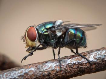
One of the most common problems presented to bovine practitioners is lameness. In many cases the lameness is chronic because it is common for beef producers to treat lameness problems with antimicrobials before presenting the animal to a veterinarian. Antimicrobial therapy usually will cure most cases of simple foot rot, and time will heal many minor injuries. So when a lameness case is presented that already has been treated without success, it is usually a more serious problem. An accurate diagnosis is required to determine a prognosis and make economical treatment or culling decisions.
Most lameness originates from problems in the foot (approximately 95 percent in rear and 99 percent in forelimb). Obviously, if swelling is present in the foot area, then this can be examined first. But because restraint techniques used to examine adult bovine feet (lifting legs in a chute, tilt tables, casting in the field, etc.) can cause further injury to upper limbs if that is the origin of the problem. I prefer to rule out upper-limb lameness before concentrating on the foot if no swelling is present.
Although shoulder and knee injuries are common lay diagnoses, these areas are rarely a problem. Disuse atrophy of muscles of the proximal forelimb from a forefoot problem can make the point of the shoulder more prominent, but this should not be confused with a swelling in this area. Also, the normal slight carpus valgus confirmation of cattle can make the carpus appear swollen at some angles of observation. Careful palpation of the upper forelimb, carpus and fetlock should rule out major problems in the upper limb.
Rear-limb lameness can be more difficult to differentiate. Stifle injuries are the second most common cause of lameness following feet problems. It is very important to diagnose these injuries prior to more involved restraint techniques because struggling of the animal can further damage this joint, turning a mild injury with a fair prognosis into a severe injury with a poor prognosis. It is common for cattle with stifle injuries, particularly cranial cruciate ligament injuries, to walk up on the toe with the heel elevated. Crepitus in the joint may be palpated during walking in gentle animals or with manipulation in a chute. Swelling of the stifle area and/or a drawer sign also are indicative of a stifle injury. Cattle with upper limb injuries are reluctant to kick, so kicking usually eliminates an upper-limb lameness and indicates a foot problem.
Advertisement
Once upper limb problems are ruled out, careful examination of the foot is indicated. Determining if one or multiple limbs are affected is important, as some diseases, such as laminitis, can affect more than one foot at times. Sometimes the effected limb will be abducted, indicating a lateral claw problem, or abducted, indicating a medial claw problem. Some problems are easily diagnosed from visual inspection, but diseases of deep structures can require further diagnostic techniques.
Hoof testers
Hoof testers can be very helpful in determining if lameness originates in the foot and which particular claw is affected. However, at times, responses to hoof testers can be difficult to interpret. Animals might move because they are uncomfortable, especially when restrained on a tilt table, and the movement often coincides with applied pressure of hoof testers. Also, animals that are excited may not respond to hoof testers, even when major lesions, like coffin-bone fractures, are present.
Four-point nerve block
If no abnormalities are found on examination and if no response is elicited with hoof testers, then a nerve block can be performed to completely rule out foot lameness. The four-point nerve block anesthetizes an area from the pastern distally. It might not completely block out the lameness, but if significant improvement is seen, then the foot should be evaluated further with radiographs. To perform the procedure, insert a 20-gauge, 1-inch needle into the dorsal aspect of the pastern, in the groove between the proximal phalanges, just distal to the fetlock. Administer 5 mls of lidocaine deep, and another 5 mls superficially. This injection is repeated on the palmar/plantar aspect of the pastern, just distal to the dewclaws. Next, palpate the nerve over the lateral aspect of the fetlock, approximately 2 cm dorsal and proximal to the dewclaw. Administer 5 mls of lidocaine over the nerve and repeat on the medial side.
Radiography
One of the best ways to evaluate the internal structures of the foot is radiography. It's my opinion that this is one of the most under-utilized tools in bovine lameness evaluations. It often is only performed when systemic anti-microbial therapy alone has failed. Many deep infections require prompt surgical debridement along with anti-microbial therapy for any chance of success. Unsuccessful antimicrobial treatment can waste valuable time and money, worsen the prognosis and remove culling options due to exacerbated weight loss, increased debilitation and antimicrobial residue problems. If significant lesions are recognized early through radiography, prognosis and treatment options can be discussed in a more timely manner. Remember that visible radiographic lesions lag behind clinical signs by approximately two weeks. So in acute cases, repeat radiographs might be necessary. However, an absence of radiographic lesions usually carries a better prognosis, so antimicrobial treatment alone might be warranted. It is worth noting that the cruciate ligaments between the digits can ossify in older, heavy animals. This will appear as smooth, sharp, slightly hook-shaped projections on the axial surface of the digits. Unless they are fractured, or appear rough or lytic, they are considered incidental findings.
Arthrocentesis
Septic arthritis of the coffin joint is a common cause of lameness in cattle. It often occurs as an extension of a Rusterholz ulcer secondary to corkscrew claw in beef cattle. A key finding on physical exam is swelling of the heel region. Radiographs might show widening of the joint space in early cases. A culture of joint fluid is the best way to choose appropriate antibiotic therapy as an adjunct to other treatment options. An 18-gauge, 1-inch needle is inserted just proximal to the coronary band at about a 45-degree angle, just lateral to the extensor process. Fluid should be placed in an EDTA tube for cytology, and sterile media for culture. In my experience, this joint is difficult to tap in cattle, even with excellent restraint. Since the coronary band area is usually swollen and inflamed, joint taps are commonly blood contaminated. Although this will alter cytology results, it should not influence culture results.
Ultrasonography
Occasionally this technique can be helpful in finding pockets of infection, and possibly tendon sheath infections/ tendon injuries. Ultrasonography also is being used to measure sole horn thickness in dairy cattle. Experience is required to interpret some lesions. It is sometimes helpful to compare the effected limb with the normal limb when there is question about the significance of a finding.
Advertisement
Related Content
Advertisement


Latest CME


Advertisement
Advertisement
Trending on dvm360
1
FDA authorizes first generic drug to combat NWS myiasis in pets
2
Weekly Vet Report: 9 Screwworm cases (including a dog), new OTC screwworm treatment authorized, & more
3
ACVIM celebrates award-winning 2026 resident research abstracts
4
dvm360® to honor the Veterinary Heroes™ during awards ceremony at Fetch Kansas City
5