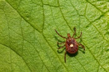
Babesia rapidly emerging parasite in the United States
Signalment: Canine, Basset Hound, 9 weeks old, female, 12 lbs.
Signalment
Canine, Basset Hound, 9 weeks old, female, 12 lbs.
Image 1
Clinical history
The puppy presents for lethargy, sleeping excessively, and pale mucous membranes. One tick has been removed.
Physical examination
The findings include rectal temperature 104.5° F, heart rate 135/min, respiratory rate 40/min, pale mucous membranes, normal capillary refill time, and normal heart and lung sounds. There is an enlarged spleen detected on abdominal palpation.
Laboratory results
A complete blood count and serum chemistry profile were performed and are outlined in Table 1, p. 15S.
Radiographic review:
Survey thoracic and abdominal radiographs were done.
Image 2
My comments:
The thoracic radiographs are normal. The abdominal radiographs show an enlarged spleen.
Ultrasound examination:
Thorough abdominal ultrasonography was performed. The puppy was positioned in dorsal recumbency for the ultrasonography.
My comments:
The liver is enlarged and shows decreased homogeneous texture. No masses noted within the liver parenchyma. The gall bladder is mildly distended, and its walls are not thickened or hyperechoic. The spleen is greatly enlarged and shows inhomogeneous texture - no masses noted. The left and right kidneys are similar in size, shape and echotexture.
Each kidney shows inhomogeneous texture. No masses or calculi were noted in either kidney. The ureter for each kidney is prominent and contains echogenic debris - a normal finding in this age of puppy. The urinary bladder is distended with urine and contains some urine sediment material - no masses or calculi noted. The left and right adrenal glands are similar in size and shape. The stomach, small intestines, and colon are normal. The pancreas shows inhomogeneous texture. One normally can expect to see small amounts of free fluid accumulated within the abdominal cavity of this age of puppy - a completely normal finding.
Image 3
Case management:
In this case, acute babesiosis is the clinical diagnosis. This case presentation is most typical of a puppy with acute babesiosis. Attending veterinarians have to collect blood and look at the blood films well before any whole blood transfusion is done.
Do you think of and do this? The preferred treatment for babesiosis is imidocarb at 6.6 mg/kg subcutaneously or intramuscularly every two weeks for three times and not clindamycin. Because of the low platelet count, I would use steroid therapy for a few days. Otherwise, supportive care and monitoring are needed.
Review on canine babesiosis
An excellent review of canine babesiosis was provided by Dr. Macintire of Auburn University at the last ACVIM Forum - Macintire DK: babesia gibsoni - an emerging disease. Proc ACVIM Forum 20:474-475, 2002. The review follows:
Babesia species are infectious organisms that affect red blood cells of vertebrate hosts. Babesia canis and Babesia gibsoni currently reside in the United States.
These organisms have traditionally been differentiated based on their appearance in stained blood smears. Babesia canis organisms are larger and appear as bilobed piriform organisms that often occur in pairs and are approximately 4-5 um in length. Babesia gibsoni organisms are smaller (1-2.5 um in diameter) and appear as round to oval or ring-shaped organisms, usually single, in red blood cells. Babesia canis is endemic in Europe, southern Africa, Asia and the Americas.
Image 4
Subtypes
There are three subtypes of Babesia canis: B. canis canis, B. canis vogeli and B. canis rossi. These strains differ in virulence, geographic location and tick vector, but are identical in appearance.
In the United States, the most common strain is B. canis vogeli, which is the least pathogenic form. Although severe hemolytic anemia, thrombocytopenia and life-threatening disease have been reported in young dogs, heavily parasitized dogs and dogs transfused with infected blood; most dogs infected with B. canis vogeli in the United States are subclinical carriers.
Although not highly pathogenic, the B. canis vogeli organism appears to be endemic in the southeastern United States, particularly among Greyhounds.
A recent study differentiating the three subspecies of Babesia canis by polymerase chain reaction and restriction enzyme analysis suggests that they may actually be closely related but distinct and separate species. The most pathogenic type, B. canis rossi, is endemic in South Africa. Babesia canis canis is found in Europe and parts of Asia and is considered intermediate in pathogenicity.
Image 5
Small Babesia organisms infecting dogs were first identified as Babesia gibsoni in dogs and jackals from India in 1910. This parasite is now considered endemic among dogs in northern Africa, the Middle East and southern Asia. In 1991, 11 dogs from Southern California were diagnosed with a small Babesia organism and developed severe hemolytic anemia and thrombocytopenia.
Based on morphologic appearance, this California organism was initially identified as Babesia gibsoni. However, recent evidence, based on PCR assays and gene sequencing, has identified at least three different small Babesia organisms that infect dogs.
Table 1
The organism initially identified as Babesia gibsoni has now been shown to be genetically distinct from small Babesia isolates originating from Japan, Sri Lanka and Malaysia. The genetic sequence of the California isolate most closely resembles Theileria species and will likely be renamed in the future.
California isolate
Experimental inoculation of dogs with the California isolate causes acute parasitemia, lethargy, anemia, thrombocytopenia and hemoglobinuria. Histopathologic lesions include diffuse nonsuppurative periportal and centrilobular hepatitis, multifocal necrotizing arteritis, membranoproliferative glomerulonephritis, reactive lymphadenopathy, diffuse erythrophagocytosis and extramedullary hematopoiesis.
In experimentally infected dogs, treatment with diminazine aceturate reduced parasitemia to undetectable levels, but parasitemia rapidly recurred following splenectomy. Parasitemia ranged from 0.9-54.8 percent in these dogs. In naturally infected dogs, parasitemia has been reported to be 5-40 percent.
Cases of naturally infected dogs in California were initially misdiagnosed as immune-mediated hemolytic anemia because the parasites were not recognized on the blood smears of infected dogs and the dogs had positive direct Coombs' tests. Treatment of the California isolate has not been rewarding. Although anti-babesial drugs may reduce circulating parasitemia, no treatment has been found that will eliminate the carrier state. Even if recovered dogs do not succumb to chronic disease, they serve as a potential reservoir for infection of other dogs.
Babesia gibsoni
In 1999, Babesia gibsoni infection was reported in nine dogs from North Carolina. Since that time, infections have been reported in dogs from Oklahoma, Alabama, Georgia, Indiana, Missouri, Wisconsin, Michigan and Florida. Almost all of the reported cases have occurred in American Pit Bull Terriers or American Staffordshire Terriers.
A recent study indicated that the DNA sequences of Babesia isolates from dogs in Oklahoma, North Carolina, Missouri, Indiana and Alabama were identical to the sequences of isolates from dogs in Japan, Malaysia and Sri Lanka but distinct for the California organism.
This small Babesia organism appears to be identical to the original pathogen from Okinawa, Japan, that is endemic in northern Africa, Middle East and southern Asia.
The parasite appears to be a rapidly emerging pathogen in the United States that is currently endemic in the American Pit Bull Terriers in diverse areas of the country east of the Mississippi River. Infected dogs develop regenerative anemia and marked thrombocytopenia within one to three weeks of infection.
Clinical signs included lethargy, fever and pallor or may be mild or inapparent in some dogs. Parasitemia persisted for three to four weeks and then became undetectable as the dogs apparently entered a carrier state.
Not as pathogenic
Initial reports of the Babesia gibsoni organism isolated from dogs in the Midwestern and Southeastern United States seem to indicate that it is not as pathogenic as the California isolate.
Although acute infection is associated with severe anemia and thrombocytopenia, many dogs survive the acute phase and become chronic carriers. A recent study reported that 55 percent of American Pit Bull Terriers tested in Alabama were subclinically infected. Dogs with subclinical infections had a lower hematocrit and platelet count compared to dogs that were negative. The tendency to relapse or exhibit signs of vasculitis, protein-losing nephropathy or hepatic failure that is seen in dogs infected with the California isolate has not been reported in dogs chronically infected with the Midwestern or Southeastern isolates.
Another Babesia species
A third species of small Babesia has been isolated from dogs in Northwest Spain in 2000. This parasite has been provisionally named Theileria annae, and closely resembles Babesia microti, a small Babesia pathogen affecting humans and rodents. All of the infected dogs in Spain exhibited intense regenerative hemolytic anemia, and some also had evidence of renal failure.
Further research is needed to characterize this new species which is genetically distinct from the California isolate and the other small Babesia organism from the United States.
Transmission
Babesia organisms are transmitted to dogs by ticks during feeding. This transmission requires two to three days. Known vectors outside the United States include Haemaphysalis bispinosa and H. longicornis. In the United States, Rhipicephalus sanguineus is the suspected vector. The risk of infection can be reduced for dogs living in endemic areas by providing aggressive tick control with a topical acaricide and flea/tick collar as well as inspecting them daily for ticks.
Transplacental transmission from dam to offspring is thought to occur because Babesia gibsoni has been detected in puppies as young as 3 days old. The incubation period is seven to 21 days. Transmission can also occur through direct blood contamination.
Blood donors should be tested negative for babesiosis. PCR analysis is currently the most sensitive assay for detecting subclinical infections. The cross reactivity between B. canis and B. gibsoni is variable. The PCR test for B. canis is specific and will not detect other species. The PCR test for B. gibsoni, however, will detect a variety of species. Blood contamination can also occur through practices such as sharing needles for vaccinations or reusing surgical instruments for tail docking or ear cropping. The organism can also be transmitted through dog fighting.
Treatment
The only drug approved for definitive treatment of babesiosis in the United States is imidocarb dipropionate (Imizol, Schering-Plough). The recommended dosage is 6.6 mg/kg intramuscularly and repeated in two weeks. Side effects of imidocarb include pain at the injection site, salivation, lacrimation, gastrointestinal signs and tremors. Pre-treatment with atropine (0.04 mg/kg subcutaneously) may prevent these cholinergic side effects. Imidocarb is very effective against B. canis but less effective against B. gibsoni. In many dogs, the degree of parasitemia is markedly reduced within 24 to 48 hours after administration, but three or four treatments at two-week intervals may be required to clear the parasitemia.
Even then, many dogs apparently develop subclinical infections and remain chronic carriers.
Diminazine aceturate is not available in the United States but can be obtained in other countries. A single injection of 3.5 mg/kg intramuscularly will clear parasitemia and improve clinical signs in dogs with B. canis. A higher dosage (5.0-7.5 mg/kg intramuscularly, repeated two to three times at two-week intervals) has been recommended for dogs with Babesia gibsoni.
Other drugs that may be effective against babesiosis include clindamycin and metronidazole. Clindamycin has been used to treat B. microti in people. Metronidazole (25-65 mg/kg daily for 10 days) resulted in clinical improvement in one group of dogs with B. gibsoni. Supportive care is required in dogs with acute or peracute disease.
A blood transfusion or Oxyglobin is indicated in dogs with severe hemolytic anemia. In one study, 85 percent of Babesia-infected dogs had a positive direct Coombs' test and 21 percent exhibited autoagglutination. The use of glucocorticoids in these cases is controversial because immunosuppression may exacerbate the parasitemia.
Use of glucocorticoids is indicated if hemolysis continues to cause severe anemia. Once the parasitemia has cleared, glucocorticoids can be tapered off fairly quickly in most cases. Other supportive measures include fluid therapy to correct dehydration and acidosis and management of concurrent diseases, such as ehrlichiosis and Lyme disease.
Newsletter
From exam room tips to practice management insights, get trusted veterinary news delivered straight to your inbox—subscribe to dvm360.


