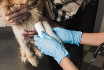
7 rehabilitation techniques to improve outcomes in critical care patients
Several effective physical rehabilitation techniques can be used in critically ill patients.
Critical care patients usually are not thought of as good candidates for physical rehabilitation. However, when rehabilitation techniques are incorporated into a treatment plan, outcomes can improve significantly. Such techniques can be used to control pain, promote healing and manage—and even prevent—complications from pneumonia, atelectasis, embolisms and pressure wounds.
Critically ill patients are often laterally recumbent for extended periods. In these cases, pneumonia, whether it is the primary disease process or secondary to aspiration, can be a fatal complication. These patients are also at risk for atelectasis, particularly if they have been physically restricted or unable to ventilate properly.
In critical care patients, breathing can also be compromised by trauma, pain, abdominal distention, pnuemothorax or hemothorax, pulmonary contusions, sedation from analgesia and severe metabolic derangements. Tidal volume, functional residual capacity and lung compliance may be reduced, causing decreased oxygen saturation and a slowdown of the healing process. Fortunately, patients at risk for these potentially life-threatening respiratory complications can be helped by rehabilitative chest physiotherapy techniques. Here are several effective physical rehabilitation techniques for use in critically ill patients.
1. Encourage movement
Increasing the depth of breathing and stimulating a cough are the best ways to encourage clearing of thick mucoid secretions from the smaller airways. You can't ask an animal to do this, but you can encourage activities that require deeper and more frequent breathing. Both walking and standing exercises can strengthen the thoracic muscles and stimulate sympathetic activity, increasing ciliary motility and decreasing mucous viscosity.
Standing or walking can be beneficial even for patients unable to do so without assistance. Use slings or harnesses that allow for mobility in a safe environment. This activity should mobilize the secretions, stimulating a cough that moves the mucus from the smaller to the larger airways where it can be coughed out or swallowed.
2. Perform chest percussion or coupage
This is an extremely effective technique for stimulating a cough response. Use the percussive force of your hands, cupping and placing them on one side of the patient's chest. Begin at the caudal aspect of the lung fields and move cranially, gently striking the chest wall in a rhythmic fashion. Proper technique is much more important than force, which should be done based on the patient's comfort level. After standing or walking exercises, percussion or coupage generally is most productive.
3. Use vibration
Vibration can help patients mobilize secretions into the larger airways. To perform this technique, lock your arms, and use your hands to vibrate the chest wall as the patient is lying down and exhaling. Perform vibration during four to six consecutive breaths.
Note that both percussion or coupage and vibration are contraindicated in patients with rib fractures, chest tubes, severe chest pain, arrhythmias or platelet counts of < 30,000/µl and should not be done over open wounds. In cases in which you are unsuccessful at stimulating a cough through patient movement, percussion or coupage techniques or vibration, try applying gentle pressure to the area of the third tracheal ring.
4. Position the patient appropriately
Proper positioning of critical care patients is important to optimize oxygen exchange. Alternating right lateral, left lateral and sternal recumbency at least every four hours can decrease both the secretion buildup in the dependent lung field and the possibility of atelectasis. Consistent rotation of the lung fields also decreases the mismatch between the alveolar ventilation and pulmonary blood flow that can occur when the amount of blood in the dependent lung field increases but the lung can't expand enough to deliver well-oxygenated blood to the body.
Note, patients with severely compromised lung fields may be unable to tolerate a specific recumbent position for an extended period. Monitor them closely for increased respiratory rate and effort, and turn them to a position that allows for optimal gas exchange.
Animals that have experienced trauma to the musculoskeletal system from an accident or surgery also can benefit from rehabilitation techniques. Maintain the limbs and joints of these patients in a neutral position to avoid the muscle fiber loss that occurs faster if the muscles are shortened. Provide padding under and in between joints to maintain air circulation and prevent moisture buildup and pressure sores. Turn the patient every four hours, and place padded doughnuts around joints if the skin looks irritated. Employ standing or assisted-standing exercises to improve circulation, neuromuscular strength and proprioception.
5. Use heat and cold therapies
Both can be used effectively in the care of critically ill patients. Thermotherapy (heat) causes vasodilation, thus increasing blood flow and oxygen delivery. It can help heal skin and surface tissues and relax muscles and connective tissues to provide temporary relief from muscle spasms. Heat should be used before massage, stretching or range-of-motion exercises. Never use it during the acute inflammatory stage. Pay extra attention to obtunded or paralyzed patients who cannot react to burns.
Cryotherapy (cold) is useful during the acute inflammatory stage. Cold packs on the affected areas cause vasoconstriction, helping to decrease hemorrhage, cellular metabolite production, histamine production, edema accumulation, cartilage-degrading enzymes and pain. Avoid cryotherapy on areas with decreased circulation or loss of sensation.
6. Try massage
Massage can promote relaxation; increase circulation, lymphatic drainage and flexibility; decrease edema and pain; and enhance proper collagen formation. There are two main types of massage:
- Effleurage is a very light stroking technique that helps loosen the connective tissue and stimulate the lymphatic system and blood flow.
- Petrissage is a deeper massage technique performed with a kneading motion to increase blood flow and oxygen delivery to tissues, and decrease stiffness, spasms and pain.
Massage is contraindicated in patients with fever, cardiovascular or hemodynamic shock or thromboembolic disease as well as over tumors, wounds or fractures.
7. Exercise the patient
Passive range-of-motion (PROM) exercise is a rehabilitation technique in which an external force is used to put a patient's joints through the full movement range and, in turn, stretch the muscles. PROM encourages healing by stimulating the nervous system, reversing the effects of chronic immobility, promoting the health of joint fluid and lining, preventing adhesions and improving soft tissue and muscle flexibility. Don't use force during these exercises; rather, apply pressure only within the patient's comfort zone. PROM must be performed properly to avoid additional damage to the joint and surrounding tissues.
Patients that are immobilized because of their critical status also are at higher risk for life-threatening thromboembolisms. These can develop when blood pools in dependent regions and creates erratic sedentary flow, circulation is compromised due to injury and clotting factors are activated by inflammation. Use rehabilitation techniques to reduce the likelihood of thrombus formation during disseminated intravascular coagulation and to limit the need for heparin therapy. Standing or assisted-standing, massage and PROM can be beneficial in resolving edema and re-establishing normal blood flow.
Conclusion
Rehabilitation techniques can improve outcomes for your critically ill patients and should be incorporated routinely into their treatment plans. Chest physiotherapy techniques, movement, percussion or coupage, vibration, proper body positioning, heat and cold therapies, massage and exercise can be used to manage pain, minimize complications and speed healing. Keep in mind, however, that using these techniques correctly and safely—particularly in critical care patients—requires the training and experience of a certified rehabilitation therapist.
Dr. Van Dyke is the founder and CEO of the Canine Rehabilitation Institute in Wellington, Fla., with locations in Fort Collins, Colo., and Annapolis Junction, Md. Pronko is with the Critical Care Unit at Colorado State University's College of Veterinary Medicine and Biomedical Sciences.
Newsletter
From exam room tips to practice management insights, get trusted veterinary news delivered straight to your inbox—subscribe to dvm360.


