

|Articles|January 1, 2013
21 tips to enhance your crystalluria interpretation
Use this list of dos and don'ts the next time you process a veterinary urine sample.
Advertisement
1 DO remember to distinguish between in vivo and in vitro crystalluria (Table 1).
2 DO remember that crystalluria cannot be reliably inferred from diagnostic urine reagent strips. Crystalluria can be confirmed only by evaluating properly collected and processed urine sediment.
Table 1: Variables influencing crystalluria
3 DO remember that fresh nonrefrigerated urine samples are more likely to reflect in vivo crystalluria.
4 DO recognize refrigeration will preserve many physical, chemical and morphologic properties of urine sediment. However, refrigeration must be used with caution when evaluating crystalluria qualitatively and quantitatively. Although refrigeration of urine samples is likely to enhance the formation of various types of crystals, in vitro crystalluria may have no relationship to events occurring in the urinary tract.
5 DON'T rely on urine samples collected and submitted by clients; unknown variables such as contamination, temperature, length of storage and changes in urine pH may result in formation or dissolution of crystals after sample collection.
6 DO evaluate the conditions of urine preservation after collection when urine contains both struvite and calcium oxalate crystals. This phenomenon could reflect both in vivo and vitro crystal formation. How so? The solubility of calcium oxalate crystals is not influenced by pH changes within the physiologic range. Thus, the calcium oxalate crystals could have formed in vivo under conditions of hypercalciuria associated with acidemia. If an acid urine sample with calcium oxalate crystals becomes contaminated with urease-producing microbes after collection, urine pH may become alkaline, resulting in struvite crystalluria.
Advertisement
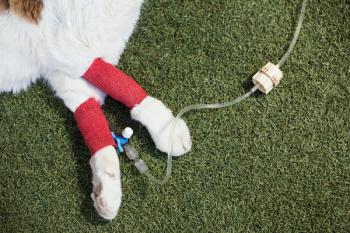
7 DO consider urine pH when interpreting crystalluria because many (but not all ) crystals tend to form and persist in certain pH ranges. Exceptions may be related to large concentrations of lithogenic substances in urine or recent in vivo or in vitro changes in urine pH. A portable pH meter will provide more accurate measurements than most diagnostic pH reagent strips (see photo above).
8 DO measure urine pH at the time of collection and again at the time of urinalysis when more than one hour is expected to elapse between the time of collection and the time of analysis. Differences between the two pH values suggest that in vitro changes have occurred and should be considered when interpreting the significance of crystalluria. This point is especially applicable when mailing urine samples to laboratories.
9 DO determine whether crystals are aggregating together because crystals that aggregate in vivo represent a greater risk for urolith formation than single crystals.
10 DO watch for different sizes of crystals. Why? Because in vivo formation of large crystals indicates that the composition of urine at the time of collection is conducive to crystal growth.
11 DO verify the composition of urine crystals of questionable identity by physical methods of quantitative mineral analysis. Contact diagnostic laboratories for details on how to submit samples.
12 DO consider whether other risk factors for urolithiasis (e.g., breed, sex, age, diet, concomitant illness) are present when interpreting the significance of crystalluria.
13 DO investigate calcium oxalate or urate crystals in fresh urine samples, especially those collected from high-risk patients. These crystals indicate that the urine formed at that time can at least sustain the crystals and may support their formation and growth.
14 DO interpret the quantity of crystals in terms of urine specific gravity values. For example, a few struvite crystals observed in urine with a specific gravity of 1.010 is quantitatively more significant than a few crystals observed in urine with a specific gravity value of 1.060. On the other hand, a concentrated urine sample is usually more conducive to crystal formation than an unconcentrated urine sample.
15 DO consider whether the patient is in a postprandial or fasting state when interpreting the significance of crystalluria. Diet-associated crystalluria is likely enhanced in the postprandial state.
16 DO consider the patient's diet when interpreting the significance of crystalluria because crystalluria may also be influenced by diet, including water intake. Why? Urine crystal formation that occurs while patients are consuming hospital diets may be dissimilar to urine crystal formation that occurs when patients are consuming diets fed at home.
17 DO recognize that crystalluria is not synonymous with the presence of uroliths. Crystalluria is often present in absence of uroliths. Conversely, uroliths can be present without concomitant microscopic crystalluria.
18 DON'T rely on microscopic evaluation of urine crystals as the sole criterion of the mineral composition of uroliths. Only quantitative analysis can provide definitive information about the mineral composition of the entire urolith. However, interpretation of crystalluria along with other clinical findings can often allow you to establish a tentative identification of the mineral composition of uroliths, especially their recently formed outermost layers.
19 DO evaluate the patient for reduction or elimination of crystals by therapy. Absence of crystals provides a useful index of the efficacy of medical protocols designed to dissolve or prevent uroliths.
20 DO repeat the urinalysis when significant crystalluria is detected in animals with otherwise normal urinary tract function. Persistent crystalluria usually represents a greater risk for urolith formation than transient crystalluria.
21 DO avoid "always or never" and "all or none" interpretations regarding the clinical significance of crystalluria.
Advertisement
Related Content
Advertisement


Latest CME


Advertisement
Advertisement
Trending on dvm360
1
Weekly Vet Report: FIP's shorter treatment window, lone star ticks driving red-meat allergies & more
2
What the new AAHA Diabetes Management Guidelines for Cats say about SGLT2 inhibitors
3
New World screwworm tracker: US case count and response update for the week of June 26, 2026
4
Considering safety when treating camels in North America
5