Equine piroplasmosis: What, how and why
What it is, how its transmittedand why this tick-borne disease might be going away.
Entomologist Glen Scoles, PhD, dissects a tick as research leader Donald Knowles, PhD, observes. All photos courtesy of Dr. Glen Scoles.Equine piroplasmosis (EP), a disease of horses and other equids, is caused by one of two protozoan parasites: Theileria equi or Babesia caballi, says Glen Scoles, PhD, a research entomologist with the U.S. Department of Agriculture (USDA) Animal Disease Research Unit (ADRU). These organisms can be transmitted by ticks or through contaminated blood from infected horses, whether transmitted iatrogenically or via blood transfusions.1 Though biologically different, the two parasites share similar pathologies, life cycles and tick vector relationships.
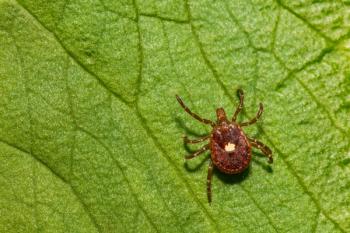
T. equi and B. caballi must undergo sexual-stage development in ticks to complete their life cycle, making ticks the definitive hosts and vectors of the disease-causing parasites. Though relatively few species of ticks can support T. equi and B. caballi, competent tick vectors in the United States include:
- Amblyomma mixtum (the Cayenne tick, formerly known as Amblyomma cajennese) is probably one of the primary U.S. vectors for T. equi.
- Dermacentor variabilis (the American dog tick) transmits T. equi.
- Dermacentor nitens (the tropical horse tick) transmits B. caballi.
- Dermacentor albipictus (the winter tick) transmits B. caballi.
An update on equine piroplasmosis
Since November 2009, nearly 300,000 U.S. horses have been tested for equine piroplasmosis (EP). To date, 262 EP-positive horses (252 Theileria equi-positive and 10 Babesia caballi-positive) have been identified. These infected horses are unrelated to the 2009-2010 T.equi outbreak on a Texas ranch where natural tick-borne transmission was determined to have occurred for at least 20 years.
Of the 262 positive horses identified (most of which were quarter horse racehorses), none showed evidence of tick-borne transmission. The racehorses, specifically, were infected by iatrogenic transmission.
Options for EP-positive horses. All EP-positive horses are placed under state quarantine and owners are offered four options:
permanent quarantine
euthanasia
export from the country
long-term quarantine with enrollment in a U.S. Department of Agriculture (USDA) treatment research program
The USDA program, introduced in February 2013, makes it possible for horses infected with T. equi to have a chance at being released from quarantine. To qualify for release, horses must complete the official treatment program, be proven cleared of the organism by a series of methods over time and test negative on all available diagnostics.
Of the 262 positive horses identified since November 2009, 162 have either died or been euthanized, 18 have been exported and 55 have been enrolled in the treatment research program. Twenty-six of the horses enrolled in the treatment program have met all of the test-negative requirements and have been released from quarantine.
In the case of the Texas ranch outbreak, 163 horses were enrolled in the treatment research program, and more than 140 horses have met all test-negative requirements and are eligible for release.
-Angela M. Pelzel-McCluskey, DVM, USDA Animal and Plant Health Inspection Services, Veterinary Services
Life cycle and transmission of T. equi and B. caballi
T. equi undergoes four stages of development. First, asexual replication occurs in the equine host's peripheral blood mononuclear cells (PBMCs), followed by asexual replication in the host's erythrocytes. Once a tick obtains erythrocytes infected with T. equi during blood feeding, the parasites sexually reproduce in the the tick's midgut, followed by a round of asexual replication in the tick's salivary glands. Sporozoites develop in the tick's salivary glands and are transferred to the horse during the feeding process, thereby infecting it with piroplasmosis.
According to Scoles, infection is transmitted in one of two ways: transtadial (or interstadial) transmission or intrastadial transmission. In the former, larval or nymphal ticks take on infected erythrocytes when they feed on an infected host. The ticks then drop off the equine host, molt, and find and feed on a new host once they reach their next development stage. In intrastadial transmission, an adult male tick feeds on an infected host before moving to another horse to transmit.
The life cycle of B. caballi is similar to that of T. equi in many respects, but there are some fundamental differences, Scoles says. For example, B. caballi does not replicated in the equine host's PBMCs, and it invades the tick's ovaries rather than its salivary glands. B. caballi is therefore transmitted when a female tick that has fed on an infected host lays infected eggs-a process called transovarial transmission. Within the tick embryo, the parasite invades the salivary glands. After the larvae hatch, the parasites develop into sporozoites that are then shed into the saliva during blood feeding to infect naive equines.
Because horses are social animals that tend to cluster together, Scoles says, male ticks, which take blood in smaller amounts than females (females engorge themselves before dropping off to lay eggs), are easily able to move from horse to horse, primarily in search of mates. Once on another horse, they may transmit the infection while feeding.
Signs
According to the USDA, equine piroplasmosis is considered an exotic disease in the United States.2 In its acute form it can present as fever, malaise, reduced appetite, increased pulse rate and respiration, anorexia, constipation followed by diarrhea, tachycardia, petechiae, splenomegaly, thrombocytopenia, and hemolytic anemia leading to hemoglobinuria and icterus-even death in some animals.
Horses are capable of carrying the parasite for long periods without showing any signs of clinical disease, and if competent tick vectors are present, they can acquire and transmit the parasite. Horses infected with T. equi never lose the infection, and while those infected with B. caballi may clear their infection in three to five years, such horses are reservoirs of infection during that period.
Diagnostics
The number of parasites present in infected horses is often too low to detect on blood smears, but there are other methods of diagnosis, Scoles says. For example, infected animals develop an immune response that can be detected using a serological assay. However, the presence of an immune response doesn't necessarily mean the horse is currently infected, Scoles says. It could just mean that the animal was infected and has cleared the infection without a change to its serology results.
That's why Scoles also uses polymerase chain reaction (PCR) assay to detect the presence of a certain parasite DNA sequence. According to Scoles, PCR is a method for chemically amplifying that sequence many times so you can get enough to detect. “PCR confirms the presence of parasite DNA,” Scoles says, “but not necessarily living parasites. However, if the animal is seropositive and positive by PCR, the combined results may confirm that you have an active parasite infection present.”
Support scientist Kathy Mason and technician Ralph Horn check ticks feeding on a horse.Outbreak in Texas
On October 2, 2009, a mare on a ranch in Kleberg County, Texas, showed clinical signs of piroplasmosis, and serologic testing conducted by the USDA detected T. equi antibodies. The remaining 359 horses on the ranch were also tested, and 292 (81%) were found to be seropositive for T. equi on initial screening.3
While previous small outbreaks had been caused by iatrogenic transmission, the high prevalence of infection on the Texas ranch and the high-quality veterinary care the horses had received (suggesting iatrogenic transmission would not be likely) led Scoles and his colleagues to believe that tick-borne transmission was the culprit in this case-a suspicion confirmed by the USDA's epidemiological investigation.
The need for a new treatment. Before the Texas outbreak, treatments for equine piroplasmosis weren't well-documented or validated, and the USDA required infected horses to be euthanized or, in some cases, permanently quarantined. One major treatment hurdle was that the USDA's definition of infection included seropositivity. Thus, a treatment that eliminated the parasite but didn't remove seropositivity would not be considered effective. With so many high-value quarter horses in danger of being euthanized in Texas, researchers explored other possibilities.
One of Scoles' colleagues at the ADRU, Massaro Ueti, PhD, a research veterinary medical officer, was tasked with developing and validating a new treatment option. Ueti and his colleagues came up with a treatment regime that used imidocarb diproprionate, an antiparasitic drug approved for the treatment of canine babesiosis that Scoles and colleagues had used to clear B. caballi in a study several years earlier.4,5
“Ueti and the rest of our team investigated whether we could clear infection in horses with imidocarb diproprionate,” Scoles says. “We demonstrated that infection could be cleared, but the horses remained seropositive for a long time.”
Tick testing. During the height of the Texas outbreak, Scoles says, USDA inspectors from the Cattle Fever Tick Eradication Program were brought in to check all of the horses on the affected Texas ranch for ticks. The inspectors shipped the collected ticks to the USDA's National Veterinary Services Lab for species identification. The lab then sent the ticks to Scoles, who separated them into groups by species and put them onto horses to see if they would transmit disease.
According to Scoles' research, the ranch had two species of competent tick vectors: A. mixtum and D. variabilis. While this was the first report that A. mixtum could transmit T. equi, D. variabilis was already known to be a vector, although Scoles believes it is an inefficient one. “It would take a lot of ticks to cause the levels of infection we saw during the outbreak,” Scoles says.
A third tick species, D. nitens, was also found on the infected horses. Scoles says that while it is known to be a vector of B. caballi, whether or not it can transmit T. equi remains unknown.
Outcomes. The treatment program developed by Ueti and his colleagues saved 163 of the ranch's horses. The Texas Animal Health Commission and USDA worked with the ranch to keep the horses quarantined during treatment. The ranch also agreed to take back many of the infected horses it had sold to people in other states and add them to the quarantine.
Amblyomma mixtum, one of the primary tick vectors for equine piroplasmosis during the Texas outbreakIatrogenic transmission
Not all U.S. outbreaks have been the result of vector-borne transmission, Scoles says: “For example, an outbreak in Florida in 2008 was caused by people transferring red blood cells between racehorses-a practice known as blood-packing (similar to blood-doping in human athletes)-in an effort to make the horses able to run faster.” The outbreak, which occurred in illegal, unsanctioned racing circles, led to the euthanasia of 20 horses.6
Eradicating equine piroplasmosis in the U.S.
Thanks to the efforts of the USDA and other organizations, the risk of equine piroplasmosis transmission by ticks is relatively low right now. Part of the reason the USDA has fought so hard to eradicate the disease has to do with its effect on the American horse industry. “If we can claim that we are ‘EP-free,' we can ship our horses anywhere in the world,” Scoles says.
He adds that prior to the Texas ranch outbreak, most outbreaks could be traced to imported animals. Up until about 2005, serological assay was used to test horses imported to the United States, a practice that yielded a fairly high rate of false negative results and allowed infected horses to enter the United States. Until the 2009 Texas outbreak, most equine piroplasmosis cases in the U.S. could be traced to just such a scenario. In fact, according to the USDA, its epidemiological investigation ultimately determined that the likely source of the Texas ranch outbreak was movement of infected animals in the 1980s or earlier from the ranch's other properties located in South America.
USDA control procedures. State veterinarians and the USDA are responsible for enforcing piroplasmosis control procedures. While Scoles says euthanasia is the simplest solution, some state veterinarians allow for quarantines in which animals are constantly monitored to ensure they are contained in tick-free premises. In fact, in cases detected since 2010, most state veterinarians have supported the long-term quarantine with treatment option for infected horses.
In the Texas outbreak, there was no indication that the ranch was involved in any illegal or unhygienic activity that would have caused the outbreak (unlike the blood-packing incident in Florida in 2008). The ranch was cooperative and allowed USDA inspectors to perform tests to determine the extent of the outbreak. And since the quarter horses were high-value animals, it was incumbent on the state veterinarian to help find a solution to the problem that didn't involve putting down more than 200 valuable horses-a mission in which Scoles, Ueti and their colleagues were highly successful.
References
1. Scoles GA, Ueti MW. Vector ecology of equine piroplasmosis. Annu Rev Entomol 2015;60:561-580.
2. Gauthier J, Harris R, James A et al., USDA, APHIS, VS, CEAH, NCRA. Equine piroplasmosis domestic pathways assessment. Other Publications in Zoonotics and Wildlife Disease 2011, Paper 181.
3. Scoles GA, Hutcheson HJ, Shlater JL, et al. Equine piroplasmosis associated with Amblyomma cajennense ticks, Texas, USA. Emerg Inf Dis 2011;17(10):1903-1905.
4. Ueti MW, Mealey RH, Kappmeyer LS, et al. Re-Emergence of the Apicomplexan Theileria equi in the United States: Elimination of persistent infection and transmission risk. PLoS One 2012;7(9):e44713.
5. Schwint ON, Ueti MW, Palmer GH, et al. Imidocarb dipropionate clears persistent Babesia caballi infection with elimination of transmission potential. Antimicrob Agents Chemther 2009;53(10): 4327-4332.
6. Short MA, Clark CK, Harvey JW, et al. Outbreak of equine piroplasmosis in Florida. J Am Vet Med Assoc 2012;240(5): 588-595.
Ed Kane, PhD, is a researcher and consultant in animal nutrition. He is an author and editor on nutrition, physiology and veterinary medicine with a background in horses, pets and livestock. Kane is based in Seattle, Washington.
Newsletter
From exam room tips to practice management insights, get trusted veterinary news delivered straight to your inbox—subscribe to dvm360.


