
|Articles|January 1, 2011
A challenging case: A dog with nonhealing corneal ulcers
Corneal ulcers usually heal rapidly with appropriate treatment, but this Labrador developed two that were slow to heal.
Advertisement
A 12-year-old 74-lb (33.7-kg) spayed female Labrador retriever was presented to the Colorado State University James L. Voss Veterinary Teaching Hospital for evaluation of a superficial corneal ulcer of the right eye of three weeks' duration. Despite medical therapy by the referring veterinarian, consisting of topical antibiotics of unknown type, atropine, and autogenous serum, the eye had not improved.
The dog had been healthy all its life and had never received any medical treatments. It was current on its vaccinations and heartworm preventive. No blood work or urinalysis had been done within the prior year.
Vital Stats
INITIAL PRESENTATION
A complete ophthalmic examination revealed a raised, pink mass (measuring 4 mm x 7 mm) of granulation-like tissue on the right corneal surface and moderate iris atrophy and nuclear sclerosis in both eyes. Mild conjunctival hyperemia was present in the right eye, and small, superficial corneal blood vessels extended from the lateral limbus to the centrally located mass. The pink mass was adjacent to a corneal area that appeared ulcerated. Fluorescein staining of the cornea confirmed that this area was a superficial corneal ulcer. The results of Schirmer tear tests and tonometry were normal in both eyes. The results of the remainder of the ophthalmic and physical examinations were normal.
Diagnosis
We diagnosed a spontaneous chronic corneal epithelial defect (SCCED) in the right eye with secondary granulation tissue formation. The SCCED diagnosis was based on the fact that the ulcer was superficial and undermined (had a surrounding lip of epithelium that was not attached to the anterior corneal stroma) and was not healing. In addition, we had ruled out other causes of nonhealing ulcers, such as keratoconjunctivitis sicca, persistent foreign bodies, infection, and eyelid anomalies such as distichiasis, ectopic cilia, and entropion.1,2
Treatment
Treatment for the ulcer consisted of applying a topical anesthetic and aggressive removal of the undermined corneal epithelium. This was done by first rubbing off the tissue with a sterile, cotton-tipped applicator followed by superficial débridement of the anterior stroma with a 3.5-mm, low-torque, diamond-tipped, motorized burr.3,4
The dog was discharged with instructions for the owner to administer topical 1% morphine ophthalmic drops (compounded by the veterinary teaching hospital's pharmacy) three times a day in the right eye.5 The morphine was used to decrease the pain that is often present after débridement. A neomycin-polymyxin-gramicidin ophthalmic ointment was also prescribed four times a day in the right eye for infection prophylaxis.
Follow-up
Two weeks later, the right cornea no longer retained fluorescein stain; however, a partially transparent scar remained in the dorsotemporal quadrant. Antibiotics were discontinued, and the defect was considered healed.
SECOND PRESENTATION
The dog was presented again about nine months later for evaluation of a superficial corneal ulcer of the left eye, which had been present for three weeks. The owner suspected that this ulcer had been caused by trauma. The referring veterinarian had treated the ulcer with an unknown topical antibiotic, atropine, and autogenous serum.
1. The left eye of the Labrador retriever in this report at the second presentation. Note the positive fluorescein stain and the ring of loose epithelium surrounding the ulcer margins. A characteristic halo of fluorescein stain is evident at the ulcer margins.
Ophthalmic examination and diagnostic testing
The ophthalmic examination revealed that the dog had moderate iris atrophy and nuclear sclerosis in both eyes. The left eye showed moderate conjunctival hyperemia, mild blepharospasm, and epiphora. A 5-mm-diameter area of fluorescein stain retention was identified on the dorsonasal quadrant of the left cornea (Figure 1). After gentle retraction of the third eyelid, a second 4-mm-x-2-mm area of fluorescein stain retention was located ventronasally (Figure 2). Both areas of ulceration were superficial and appeared to have loose epithelium at their edges. The results of Schirmer tear tests and tonometry were normal in both eyes, and the remainder of the physical examination was unremarkable.
2. The left eye of the Labrador retriever in this report at the second presentation. A second area of ulceration is evident with fluorescein stain at the ventral cornea surface after slightly retracting the third eyelid.
Diagnosis
SCCED of the left eye was diagnosed based on the clinical signs of this and the previous nonhealing chronic, superficial, corneal ulcer with loose corneal epithelial edges and the fact that both ulcers had been refractory to traditional corneal ulcer treatments. In addition, as was done during the previous episode of ulceration, other underlying causes for delayed healing were ruled out by the ophthalmic examination. Further evidence that supported the diagnosis of SCCED in this dog were the characteristic staining patterns, which consisted of poorly attached areas of corneal epithelium at the perimeter of the ulcers, under which the fluorescein stain leaked, leading to a dull-green halo of stain around the brighter-green staining center of the ulcer.1,2,6,7
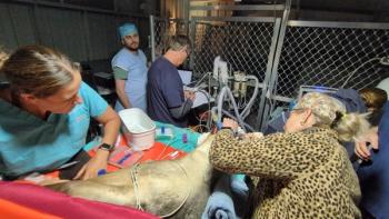
3. The dog in this report undergoing mechanical corneal débridement using a sterile cotton-tipped applicator soaked in dilute povidone-iodine solution.
Treatment
After manually restraining the dog and applying several drops of topical proparacaine to the left cornea, the loose epithelium was mechanically débrided by using a sterile, cotton-tipped applicator soaked in dilute (1:50) povidone-iodine solution in a gentle circular motion (Figure 3). During the débridement, most of the loose epithelium that joined the two areas that had previously retained stain was removed. This procedure appeared to increase the area of corneal ulceration to about half of the corneal surface (Figure 4), but actually, it made it evident that the epithelium between the two apparent ulcers had not been firmly attached epithelium and needed to be removed to allow for proper healing.
4. A spontaneous chronic corneal epithelial defect in the dog in this report after mechanical débridement. The ulcer appears to be much larger because of the removal of nonadherent diseased epithelium.
After débridement, a superficial grid keratotomy was performed by using a 22-ga needle in a checkerboard pattern.8 The dog was sent home with topical 1% morphine ophthalmic drops to be administered three times daily to treat pain,5 neomycin-polymyxin-gramicidin ophthalmic ointment to be administered three times daily, and an Elizabethan collar to be worn until the dog was rechecked in seven days.
One-week recheck
One week later, the left eye had increased periocular tear staining, mild blepharospasm, mildly injected conjunctiva, and a 2-mm-x-4-mm area of positive fluorescein uptake in the dorsal portion of the cornea. The results of the remainder of the ophthalmic examination were normal. The corneal ulcer appeared to be decreasing in size and there was no lip of loose epithelium, so the antibiotic ophthalmic ointment was continued and the topical morphine was discontinued.
Three-week recheck
At a three-week recheck appointment, the left eye still had moderate blepharospasm and mildly injected conjunctiva. The dorsal cornea had a 4-mm-x-1-mm area of positive fluorescein uptake, and a lip of loose epithelium was present around the ulcerated area. The loose epithelium indicated that the ulcer had not healed and, in fact, had again become undermined.
Advertisement
We again applied a topical anesthetic and débrided the ulcer with a sterile cotton-tipped applicator soaked in dilute povidone-iodine solution. This time the 3.5-mm, diamond-tipped motorized burr was applied to the surface of the corneal ulcer to provide additional epithelial débridement and to remove a thin area of the underlying corneal stroma, which would improve the likelihood of good epithelial adherence (Figure 5).4
5. The dog in this report undergoing corneal débridement with a diamond burr to treat a spontaneous chronic corneal epithelial defect in the left eye.
The dog's therapy of topical 1% morphine solution administered three times a day for pain management was reinstituted and the antibiotic ophthalmic ointment was continued. The owner declined placement of a bandage contact lens to protect the cornea and help alleviate pain.
A two-week recheck appointment was scheduled, but the owner instead returned in three weeks.
Follow-up
At the recheck appointment, the dog showed no clinical signs of ocular discomfort. No areas of fluorescein dye uptake were present on either cornea. A small area of granulation tissue was present at the site of the ulcer in the left eye. Medications were discontinued, and no rechecks were scheduled. The client was informed of the potential for the SCCED to recur in either the same or the contralateral eye.
The dog had no further ocular lesions. However, it was euthanized eight months later because of an unrelated disease.
DISCUSSION
Corneal ulcers are one of the most common ophthalmic diseases seen in veterinary private practice. The affected patient may present with a history of suspected trauma to the eye or without any apparent cause. The results of a comprehensive ophthalmic examination as well as the dog's signalment and history are important factors to take into consideration when determining the underlying cause and instituting appropriate therapy. A routine, uncomplicated corneal ulcer generally heals quickly, and thus, an ulcer that does not heal quickly should be further evaluated to determine the underlying cause.
SCCED has also been called indolent ulcer, refractory ulcer, boxer ulcer, persistent corneal erosion, recurrent ulcer, and nonhealing erosion.1,2,6 SCCED is a unique form of superficial corneal ulceration that fails to heal through a normal wound healing process.6,9 This delayed healing time usually lasts more than 14 days.
Clinical signs
Clinical signs of this disorder are those indicative of pain, including blepharospasm, photophobia, and epiphora. An ophthalmic examination usually reveals mild corneal edema in the area of the defect, and a lip or roll of corneal epithelium often surrounds the stained area. Fluorescein staining is positive but may look dull or appear dull around the edges of the defect where the stain dissects under the unattached epithelial lip. Many cases (58% to 64%) of SCCED have some degree of superficial corneal neovascularization, and most SCCEDs are located in the axial or paraxial cornea.7
Signalment and history
SCCEDs occur most commonly in dogs, although they are reported in cats and horses.9 They are more common in middle-aged dogs of either sex. Although they were originally described in Boxers,10 SCCEDs have since been documented to occur in most dog breeds.9 Boxers are still thought to be overrepresented.
Although most of these ulcers occur secondarily to trauma, they may occur spontaneously and without apparent cause.9
Pathophysiology
Healing of an uncomplicated superficial corneal ulcer is rapid, and most heal in three to five days.9 The cornea undergoes healing via epithelial sliding to cover the ulcerated area and mitosis helps restore the abnormally thin corneal epithelium to its normal thickness.6 If the epithelium does not cover the ulcer and adhere to the underlying stroma properly, it becomes indolent.
The pathophysiology of SCCEDs remains unclear. An early ultrastructural investigation of affected corneas suggested that a defect in the epithelial basement membrane with associated basal-cell abnormalities at the ulcer site may prevent corneal reepithelialization.1
Abnormal epithelial structure. More recent studies have used standard techniques such as immunohistochemistry and more sophisticated, quantitative microscopy techniques to examine SCCEDs in dogs. In one study, 48 corneal samples from dogs with SCCEDs taken during therapeutic superficial keratectomies demonstrated that the corneal tissue adjacent to the ulcerated area had epithelium that was not only poorly attached to the underlying stroma but also had abnormal epithelial structure.11 The adhesion complexes between the epithelium and its basement membrane were often deficient or absent in the areas of and surrounding the ulceration.11 Particularly interesting was the presence of an abnormal, acellular zone consisting of collagen fibrils and amorphous material in the superficial corneal stroma of all the samples.11 In samples of normal cornea surrounding the ulcer in these affected dogs, no basement membrane abnormalities were present.11
Acellular barrier. The epithelial changes seen in the SCCED dogs were not seen in dogs that had experimentally induced chronic ulcers.12 The investigators suggested that the acellular zone in the superficial stroma plays an important role in the refractory nature of these ulcers in that it presents a barrier to the reformation of the corneal adhesion complexes and to the formation of normal basement membranes.12 They also concluded that this disease is not analogous to chronic superficial corneal erosions in people, which are thought to be a form of corneal dystrophy.12,13
Substance P. Recent studies have also evaluated the role of substance P in dogs with indolent ulcers. Substance P is a neuropeptide that is found in nearly all canine corneal nerves.7,14 Apparently, a dense abnormal plexus of substance P is present around the periphery of the erosion in dogs with SCCED.7 That suggests that substance P may play a possible, yet unknown, role in the pathophysiology of this disease.7
Treatment
Numerous strategies, including medical and surgical options, have been proposed and investigated to promote the healing of SCCEDs. Conventional, conservative medical treatment consists of the administration of prophylactic topical antibiotics. It is usually ineffective as it fails to remove or ameliorate the cause of the problem.9 Thus, effective therapy must remove the loose epithelium that is not adhering to the stroma, promote the normal adhesion of the new epithelium to the underlying stroma, and protect the delicate, newly forming epithelium.
Débridement. Débridement with a cotton-tipped applicator is an effective way of removing nonattached epithelium. In most dogs, the débriding can be done without sedation if a topical anesthetic is applied. It is important to remove all the loose epithelium even though this procedure generally creates a defect that is much larger than the original ulcer and in some cases the entire corneal surface becomes denuded of epithelium. Normal epithelium cannot be removed with a cotton-tipped applicator, so if the epithelium comes off, it is unhealthy. In the case presented here, we used a cotton-tipped applicator that had been dipped in dilute povidone-iodine solution to kill any organisms that might be adhering to the ulcer.
Keratotomy. Once the ulcer has been débrided, promotion of the adherence of the newly forming epithelium will increase the healing ability of the cornea.9 For example, use of a hypodermic needle to make small punctures (anterior stromal micropuncture, punctate keratotomy) or linear scratches (grid keratotomy) in the anterior stroma has been extensively used to promote adherence,8 and the success rate of these keratotomy techniques is about 80%.9 These methods physically provide a surface in the stroma for epithelial adherence and also increase collagen IV and laminin, molecules that are important in epithelial adhesion.12
Surgical keratectomy. Surgical, superficial keratectomy removes the nonhealing epithelium and anterior stroma more extensively than grid keratotomy does and has been reported to have a 100% success rate, but it requires general anesthetic and a surgical microscope to complete. It often leads to corneal scarring.8,9
Diamond burr ketatectomy. Diamond burr polishing of the anterior stroma is a method of removing loose epithelium and the anterior stroma that has been used in human medicine for several years to treat recurrent corneal erosions.15 The studies of this technique in people show that it is a safe, convenient, inexpensive, and effective method of débridement.16 The diamond burr is being used in dogs as well. A retrospective study comparing the diamond burr superficial keratectomy with two methods of grid keratotomy suggested that the diamond burr method allows for faster healing than either method of grid keratotomy does.4 In addition, the procedure had low complication rates.
We chose to treat the dog in this report with this method after the failure of the grid method in the left eye, and it proved to be noninvasive, easy-to-perform, and effective. The diamond burr was used in the right eye but not used initially in the left eye based solely on ophthalmologist preference.
Corneal protection. Methods of protecting the newly forming epithelium that have been described include bandaging contact lens placement, temporary tarsorrhaphies, and third eyelid flaps. These methods not only protect the cornea but also may help relieve discomfort associated with these procedures.
Tissue glue. A technique involving placement of a thin layer of topical cyanoacrylate tissue glue (Tissuemend II—Veterinary Products Laboratories) over the ulcerated area after débridement has been reported.17 The glue remains in place until the epithelium grows under it at which time it is elevated off the corneal surface. This technique appears to be effective, with healing times that range between two and eight weeks.17
Topical medical therapy. In addition to topical antibiotics that act only to prevent infection of uninfected corneal ulcers but do nothing to heal them, topical medical therapies that promote healing have been investigated. These include polysulfated glycosaminoglycans and aprotinin (which inhibit proteolytic enzymes),18,19 epidermal growth factor,20 substance P,7 and fibronectin.21 Although many of these substances promote healing, none is considered a standard therapy.
A recent study of clinical cases of dogs with SCCEDs demonstrated that topical tetracycline (Terramycin Ophthalmic Ointment—Pfizer Animal Health) significantly shortened healing times compared with a placebo.22 It was hypothesized that increasing the expression of growth factors and signaling targets increased the corneal epithelial cell migration, which promoted healing. The authors concluded that this method would be a safe, effective, and relatively inexpensive way of increasing the ulcers' healing ability.22
The authors' approach. Although much is still unknown about the pathophysiology of SCCEDs and the most effective treatment regimen is under debate, the high rates of healing we have achieved using the diamond burr technique has led us to use this as our first-line therapy (directly after removing the epithelial edges with a cotton-tipped applicator). If this procedure fails after two débridements, we will then use the more invasive grid keratotomy technique once before we resort to performing a superficial keratectomy.
Concurrent with the débridement and diamond burr method, prophylactic topical antibiotics should be administered three to four times a day and the dog should wear an Elizabethan collar at all times to prevent self-trauma until the defect has healed. Even occasional rubbing of the eye may inhibit healing.
Prognosis
With appropriate treatment and lack of complications, SCCEDs should heal in about two weeks. It is reported that about 50% of patients will have a recurrence in the same or opposite eye—as occurred in the dog in this report—so it is important to inform clients about the potential for the indolent ulcer to recur.5 It is also important to inform clients about the delayed healing process and the potential need for multiple treatments.
Kyshia T. Davis, DVM
Juliet R. Gionfriddo, DVM, MS, DACVO
Department of Clinical Sciences
College of Veterinary Medicine and Biomedical Sciences
Colorado State University
Fort Collins, CO 80538
REFERENCES
1. Kirschner SE, Niyo Y, Betts DM. Idiopathic persistent corneal erosions: clinical and pathological findings in 18 dogs. J Am Anim Hosp Assoc 1989;25:84-90.
2. Maggs D. Cornea and sclera. In: Maggs D, Miller P, Ofri R, eds. Slatter's fundamentals of veterinary ophthalmology. 4th ed. St. Louis, Mo: Saunders Elsevier, 2008;175-202.
3. Soong HK, Farjo Q, Meyer RF, et al. Diamond burr superficial keratectomy for recurrent corneal erosions. Br J Ophthalmol 2002;86:296-298.
4. Sila GH, et al. A retrospective evaluation of the diamond burr superficial keratectomy in the treatment of spontaneous chronic corneal erosions in dogs from 2006 to 2008, in Proceedings. 40th Annu Conf Amer Coll Vet Ophthalmol 2009;73.
5. Stiles J, Honda CN, Krohne SG, et al. Effect of topical administration of 1% morphine sulfate solution on signs of pain and corneal wound healing in dogs. Am J Vet Res 2003;64(7):813-818.
6. Bentley E. Spontaneous chronic corneal epithelial defects in dogs: a review. J Am Anim Hosp Assoc 2005;41:158-165.
7. Murphy CJ, Marfurt CF, McDermott A, et al. Spontaneous chronic corneal epithelial defects (SCCED) in dogs: clinical features, innervation, and effect of topical SP, with or without IGF-1. Invest Ophthalmol Vis Sci 2001;42(10):2252-2261.
8. Stanley RG, Hardman C, Johnson BW. Results of grid keratotomy, superficial keratectomy and debridement for the management of persistent corneal ulcers in 92 dogs. Vet Ophthalmol 1998;1:233-238.
9. Bentley E, et al. Diseases and surgery of the canine cornea and sclera. In: Gelatt KN, ed. Veterinary ophthalmology. 4th ed. Ames, Iowa:Blackwell Publishing, 2007;690-752.
10. Gelatt K, Samuelson D. Recurrent corneal erosions and epithelial dystrophy in the Boxer dog. J Am Anim Hosp Assoc 1982;19:453-460.
11. Bentley E, Abrams GA, Covitz D, et al. Morphology and immunohistochemistry of spontaneous chronic corneal defect (SCCED) in dogs. Invest Ophthalmol Vis Sci 2001;42(10):2262-2269.
12. Bentley E, Campbell S, Woo HM, et al. The effect of chronic corneal epithelial debridement on epithelial and stromal morphology in dogs. Invest Ophthalmol Vis Sci 2002;43:2136-2142.
13. Ramamurthi S, Rahman MQ, Dutton GN, et al. Pathogenesis, clinical features and management of recurrent corneal erosions. Eye (Lond) 2006;20:635-644.
14. Marfurt CF, et al. Morphology and neurochemistry of canine corneal innervations. Invest Ophthalmol Vis Sci 2001;42:2242-2251.
15. Aldave AJ, Kamal KM, Vo RC, et al. Epithelial débridement and Bowman's layer polishing for visually significant epithelial irregularity and recurrent corneal erosions. Cornea 2009;28:1085-1090.
16. Wong VW, Chi SC, Lam DS. Diamond burr polishing for recurrent corneal erosions: results from a prospective randomized controlled trial. Cornea 2009;28:152-156.
17. Bromberg NM. Cyanoacrylate tissue adhesive for treatment of refractory corneal ulceration. Vet Ophthalmol 2002;5:55-60.
18. Miller W. Using polysulfated glycosaminoglycan to treat persistent corneal erosions in dogs. Vet Med 1996;91:916-922.
19. Morgan R, Abrams K. A comparison of six differenttherapies for persistent corneal erosions in dogs and cats. Vet Comp Ophthalmol 1994;4:38-43.
20. Kirschner SE, Brazzell RK, Stern ME, et al. The use of topical epidermal growth factor for the treatment of nonhealing corneal erosions in dogs. J Am Anim Hosp Assoc 1991;27:449-452.
21. Ollivier FJ, Brooks DE, Kallberg ME, et al. Evaluation of various compounds to inhibit activity of matrix metalloproteinases in the tear film of horses with ulcerative keratitis. Am J Vet Res 2003;64(9):1081-1087.
22. Chandler HL, Gemensky-Metzler AJ, Bras ID, et al. In vivo effects of adjunctive tetracycline treatment on refractory corneal ulcers in dogs. J Am Vet Med Assoc 2010;237(4):378-386.
Newsletter
From exam room tips to practice management insights, get trusted veterinary news delivered straight to your inbox—subscribe to dvm360.
Advertisement
Related Content
Advertisement


Advertisement
Advertisement
Trending on dvm360
1
Key takeaways from AAHA’s 2026 oncology guidelines
2
Wildlife linked to H5N1 spread in Wisconsin dairy farm
3
Strategic design of geriatric pet programs in veterinary practice: Part 2
4
The Bond Factor: Why stronger connections are driving technology adoption in veterinary care
5