Classifying tooth resorption in cats and dogs
Understanding how tooth resorption is diagnosed and classified enables you to provide better patient care and to increase patient comfort.
Tooth resorption is a centuries-old disease that continues to puzzle the veterinary profession. Although its development may seem unpredictable at times, resorption is a common condition that veterinarians and technicians must understand to provide optimal care. At a recent Fetch dvm360® conference, Benita Altier, LVT, VTS (Dentistry), detailed the diagnosis and classification of tooth resorption in cats and dogs.
The word resorb, by definition, means to break down and assimiate (absorb) something, Altier said. The tooth and alveolar bone are 2 distinct structures held together by periodontal ligament fibers. Tooth structures resorb via several mechanisms, progressing from internal or external locations on the tooth. Resorption can be inflammatory or noninflammatory in nature.
Altier, who owns the veterinary dentistry consultancy Pawsitive Dental Education in Easton, Washington, and is president-elect of the Academy of Veterinary Dental Technicians, noted that the pathologic process involved in tooth resorption is not fully under-stood. Even without a concise understanding of genetic or environmental factors that make an animal susceptible to tooth resorption, researchers do have a clear image of what occurs once the disease is initiated. Classification systems have been established for cats and dogs to map the locations of lesions, the severity of disease, and treatment options.


The most critical component of diagnosis
Altier was adamant that the best—and often only—way to diagnose tooth resorption is through a comprehensive dental evaluation that includes intraoral radiographs. She added that full-mouth dental radiographs should be obtained every time a pet is anesthetized for dentistry as things can change rapidly from year to year.
“We cannot clinically evaluate a pet until it is under anesthesia,” she said, adding that the rates of pets anesthetized annually for dentistry are extremely low. “Unfortunately, we know that not all pets presented to the veterinary hospital receive diagnosis and treatment, so many, many cats and dogs are suffering with tooth resorption left untreated and painful.”
In addition to radiographs, Altier recommends that each tooth be examined with a dental explorer for signs of mobility, red gingival mounds, and broken crowns. From a strictly visual standpoint, tooth resorption may present as gingival redness focal to a single tooth. This is a red flag, she warned, and in these cases the client should be informed that tooth resorption is likely. However, you don’t need direct evidence of resorption to anesthetize a patient. “Just like we don’t need evidence of disease to decide to recommend routine blood testing, in dentistry, we are screening for everything,” she said.
Classifying tooth resorption
Cats
The overall incidence of feline tooth resorption varies widely among published studies, from 20% to 75%, Altier explained. Despite the wide range, she feels strongly that if you own 2 cats, it is very likely that 1 of them will develop tooth resorption.
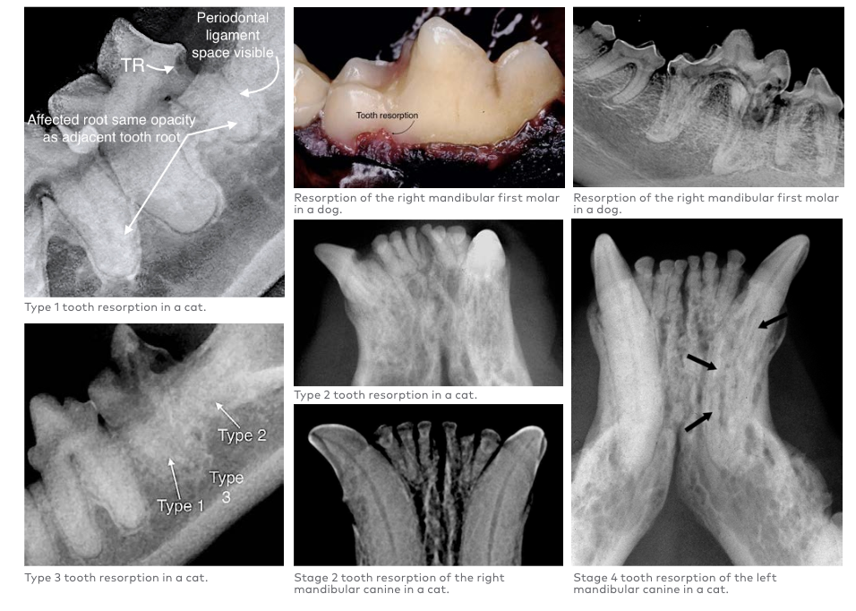
“It might seem like semantics to classify tooth resorption individually, but it is important,” Altier noted. During a dental examination, veterinary professionals should record a diagnosis for each tooth based on clinical and radiographic evidence. When assessing a feline patient, both type and classification of the disease should be documented.1 Treatment is dictated by the type of resorption present. Three types of tooth resorption are recognized in cats:
- Type 1 (T1): The tooth retains normal density and the periodontal ligament space is unchanged. Resorption tends to originate at the cementoenamel junction and will destroy the tooth in a coronal direction, an apical direction, or both.
- Type 2 (T2): Radiography will show some narrowing of the periodontal ligament space and decreased radiopacity. It is also common for the root structure to transform into a bone substance that has the same density as the adjacent bone.
- Type 3 (T3): T3 resorption occurs in multirooted teeth and presents as a combination of T1 and T2.
Stages indicate the progressive nature of the disease and also the location and extent of the lesions. Five stages of tooth resorption are recognized in cats:
- Stage 1 (TR1): Stage 1 resorption presents with only mild clinical evidence of hard tissue loss and is rarely detected, especially if the lesion is hidden behind soft tissue.
- Stage 2 (TR2): Some radiographic evidence is detectable, such as a change in the dentin. Granulation tissue may be seen creeping into even a pinpoint-sized hole. The pulp is not affected, but the tooth is painful.
- Stage 3 (TR3): Lesions become recognizable. The pulp has been invaded and the tooth is extremely painful. The integrity of the tooth is intact, and the crown has not broken off.
- Stage 4 (TR4): Stage 4 is divided into 3 subcategories. In stage TR4a, the crown and root are equally affected, there is extensive damage to the cementoenamel pulp, and much of the tooth’s integrity is lost. In TR4b, most of the damage is present in the crown, and in TR4c the root is affected more severely than the crown.
- Stage 5 (TR5): Total resorption results in loss of the crown, with roots muddled into the bone.
Altier is not familiar with any studies that provide a definitive answer on how long it takes to advance from stage 1 to stage 5. “The progression is most likely specific to that patient and that tooth,” she said, “but in my experience, it takes months to years for a tooth to go from beginning to end-stage resorption.”
Dogs
Because much of the veterinary research on tooth resorption has focused on cats, less is known about the pathogenesis of the condition in dogs (in which it is sometimes called idiopathic root resorption). However, a 2010 study from the University of California, Davis (UC Davis) provided an unprecedented look into how the disease progresses in dogs.2 The 2 most common types of tooth resorption were detected in 120 of the 224 (53.6%) study dogs and in 943 of 8478 (11.1%) teeth studied. External replacement resorption was found in 34.4% of dogs and external inflammatory resorption in 25.9%.
Based on the findings from this study, as well as the similarities in how tooth resorption develops in humans and dogs, the classification of canine tooth resorption mimics the system used in human dentistry.2,3 Categories are listed in the order of most common occurrence in the UC Davis study dogs2:
- External replacement resorption: This is the most commonly occurring tooth resorption in dogs (34.4%). Radiographs reveal an indiscernible periodontal ligament space and remodeling of root structure into the alveolar bone.
- External inflammatory resorption: Nearly 26% of the study dogs were found to have external inflammatory resorption, in which inflammation is thought to be the initiating factor. Radiographs may show areas of periapical lucency around resorbing tooth roots.
- External cervical root surface resorption: Just 0.3% of study dogs fell under this classification. Radiographs show the progression of these lesions beginning at the cervical area of the tooth where cementum meets enamel.
- External surface resorption:Only 0.2% of study teeth were affected by external surface resorption, a condition often unaccompanied by clinical signs and rarely apparent on radiographs. When it is seen on radiographs, it appears as shallow discontinuances of the tooth structure on the lateral edges of the root, and it only involves the cementum and dentin.
- Internal inflammatory resorption: This was a very rare finding in study dogs (0.1%). These oval-shaped enlargements, thought to result from endodontic disease, are often located in the cervical area of the root canal.
- Internal surface resorption: This type of resorption was found in only 1 study dog that did not have concurrent periodontal or endodontic disease. Mild trauma may be the initiating factor causing these oval-shaped enlargements found in the apical third of the root canal.
- Internal replacement resorption: Not found in any study dogs, internal replacement resorption is a progressive condition that typically presents as an uneven expansion of tunnel-like areas near the coronal segment of a root fracture.
The bottom line
Educating pet owners well and often about the importance of yearly dental evaluations is the best way to advocate for your patients and help those that may be suffering in silence. “Like any dental disease, tooth resorption is progressive,” Altier said. “And without treatment, it will never get any better than it is today. You have to intervene."
References
- AVDC nomenclature. Teeth abnormalities and related procedures. American Veterinary Dental College. Accessed September 19, 2020. https://avdc.org/avdc-nomenclature/
- Peralta S, Verstraete F, Kass P. Radiographic evaluation of the types of tooth resorption in dogs. Am J Vet Res. 2010;71:784-793. doi:10.2460/ajvr.71.7.784
- Andreasen FM, Andreasen JO. Luxation injuries of permanent teeth: general findings. In: Andreasen JO, Andreasen FM, Andreason L, eds. Textbook and Color Atlas of Traumatic Injuries to the Teeth.4th ed. Blackwell Munksgaard, 2007; 372-403.