Which is it? Acute renal failure vs. chronic kidney disease (Proceedings)
Renal failure results when approximately three fourths of the nephrons of both kidneys cease to function.
Renal failure results when approximately three fourths of the nephrons of both kidneys cease to function. Acute renal failure (ARF) results from an abrupt decline in renal function, and is usually caused by an ischemic or toxic insult to the kidneys. Ischemic or toxicant-induced injury most frequently results in damage to the metabolically active proximal tubular and thick ascending loop of Henle's epithelial cells, causing impaired regulation of water and solute balance. Nephrotoxicants interfere with essential tubular cell functions and cause cellular injury, swelling, and death. Renal ischemia causes cellular hypoxia and substrate insufficiency, which leads to ATP depletion and cellular swelling and death. Vasoconstriction secondary to toxic or ischemic tubular epithelial injury further decreases glomerular filtration. It is important to note that tubular lesions and dysfunction caused by toxic and ischemic insults may be reversible.
In contrast, nephron damage associated with chronic renal failure (CRF) is usually irreversible. Whether the underlying disease process primarily affects glomeruli, tubules, interstitial tissue, or renal vasculature, irreversible damage to any portion of the nephron renders the entire nephron nonfunctional. Healing of irreversibly damaged nephrons occurs by replacement fibrosis, and therefore a specific etiology is rarely determined once end-stage kidney damage is present. Chronic renal failure occurs over a period of months or years and is a leading cause of death in dogs and cats. Improvement of renal function is usually not possible in cats and dogs with CRF; therefore treatment is aimed at reducing renal workload and the clinical signs associated with the decreased renal function, as well as preventing progression of the renal lesions.
Many different and sometimes confusing terms are used to describe renal function and its deterioration. Renal disease implies that renal lesions are present; it does not qualify the etiology, severity, or distribution of the lesions nor the degree of renal function. Renal reserve may be thought of as the percentage of nephrons not necessary to maintain normal renal function. Although it probably varies from animal to animal, it is greater than 50% in normal cats and dogs. Renal insufficiency begins when renal reserve is lost. Animals with renal insufficiency appear outwardly normal but have reduced capacity to compensate for stresses such as infection or dehydration. Azotemia is increased concentrations of urea nitrogen, creatinine, and other nonproteinaceous nitrogenous waste products in the blood. Renal azotemia denotes azotemia caused by renal parenchymal lesions. Renal failure is a state of decreased renal function that allows persistent abnormalities (azotemia and inability to concentrate urine) to exist; it is a term used to indicate a level of organ function rather than a specific disease entity. Uremia is the presence of urine in the blood. Uremia may occur secondary to renal failure or postrenal disorders, including urethral obstruction and urinary bladder rupture. The uremic syndrome is a constellation of clinical signs, including anemia, gastroenteritis, acidosis, pneumonitis, osteodystrophy, and encephalopathy, which occur secondary to uremia. Recently, a staging system for chronic kidney disease in dogs and cats has been adapted that helps alleviate some of the above confusion.
Clinical features and diagnosis
Clinical signs of renal failure are often nonspecific and include lethargy, depression, anorexia, vomiting, diarrhea, and dehydration; occasionally uremic breath and/or oral ulcers may be present. A diagnosis of renal failure is confirmed when azotemia with concurrent isosthenuria or minimally concentrated urine is persistent. Prerenal dehydration and azotemia superimposed on an inability to concentrate urine (e.g., Addison's disease or overzealous use of furosemide) initially mimics renal failure; however in this case, volume replacement results in resolution of the azotemia.
Acute renal failure occurs within hours or days of exposure to the insult. Unique clinical signs and clinicopathologic findings associated with ARF include enlarged or swollen kidneys, high hematocrit, good body condition, normoglycemic glucosuria, an active urine sediment (e.g., granular casts and renal epithelial cells), and relatively severe hyperkalemia and metabolic acidosis (especially in the face of oliguria). Clinical signs associated with ARF tend to be severe when compared with those of a patient with CRF and the same magnitude of azotemia. Renal ultrasonography findings in dogs and cats with ARF are usually nonspecific with diffuse normal to slightly hyperechoic renal cortical echotexture. In patients with calcium oxalate nephrosis associated with ethylene glycol ingestion, the renal cortices can be very echodense. Histopathologic examination of renal cortical biopsies from patients with ARF will reveal varying degrees of tubular necrosis. Evidence of tubular epithelial regeneration can be observed as early as three days after the acute insult and is a positive prognostic indicator.
In contrast to ARF, CRF occurs over a period of months or years, and its clinical signs are often relatively mild for the magnitude of the azotemia. Unique signs of CRF include a long-standing history of weight loss and polydipsia-polyuria, poor body condition, nonregenerative anemia and small and irregular kidneys. A diagnosis of CRF is usually based on a combination of compatible historical, physical examination, clinicopathologic and imaging findings. Plain film radiographs can confirm the presence of small kidneys. Renal ultrasonography will usually reveal diffusely echo dense renal cortices with loss of the normal cortico-medullary boundary. The increased cortical echogenicity is caused by fibrous scar tissue replacement of irreversibly damaged nephrons. Radiology and ultrasonography can also help rule out potentially treatable causes of CRF like pyelonephritis and renal urolithiasis. Renal biopsy is not routinely performed on CRF patients unless the diagnosis is in question. Renal histopathology will show some combination of loss of tubules with replacement fibrosis and mineralization, glomerulosclerosis and glomerular atrophy, and foci of mononuclear cells (small lymphocytes, plasma cells, and macrophages) within the interstitium associated with fibrous scar tissue replacement.
Treatment for ARF
A list of treatment guidelines for ARF is presented in Table 1. Identification and correction of any prerenal or postrenal abnormalities is essential. Fluid deficits should be replaced intravenously within 4-6 hours with either 0.45% saline in 2.5% dextrose or normal saline solutions. Maintenance and continuing fluid loss needs should be provided over a 24-hour period using 0.45% saline in 2.5% dextrose to prevent potential worsening of hypernatremia and hyperkalemia. Oliguria is common in patients with ARF and was once thought to be a hallmark of the syndrome; however nonoliguric ARF is being recognized with increasing frequency; therefore urine production should be quantitated so that maintenance fluid needs can be properly assessed. Since approximately two thirds of normal maintenance fluid needs are the result of fluid loss in urine, oliguric and nonoliguric patients can have large variations in their fluid needs. If indwelling urinary catheters are used to measure urine volume, strict aseptic technique and closed collection systems must be used. Uremic patients have depressed cellular immunity and phagocytic function, and infection is a leading cause of death. Intermittent urinary bladder catheterization is usually preferable over indwelling catheterization for timed urine collections.


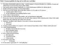
Table 1. Treatment guidelines for dogs and cats with acute renal failure
During the period of rehydration, the acid-base and electrolyte status should be evaluated and treated. Metabolic acidosis and hyperkalemia are common in patients with oliguric ARF; the acidosis is usually partially compensated for by a respiratory alkalosis. Bicarbonate therapy should be reserved for patients whose blood pH is 7.15 or less. Overzealous sodium bicarbonate therapy can create ionized calcium deficits and sodium excesses, which may contribute to hypervolemia in the oliguric patient. Hyperkalemia can cause cardiac conduction abnormalities, and is the most life-threatening electrolyte disturbance that occurs in dogs and cats with ARF.
If signs of overhydration are not present and oliguria persists after apparent rehydration, mild volume expansion with 3% to 5% of the patient's body weight in fluid may be initiated, since dehydration of this magnitude is difficult to detect clinically. Monitoring body weight, plasma total solids, hematocrit, and central venous pressure will help protect against overhydration. When fluid therapy alone fails to induce diuresis, either mannitol or furosemide is the therapeutic strategy of choice. If one regimen is ineffective, the other may be tried. Furosemide therapy is a better choice for over hydrated patients; however, it appears that furosemide treatment is more efficacious in ischemic ARF compared to toxicant-induced ARF. Also, furosemide may potentiate gentamicin-induced nephrotoxicosis. Whether or not diuresis occurs, maintenance fluid requirements should be derived from the volume of urine produced. If diuresis occurs, polyionic solutions (e.g., Normosol or lactated Ringer's) should be used for maintenance fluid requirements; potassium supplementation is often necessary and should be determined by measuring serum potassium concentrations.
Treatment for CRF
In the patient with CRF, regenerative and compensatory nephron changes have had time to occur, yet the presence of renal failure indicates the inadequacy of these compensatory processes. Even though CRF is usually irreversible, with proper treatment (Table 2) the severity of clinical signs can generally be reduced. In addition, treatment is directed at several disorders that may contribute to progression of renal failure (e.g., systemic hypertension, and soft tissue mineralization).


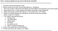
Table 2. Treatment guidelines for dogs and cats with chronic renal failure
Reduction of dietary protein intake has long been the cornerstone of management in dogs and cats with CRF. Ideally, dietary protein reduction allows all essential amino acid requirements to be met without excesses by feeding lowered quantities of high-biologic value protein, and it results in decreased need for renal clearance of phosphorus, urea, and other nitrogenous metabolites. When feeding protein-reduced diets, it must be kept in mind that the energy requirements of the body have a higher priority than does protein anabolism; therefore, if the available carbohydrates and fats are insufficient to meet caloric requirements, endogenous proteins are often broken down as a source of energy. Catabolism of endogenous proteins for energy increases the nitrogenous waste the kidney must excrete and exacerbates the clinical signs of renal failure.
Researchers have established that minimum protein requirements for dogs and cats with CRF are higher than those of normal dogs and cats. Ideally, dogs and cats with CRF should receive a minimum of 2 to 2.2 g and 3.3 to 3.5 g of protein per kilogram per day, respectively. A good recommendation for dietary protein reduction is to feed the maximum amount of high-biologic-value protein that the animal can tolerate at his/her level of renal function. A favorable response to reduced dietary protein is a stable body weight and serum creatinine and albumin concentrations, with decreasing serum urea nitrogen and phosphorus concentrations.
Management of the hyperphosphatemia that occurs in CRF is closely related to dietary protein reduction, inasmuch as protein-reduced diets are also phosphorus-reduced. In addition to feeding a phosphorus-restricted diet, administration of enteric phosphate binders such as calcium acetate or aluminum hydroxide help combat hyperphosphatemia. Enteric phosphate binders do not directly lower plasma phosphorus concentration but bind phosphates in the intestinal tract and prevent their absorption. Enteric phosphate binders are generally ineffective if dietary phosphorus intake is not reduced. The use of ultra low-dose calcitriol to further reduce phosphorus and parathyroid hormone concentrations remains controversial. Calcitriol treatment requires extensive monitoring and should only be initiated after hyperparathyroidism has been documented.
Vomiting and anorexia are common in dogs and cats with CRF and can often result in decreased caloric intake and dehydration. Causes of vomiting and anorexia include: (1) stimulation of the chemoreceptor trigger zone by uremic toxins, (2) decreased excretion of gastrin resulting in increased gastric acid secretion (serum gastrin concentrations in dogs and cats with renal failure may be as high as five and twenty times the normal concentrations, respectively), and (3) gastrointestinal irritation secondary to uremic vasculitis. Vomiting may be treated with trimethobenzamide or metoclopramide, which block the chemoreceptor trigger zone, or with chlorpromazine, which blocks the emetic center. Metoclopramide also increases gastric motility and emptying without causing gastric acid secretion and is the drug of choice for vomiting associated with renal failure. Chlorpromazine may cause hypotension and decreased renal blood flow, and therefore should only be used if other antiemetics are ineffective. H2-receptor blockers have been shown to effectively decrease gastric acid secretion, which may attenuate vomiting in dogs and cats with CRF.
The nonregenerative anemia observed in animals with CRF is the result of a combination of decreased erythropoietin production, shortened red cell survival, gastrointestinal blood loss, and the effects of uremic toxins such as parathyroid hormone on erythropoiesis. Anabolic steroids are thought to promote red cell production and a positive nitrogen balance, however several months of treatment are usually required before a response is observed and benefits are usually minimal. Short-term studies in uremic dogs treated with anabolic steroids have failed to demonstrate any benefit with regard to body weight, serum albumin concentration, nitrogen balance, or muscle mass. In contrast, studies assessing the effects of recombinant human erythropoietin (r- HuEPO) treatment on anemia in dogs and cats with CRF have been generally successful; however, the cost of treatment is high for medium and large dogs. Although not approved for use in dogs and cats, 100 units of r-HuEPO per kilogram of body weight subcutaneously three times weekly has been used successfully. The dose interval is lengthened once a target packed cell volume (PCV) is achieved (PCV of 30-35% in cats and 35-40% in dogs). This treatment, in addition to increasing PCV, results in increased appetite, weight gain, increased strength, and an improved sense of well-being. It should be noted however, that the potential for antibody formation in dogs and cats treated with r- HuEPO exists. Most studies suggest that approximately 30% of dogs and cats treated with r-HuEPO will develop anti-r-HuEPO antibodies. If antibodies are produced against r-HuEPO, they may also react with endogenous erythropoietin, making the patient transfusion-dependent. In addition, iron supplementation PO may be necessary during r-HuEPO treatment because of rapid initiation of erythropoiesis and marginal depletion of iron stores in patients with CRF.

Urinalysis offers a noninasive, rapid screening for canine cancer detection
February 9th 2024This is the first rapid test using urine developed by the Virginia Tech College of Engineering, College of Agriculture and Life Sciences, and the Virginia-Maryland College of Veterinary Medicine
Read More


Sweet pee new remedy in feline diabetes
November 9th 2023A novel class of drugs normalizes blood glucose in type 2 diabetic cats by dumping sugar into urine rather than modulating glucose uptake in the tissues but patient selection and close monitoring are crucial to using them safely
Read More

