When the blood pressure bottoms out (Proceedings)
Sustained hypotension is a life threatening situation where the body's major organs (kidney, liver, brain, and heart) can experience irreversible damage from inadequate perfusion pressure. Veterinary technicians may encounter hypotension frequently when caring for emergency and critical care patients, as well as anesthetized or post operative patients who are frequently at risk of systemic hypotension.
Sustained hypotension is a life threatening situation where the body's major organs (kidney, liver, brain, and heart) can experience irreversible damage from inadequate perfusion pressure. Veterinary technicians may encounter hypotension frequently when caring for emergency and critical care patients, as well as anesthetized or post operative patients who are frequently at risk of systemic hypotension.
Hypotension is not a disease process in itself but rather a failure of normal regulatory mechanisms. Regulation of blood pressure is controlled by neural response, hormonal actions, and local metabolite release such as baroreceptor activation, catecholamines and anti-diruretic hormone, and nitric oxide respectively. Hypotension can result from many underlying conditions and causes are generally referred to as one of three categories: decreased preload, decreased cardiac output, and decreased systemic vascular resistance. Some of the most common causes are hypovolemia, cardiogenic shock, sepsis and anesthesia respectively.
Physiology of blood pressure
Blood pressure is the product of cardiac output and systemic vascular resistance. Cardiac output is the product of heart rate and stroke volume. Vascular resistance refers to the ability of blood to flow through peripheral vasculature and is affected by vasomotor tone of vessels and the viscosity of the blood. Stroke volume is affected by preload, contractility, and afterload.
Blood pressure is broken down into three different measurements and is measured in mmHG. Systolic arterial pressure (SAP) is the pressure generated by the left ventricle in full contraction (systole). Diastolic arterial pressure (DAP) represents the lowest pressure of arteries during cardiac filling (diastole). Mean arterial pressure (MAP) is the average of pressure in the arteries during the entire cardiac cycle. MAP is more affected by DAP because most of the cardiac cycle is spent in diastole. Adequate perfusion of the body's organs is judged by MAP, but coronary artery perfusion actually takes place during diastole. MAP can be roughly approximated by adding one third of SAP value to two thirds DAP value.
Hypotension is typically defined as MAP <60mmHg or SAP <80mmHg. At lesser pressures, the kidney and brain are known to be compromised in glomerular filtration rate and cerebral perfusion. Periods of hypotension greater than 15-30 minutes (prolonged hypotension) results in damage to nephrons and cellular death.

Table 1: Normal Blood Pressure Values
Methods of measuring blood pressure
Direct (invasive) blood pressure measurement is the most accurate method of assessment and one method is becoming a common standard of care. An arterial catheter must be placed and maintained, and typical sites for placement are the dorsal pedal and femoral arteries. The arterial line is attached to a sterile pressure transducer and connected to a multiparameter monitor. The transducer is kept at heart level and allows a continual flush (usually 3ml/hr) of heparinized saline to maintain patency. The system interprets the physical pressure received at the transducer to an electrical signal and displays a waveform on the monitor. Advantages of direct arterial pressure monitoring are accuracy, continuous measurement, alarm limit notification, and the ability to use the arterial catheter for repeated blood gas analysis. Disadvantages include the cost of patient monitor and transducer, care and risk of indwelling catheter (hemorrhage, thrombosis, infection, tissue necrosis), and possible misuse of arterial line (injection of medication). Inaccurate readings may result from using compliant tubing, kinks of the catheter, and clot formation or air bubbles within the catheter. Direct pressure monitoring yields measurements of SAP, DAP, and MAP.
Telemetric blood pressure monitoring is another invasive method that has been utilized in laboratory settings but has not yet been implemented in hospitalized patients. A foreseeable use of this method would be long term patient monitoring within the home. It involves implanting a femoral artery catheter and a subcutaneous device that transmits digital data to a computer.
Indirect blood pressure measurement is performed using Doppler, oscillometric, or photoplethysmography mechanisms. These methods measure blood flow through an artery by occluding the arterial flow with a circumferential cuff and measuring the pressure once blood flow returns as the cuff is slowly deflated. Common sites include the base of the tail and the metacarpal and metatarsal area. All sites must be carefully evaluated for a properly sized cuff (40% diameter of extremity) and fit just snuggly. Cuffs too large or small may produce values erroneously low and high respectively. Likewise, cuffs too tight or loose may alter values similarly. These methods are not accurate in very small animals and are least accurate in states of hypotension.
Doppler flow ultrasound involves a piezoelectric crystal (probe) held in place over an artery and a sphygmomanometer gauge. The frequency of the signal reflected by the arterial blood is changed into an audible sound. Gel is placed on the concave side of the probe and the probe is positioned over a palpable pulse. The contact of the probe should be firm enough to hear a clear pulse and to avoid sounds of movement but yet gentle enough to avoid constricting blood flow. Typical sites are the base of the tail and the metacarpal and metatarsal area. Personal preference will dictate whether the probe is held in place by hand, directly distal to the cuff, or taped directly under the cuff. Due to cuff inflation moving the probe or the cuff pressure occluding the artery too much, the author prefers to hold the probe by hand. Operator experience can affect the readings obtained. Once the probe is in place and a consistent pulse is heard, the cuff is inflated past the point where the pulse sound is lost. The relief valve is then opened to slowly release cuff pressure. The first returning sound of the pulse corresponds to systolic arterial pressure. One study recently compared Doppler readings with direct blood pressure readings in anesthetized cats and found the readings to more closely correlate MAP. It is generally accepted that Doppler readings on cats underestimate systolic pressure by 10-15mmHg.
Oscillometric method of measuring blood pressure utilizes a monitoring unit that performs repeated inflation and deflation of the cuff. A transducer senses changes in the pulses present in the limb. Many machine types calculate systolic and diastolic values after determining the peak amplitude of the oscillations which corresponds to MAP. Heart rate is also determined from the number of oscillations during a minute. Inaccurate readings may result from patient movement, inappropriate cuff selection or placement, or if significant arrythmias are present. Depending on the brand of monitor, readings may not be reliable on cats or very small animals.
Photoplethysmography is designed for human patients to use on a finger where it senses blood volume changing under the cuff in a cyclic pattern. The tail base is a possible location but use of this method has not come into practice yet despite being determined accurate.
Management of poor blood pressure
When caring for an anesthetized or critically ill patient frequent blood pressure monitoring is essential. In both instances, accuracy and trends are important as well as closely monitoring other vital signs such as heart rate, mucous membrane color, capillary refill time, hydration status, ongoing fluid loss, central venous pressure (CVP) if available, urine output, and depth of anesthesia.
Specifically considering anesthetized patients, if a hypotensive reading is obtained the patient must be checked first. Many pre-medications and all inhalant anesthetic agents are potent vasodilators. In excessively deep states of anesthesia, bradycardia will exacerbate existing peripheral vasodilation and result in hypotension. If the patient is in crisis, turn the gas off immediately and flush pure oxygen several times to flush out the system. And if bradycardia persists and is causing low cardiac output atropine at 0.02-0.04 mg/kg IV or glycopyrrolate at 0.005-0.01 mg/kg IV would be indicated. It would be inappropriate to use glycopyrrolate in emergency situations due to the slower onset of action. If the patient is not in jeopardy, the blood pressure reading should be confirmed. The cuff should be checked for correct fit and repeat measurements performed. Moving the cuff to a new location is wise to determine accuracy and rule out an erroneous reading. When monitoring direct arterial pressures the catheter should be evaluated, flushed, and zeroed appropriately. If repeat readings confirm hypotension, assessment of the patient should continue. Confirming the hypotension as true would be done at the same time as evaluating the anesthesia plane and lowering it to see if it is a contributing factor.
The patient must be thoroughly evaluated to assess fluid status and recognize any subclinical dehydration or current hypovolemia. The surgeon should be asked if blood loss is appropriate or if excessive hemorrhage has occurred. A crystalloid fluid bolus of 10-20 ml/kg (depending on clinician) over 15 minutes would be the next supportive measure for most patients. However, if the patient suffers from heart failure or anuric renal failure heavy fluid therapy and bolus administration would be contraindicated. If the blood pressure shows no improvement from a fluid bolus, a repeat bolus of crystalloids would be indicated. Placement of another IV catheter is often necessary. Additionally, if a jugular catheter is present a CVP should be measure which will provide valuable information about the patient's fluid status and specifically the pre-load. Often clinicians will choose to continue fluid boluses a third time until the CVP reaches 8-10 cm H2O in dogs and 5-7 cm H2O in cats. At those values the patient's volume is likely adequate and treatment would then move to colloid fluids and drug therapy to support blood pressure.
If the patient is still hypotensive all drugs used for anesthesia should be evaluated again. Treatment of perioperative hypotension can require judgment calls and extensive experience to know which drugs to continue and which to remove. Reversal of narcotics used in pre-med or intraop may sometimes be indicated. However, depending on the narcotic used, the drug may be more cadiovascularly sparing than the inhalant agent. Lowering or stopping the gas entirely may be preferable. Critical patients may be able to be maintained on injectable drug protocols alone given as a continuous rate infusion.
Performing a PCV may be helpful to determine if blood products or colloids are indicated. As another supporting treatment of volume status either natural or synthetic colloids will expand intravascular volume quicker and last longer than crystalloid fluids alone. Packed red blood cells (at recommended dosages) and Hetastarch at 5ml/kg IV may be administered. Once the patient's fluid volume has been expanded and if hypotension persists drug therapies will be considered.
When considering a critically ill patient specifically, many of the above steps in treating hypotension will be the same. A repeating trend of hypotensive readings should be given complete attention whereas a single low blood pressure value should first be confirmed as true. Once hypotension has been confirmed any sedation or pain medications being used would be examined and either decreased, discontinued, or reversed if thought to be contributing. The patient's volume status would be considered next and most likely there will be more information available to guide therapy when compared to the average anesthesia patient. Most critical care patients will have direct arterial and central venous pressures monitored as well as "ins and outs" monitored (all fluid input, urine output, and other ongoing fluid losses). All of those values will help guide the decision to push more fluid volume or reach for pharmacological support.
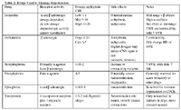
When selecting drug therapy the clinician will try to determine if the primary condition is more directly related to vascular volume, cardiac function, or decreased vascular resistance. Drugs typically selected may be naturally occurring catecholamines (dopamine, epinephrine, norepinephrine), positive inotrope (dobutamine), or synthetic form of the naturally occurring (ADH) antidiuretic hormone (vasopressin). The effects of these drugs are determined by the receptors stimulated and sometimes by dosage range. Thus these drugs will achieve different effects in varying degrees depending on whether alpha, beta, a combination of both, or multiple vasopressor receptors are stimulated. It is generally accepted that in cases where it is unknown whether poor cardiac output or vasodilation condition is the underlying cause of hypotension it is a safer option to treat with a agonist.

Table 2: Drugs Used to Manage Hypotension