Update on treatment of Malassezia dermatitis (Proceedings)
Malassezia pachydermatis is a commensal yeast organism that is a normal resident of the skin, mucosa, and ear canals. In dogs and cats, Malassezia pachydermatis colonizes the skin soon after birth, and is the primary yeast species associated with skin and ear disease.
Malassezia pachydermatis is a commensal yeast organism that is a normal resident of the skin, mucosa, and ear canals. In dogs and cats, Malassezia pachydermatis colonizes the skin soon after birth, and is the primary yeast species associated with skin and ear disease. Malassezia are part of the normal cutaneous flora on healthy dogs and cats, with the principal carriage sites being the mucocutaneous areas, ears and interdigital regions. The diagnosis of Malassezia dermatitis is based upon history, dermatological examination, cytological results, and response to therapy. Unlike its relatively commonplace occurrence in the dog, generalized Malassezia dermatitis in the cat is often associated with more serious underlying conditions, such as paraneoplastic syndrome.
Pathogenesis
The pathogenesis of Malassezia dermatitis is multi-factorial. Malassezia yeast normally colonize the skin and external ear canals of animals in very low numbers, but in a diseased state, alterations to the skin contribute to increased susceptibility to infection. Alteration in surface lipids, increased moisture and humidity, increased staphylococcus numbers and/or disruption of the stratum corneum barrier function encourages overgrowth of the yeast organism. Primary diseases that can cause these changes include endocrine disorders, allergic disease, parasitic disease, metabolic disease (ex. superficial necrolytic dermatitis in dogs), and thymoma-associated dermatoses in cats.
Clinical Presentation in Dogs
Pruritus and erythema are the classic signs of Malassezia dermatitis, with an array of secondary lesions that can occur, which include excoriations, hyperpigmentation, lichenification, crusts, scale, maceration and intertrigo. It closely resembles staphylococcal pyoderma, and cytology MUST be performed to determine if one or both are present. Malassezia dermatitis can cause some dogs to be more dry and flaky (seborrhea sicca) while others will be more greasy (seborrhea oleosa). The periocular skin, perioral skin, interdigital spaces, claw folds, axilla, groin, body folds, ventral neck, and flexure surfaces of the limbs are commonly affected areas.
Clinical Presentation in Cats
Although extremely common in the dog, Malassezia dermatitis is unusual in the cat, and causes variable pruritus. It has been seen in inflammatory dermatoses such as atopic dermatitis, adverse food reaction, and ectoparasitism. However, cats with paraneoplastic skin diseases, such as paraneoplastic alopecia (secondary to pancreatic or hepatobiliary carcinoma) and thymoma associated dermatoses are commonly found to have secondary Malassezia dermatitis.
Diagnosing Malassezia Dermatitis and Otitis
In the canine, the presence of ≥1 yeast/high power field (HPF) (400X) on the skin and ≥5 yeast/HPF in the ears are generally considered abnormal. However, some dogs can mount a hypersensitivity response to extremely low numbers of Malassezia pachydermatis, resulting in a pathological effect despite the presence of a "normal" amount of yeast organisms, making these numbers a guideline only.
Skin cytology will reveal oval, elongated cells of 3-5 µm in diameter with a typical single polar budding that gives them their characteristic footprint, or peanut, appearance. Cytological collection methods include adhesive tape stripping, gentle superficial skin scrapings on dry skin, and direct impression smears with glass slides onto greasy skin. Obtaining exudate with cotton tipped swabs and rolling the exudate on glass slides is the preferred collection method for otitis exudate. A cotton tipped swab or metal spatula can be used to scrape the claw fold, with the resulting exudate rolled onto a glass slide when evaluating for Malassezia nail bed infections (paronychia).Although Malassezia organisms can be demonstrated on histopathology, the sensitivity is less than that of cytology, and the lack of organisms on a biopsy sample does not rule out the possibility of their presence on the patient.
Antifungal Therapy
The antifungal therapy chosen for a patient with Malassezia dermatitis or otitis should be based upon the severity of the infection, the health status of the patient, and the expectations of the pet owner in regards to labor and side effects. Appropriate diagnosis and control of the primary disease is also critical in preventing relapse.
Treatment of Malassezia Otitis (Cat and Dog)
Treatment of Malassezia otitis externa classically involves appropriately cleaning the ear with an effective ear cleaner and using an antifungal eardrop. Cleaning the ear removes exudate, allowing eardrops to be more effective, can change the environmental pH so it is less suitable for yeast overgrowth, and some ear cleaners even have antifungal properties, such as Epiotic® (Virbac) and DermaPet Skin/Ear Cleanser® (DermaPet). Two weeks of therapy with medicated eardrops are typically utilized in Malassezia otitis cases, ideally with a cytology recheck to evaluate response to treatment.
Many antifungal otic preparations are available on the market and include miconazole, clotrimazole, thiabendazole, and nystatin. Miconazole is the author's otic treatment of choice.
In cases of Malassezia otitis media, oral therapies as outlined for treatment of Malassezia dermatitis are typically utilized in addition to topical therapy, and it is usually recommended to perform an anesthetic ear flush to clean out exudate and infection in the middle ear. These cases can often benefit from a veterinary dermatologist with the capability of performing video otoscopy. Treatment is often recommend for 4-8 weeks based on response to therapy, with rechecks advised every 2 weeks so it can be determined how the patient is responding and when treatment can be discontinued.
Topical Therapy of Malassezia Dermatitis (Dog and Cat)
There are many great shampoos, wipes, sprays, lotions and leave-on conditioners on the market that contain antifungal agents such as selenium sulfide, miconazole, ketoconazole, clotrimazole and chlorhexidine.
These topicals are typically used to treat localized or mild generalized infections, as adjunctive therapy when oral systemic treatment is needed, and for prevention of future infections in animals that are prone to recurrent yeast infections. The frequency of bathing depends on the patient and case, but it is not uncommon to recommend once-twice weekly topical therapy, and up to daily if using the leave on conditioners. Selenium sulfide containing shampoos should NOT be used in cats.
Systemic Treatment af Malassezia Dermatitis in the Canine
The oral medications that have been found effective for treating canine Malassezia dermatitis are ketoconazole, itraconazole, fluconazole and terbinafine. Lufenuron and griseofulvin have been found to NOT be effective treatments, and therefore SHOULD NOT be used.
Ketoconazole (Nizoral®, Jannsen) and itraconazole (Sporonox®, Jannsen) at 5mg/kg/day are extremely effective treatments. Many cases will need an initial treatment duration of 2-3 weeks, with some cases requiring more protracted treatment. Patients should be rechecked to repeat cytology and evaluate response to therapy to determine if changes or continuation are needed. Because itraconazole stays in the stratum corneum for a prolonged period of time, pulse therapy can be employed. A study by Pinchbeck, et al at the Ohio State University found that dogs treated with 5mg/kg/day itraconazole for 2 days followed by 5 days without treatment for 3 cycles (i.e. 3 weeks) responded just as well as dogs who received the medication at 5mg/kg/day for 21 consecutive days.
Fluconazole (Diflucan®, Pfizer) and terbinafine (Lamisil®, Novartis) are two other options for patients with Malassezia dermatitis. The dose for fluconazole is 2.5-10mg/kg/day, with the author usually aiming for 5mg/kg/day. The dose for terbinafine is 30mg/kg/day. Fluconazole use has greatly increased in recent years due the introduction of an inexpensive generic and its wide safety margin. It is a quite safe and effective treatment, however, anecdotal evidence suggests that it is not as clinically effective as the other azoles in some cases. Terbinafine, although now available as a less expensive generic, is not commonly used for Malassezia dermatitis, likely because of the safety, efficacy, and comfort with the azoles. The author has used it successfully in a handful of cases that have not responded to or didn't tolerate the azoles.
**Regardless of which oral treatment is started, recheck in 2-3 weeks while the patient is still receiving treatment, to assess response to therapy.**
The main side effect of all these medications is gastrointestinal, with inappetance, vomiting and diarrhea most commonly seen. However, the azoles can cause adverse reactions in the liver, drug reactions and have serious drug interactions. They are metabolized by the P450 liver enzyme, and will therefore interact with other drugs metabolized by the same system (ex. cyclosporine). A drug-triggered vasculitis has been reported in a low percentage of patients on itraconazole at doses of ≥10mg/kg/daily. Caution should be taken before using itraconazole and ketoconazole in dogs with history of liver disease, fluconazole in patients with history of renal disease, and it is generally recommended to check bloodwork in aged or debilitated dogs prior to using.
It is recommended to administer ketoconazole, itraconazole capsules, fluconazole and terbinafine with food, whereas the recommendation for liquid itraconazole (Sporonox®) is one hour before or two hours after a meal for optimal absorption. Typically compounded itraconazole liquid is not as effective as Sporonox® liquid, and its use should be avoided.
Systemic Treatment of Malassezia Dermatitis in the Feline
Ketoconazole has been associated with severe gastrointestinal upset as well as hepatotoxicity in cats, and its use should be avoided. Limited information is available about treating Malassezia dermatitis in cats with terbinafine, although its successful use has been reported in the treatment of dermatophytosis in cats.


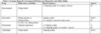
Table 1: Systemic Drugs For Treatment Of Malassezia Dermatitis And Otitis Media
Itraconazole and fluconazole tend to be safe, well-tolerated and effective treatments for feline Malassezia dermatitis.


