Understanding and treating inappropriate elimination (Proceedings)
The most common behavior problem of cats is inappropriate elimination (IE).
The most common behavior problem of cats is inappropriate elimination (IE). It is the cause of owners taking drastic measures including banning the cat to the outdoors, abandonment, surrender to a shelter, and euthanasia. It is important that a simple cookbook answer not be used for these cats as is illustrated by the approach that I use.
Most IE is driven by a behavior disorder. However, there is almost always something that initiates the problem. Sometimes IE begins with a stress-producing or insecurity-producing event. Sometimes it begins due to a medical condition. We need to try to find the initiating event and deal with it. If we can do so, treatment for the initiating event is done simultaneously with treatment for the behavioral aspect. However, sometimes the initiating event is resolved, and we only left with the behavioral aspect. But, failing to look for the initiating event can be a disastrous oversight when dealing with this problem.
Underlying medical causes
Although many cats present with behavior-driven IE, this behavior may originate in several physical abnormalities. It is important to address these before proceeding to behavior modification techniques. History is sufficient for some; specific tests are needed for others.
1. Cystitis
Bladder inflammation, whether sterile or bacterial cystitis, frequently results in inappropriate urination. These cats typically have one or more clinical signs of dysuria, pollakiuria, increased frequency of urination, and hematuria. However, these may be present and missed by owners. Urinalysis usually reveals bacturia, hematuria, and crystalluria although some affected cats will have a normal urinalysis. A urine culture is the most sensitive way of detecting bacturia. Bladder ultrasound can detect chronic cystitis (thickened, irregular bladder walls) as well as uroliths.
2. Pain
Certain types of pain may be manifested when the cat goes to the litter box and positions to urinate or defecate. Most notable is pain from the lumbar spine, lumbosacral junction, hips, and knees. Uroliths in the bladder can also create pain that is manifested when the cat urinates. The discomfort associated with constipation can also be litter box related. These cats' histories may include lameness, reluctance to jump or run, failure to raise the tail when petted, increased sedentary lifestyle, hiding, personality change (especially aggression), reluctance to being picked up, sitting or lying down slowly, a hunched up posture of the back, standing (instead of squatting) in the litter box when urinating or defecating) and protecting a body part. Radiographs of these areas are diagnostic. Litter box-related pain can also be generated by ingrown toenails, especially in older cats, and clumping litter that sticks to the hair on the ventral surface of the feet. These can be detected by examination of the feet. Impacted or infected anal sacs will become painful when the cat defecates, sometimes resulting in a litter-box-pain association. Anal sac palpation or expression is diagnostic.
3. Polyuria
Polyuria may cause a cat to urinate inappropriately. Common causes of polyuria in cats include diabetes mellitus, renal disease, and hyperthyroidism. Blood panels that include glucose, creatinine, BUN, and total T4 are diagnostic.
The work up
The ideal workup consists of directed history taking, physical examination, a blood chemistry panel including total T4, radiographs of the abdomen, lumbar spine, hips, and knees, bladder ultrasound, urinalysis, urine culture, and anal sac palpation/palpation. When possible, the minimal workup should consist of directed history taking, physical examination, anal sac palpation/expression, radiographs, and urinalysis. Your clinical judgment and the owner's motivation and finances will direct how extensive your workup can be.
Litter box issues
Cats will avoid litter boxes under several situations that include:
• Too few litter boxes for the number of cats in the household. The "official" recommendation is one more litter box than the number of cats. This is not practical for many cat owners, but it is a rule that should be imposed, or at least discussed.
• Hooded litter boxes. Although there are many advantages to hooded litter boxes (for owners, at least) odors are trapped within the litter box making it undesirable to many cats. Hoods should be removed, at least until the problem is resolved.
• Type of litter. Clumping litter best simulates sand or soil and is preferred by most cats. It is my litter-type of choice unless it is heavily scented or deodorized. When IE is occurring, avoid clumping litters for "multiple cats" due to the odors that may offend cats. If the owner objects to dusty clumping litter, recommend that a higher quality product be purchased. Synthetic litters made from wheat, newspaper, etc. can result in substrate aversion. Although well accepted by many cats, I recommend avoiding them in IE situations.
• Recent change of litter type or brand. Cats are creatures of habit. A change in the litter type or a different brand of the same type can result in IE.
• Dirty litter box. The litter box should be scooped each day (several times if used by multiple cats) and dumped and scrubbed once per month.
• Location. Cats often avoid litter boxes that are in high traffic areas and prefer those in somewhat secluded locations.
• Litter box liner. Some cats do not like these liners. When IE is occurring, remove them.
• Side height. Cats with arthritis can have difficulty climbing in or out of litter boxes with sides that are 4 inches or more high.
Household issues
The smell of urine will attract the cat so it is imperative that it be removed from carpet, other flooring, furniture, and bedding. If the item is washable, do so with hot water and bleach if possible. It is important to find all of the areas in carpet; this can be accomplished with the use of a black light. Most owners find more areas than they were aware of. Treating the pad is paramount. To do so, pour ½ cup of water on the area and spray the carpet with Zero Odor®. This is the most effective odor removal product that I have used. Zero Odor® can also be used in and around the litter box to remove lingering odors.
The pheromone Feliway® can be used to mark the cat's "good zones." As a rule, cats will not urinate or defecate in their "good zones." It should be sprayed on the locations of IE; alternatively, the Feliway® Diffuser can be used when many areas need to be treated in the same room.
The behavior aspects
Inappropriate elimination may be a primary behavior disorder or a secondary behavior disorder initiated by the diseases previously discussed. Obviously, if another primary disease is present, it must be addressed. However, specific measures must be taken to address the behavioral component. Situations that cause stress and insecurity need to be explored. Common causes include:
• A new pet (especially a puppy or kitten) or person in the household.
• A pet or person who left the household.
• New carpet, drapes, furniture.
• Rearrangement of furniture.
• Cat moved to new home.
• Harassment of the cat by other cats, dogs, or children.
• Too many cats in the household. For each additional cat there is a 10% increase in likelihood that IE will occur. If the owner has 8 cats, there is an 80% chance of IE problems.
Solutions
There are many aspects to dealing with this problem. Piecemealing the treatment protocol is much less effective than using all of your tools at once.
1. Diagnose and treat underlying diseases.
2. Identify stress or insecurity producing situations. Elimination of any of these situations (when possible) is very beneficial to successful treatment.
3. Correct litter box issues. Addition of Cat Attract Litter Additive® to the litter can make the odor in the litter box attractive instead of offensive to the cat.
4. Find all areas of IE with a black light. Remove the odor in these areas with Zero Odor®.
5. Create "good zones" with Feliway® to discourage the cat from returning to previously soiled areas.
6. Put the cat on a psychoactive drug. My first choice is buspirone (BuSpar®). If it is used with all of the above for 2 weeks without response, I change to clomipramine (Clomicalm®). If that is not successful, I use fluoxetine (Prozac®).
Prognosis
There are three factors that influence prognosis. 1) Identifying and elimination of stress or insecurity-producing situations improves the prognosis significantly. 2) Duration is also extremely important. If I begin working with a cat with IE with less than 1 month duration, I give a good prognosis. If IE has been present more than 6 months, the prognosis is poor. If it has been occurring more than one year, I do not recommend treatment. 3) Client compliance/motivation is critical. Some clients just want a quick fix with little expense or effort on their part. This attitude will almost always result in failure.
Products and sources
• Black light: Streamlight Twin Task 3C, www.4lessdepot.com/detail_streamlight_tt3c.html.
• Zero Odor, Zero Odor, LLC, Pound Ridge, NY; 914-764-1566 or www.zeroodorstore.com.
• Feliway, Ceva Animal Health, Lenexa, KS. www.feliway.com.
• Cat Attract Litter Additive, Precious Cat, Inc., Englewood, CO.; www.preciouscat.com.
• BuSpar, Bristol-Meyers Squibb Co., Princeton, NJ.
• Clomicalm, Novartis Animal Health, Inc., Greensboro, NC.
• Prozac, Eli Lilly and Company, Indianapolis, IN.


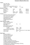
Inappropriate Elimination History Form
Orogastric tube feeding
Orogastric tube feeding permits the instillation of substantial amounts of high quality food into the cat's stomach in a few seconds. The tube is passed but not left indwelling. It may be performed several times per day, if needed. This is a technique that trained veterinary technicians should be able to perform easily. However, it is not a procedure that owners should be taught except under very unusual circumstances.
The equipment includes an 18 Fr. feline orogastric tube, a feline mouth speculum, and two 60 cc catheter tipped syringes. (The tube and speculum are available from DVM Solutions, 1-866-373-9627.) The author's preferred feeding product is Maximum-Calorie (Iams). A 10 pound cat needs about 160 ml per 24 hours. One can is put in a microwavable container with 15 ml of water. After heating to about body temperature, the mixture is stirred well. Then, it is ready for feeding. What is not fed is placed in a refrigerator for storage up to 5 days. The next feeding is preceded by heating the food in a microwave to about body temperature. Failure to heat the food will result in very poor syringability and vomiting by the cat.
Feeding should occur on a gradually increasing schedule. A 10 pound cat should receive about 25-30 ml per feeding the first day. Depending upon what time of day feeding begins, it should get either one or two feedings that day. If there is no vomiting, it should get two feedings of about 50 ml twice the second day then about 80 ml twice per day thereafter.
Loading syringes
The food is removed from the refrigerator and warmed in a microwave oven. The syringes are filled with the calculated amounts. The tip of the orogastric tube is dipped into the food for lubrication. Both syringes are loaded and placed on the feeding table.
Routine feeding
The cat's front and rear legs are held by one technician. The index finger is placed between the carpi, and the hand is closed resulting in wrapping the thumb around one leg and the other 3 fingers around the other. The same approach is taken for the rear legs at the level of the hock. The cat is held in an upright position so gravity facilitates the food staying in the stomach. The mouth speculum is placed in the cat's mouth so its canine teeth are in the rectangular holes. The tube is advanced through the oropharynx. The tube is passed so only about 2-4 inches are visible. This assures that it is in the stomach. The first syringe is attached to the tube and emptied. To empty this large syringe, the base of the plunger is placed against one's sternum, and the plunger is pushed into the barrel by leaning forward. Be sure to hold the flange of the tube and the barrel of the syringe as the food is injected. Be sure not to pull the tube out of the stomach as the food is injected, as the esophagus will not hold 60 cc of food. The first syringe is removed, but the tube is not. The second syringe is attached and emptied in the same manner. The tube is removed with the syringe attached. This prevents food from dribbling into the pharynx during tube removal.
About 10% of the time a third person is needed for restraint. This occurs because the cat is resistant to feeding and is strong enough to pull its head out of one's hand or the cat is especially large. Note that the third person uses both hands to hold the cat's neck so it cannot continue to pull away. This permits easy and efficient feeding.
Problems and special situations
There are some situations that require caution. Cats that are fractious should not be fed with an orogastric tube because there is increased risk of passing the tube into the trachea and infusing food into the lungs. If the medical situation permits, the fractious cat can be given a low dose of acepromazine, diazepam, or other sedative then fed. However, do not sedate the cat so the swallowing reflex is diminished. Cats with intrathoracic disease (heart failure, pulmonary edema, pleural effusion, etc.) that compromises respiration should not be fed with an orogastric tube as the stress of this procedure could decompensate them. Cats with nasal congestion (upper respiratory infections) should be fed with caution. Cats are nasal breathers and become anxious when forced to breath through their mouths. If the nasal passages are blocked and an orogastric tube is passed, they can panic and die of respiratory failure.
If the cat is very small, proper tube depth can be determined by placing the tip of the tube at the level of the last rib and marking the tube at the level of the cat's nose. The 18 Fr. orogastric tube is too large for kittens less than 6 months of age. For those cats use an 12 Fr. rubber tube (Sovereign Feeding Tube and Urethral Catheter). However, the food (Maximum Calorie) needs to be poured through a kitchen strainer to remove particles that will obstruct this smaller tube. It may also be necessary to add more water than described above. However, the more water is added the more the quantity of food is needed.
Esophagostomy tube placement
Indications
The purpose of the esophagostomy tube is to permit feeding for long periods of time in anorectic cats. It is placed in the esophagus, not in the stomach. If it is in the stomach the lower esophageal sphincter will be open permitting reflux of gastric acids that will cause esophagitis.
Surgical procedure
Esophagostomy tubes are available from DVM Solutions, 1-866-373-9627.
Anesthesia should be induced with either propofol or an anesthetic gas (sevoflurane, isoflurane, or halothane). The cat should be intubated and maintained on a gas anesthetic.
Prep the left side of the cat's neck because the esophagus is on the left at this level. The tip of the Global tube is cut off so food flows out its end. A stylet made from a coat hanger is inserted into the tube so the tip of the coat hanger is at the tip of the e-tube. It should not protrude from the end. A curved 5-7" forceps is inserted through the mouth into the esophagus. The tip of the hemostat is seen pointing laterally. Go nearly to the point of the shoulder if your hemostat is long enough.
Position the tip of the hemostat so it is about 2 cm dorsal to the jugular vein. Cut over the tip of the hemostat until it is exposed. A final cut of transparent esophageal lining is needed. Grab the stylet-containing tube and pull it about 2-3 cm into the esophagus. Release the forceps and remove them from the mouth as you redirect the tube in a caudal direction. Traction on the cat's head in a cranial direction aids in this step. Advance the tube and stylet about 2 cm, and then withdraw the stylet as the tube is further advanced into the esophagus. Advance it so about 8 cm of tube is visible.
Using soft, 3-0 or 4-0, non-dissolvable suture material, create a purse string suture around the tube. Tie the suture in the center so about 20 cm ends are remaining. Using these ends make a Chinese finger trap to secure the tube. To do so, pass the ends of the suture around the tube in opposite directions then tie them off, being careful not to crimp the tube with the knots. Repeat this two more times, moving up the tube 2-3 mm with each knot.
Spray the hairless area with 3M No Sting barrier spray. (This product is available through veterinary distributors that carry products made by 3M Animal Health.) Hold the cat's head over the edge of the table to facilitate the taping procedure. Make a gentle curve with the tube so the plastic fitting is at the dorsal midline and directed caudally. Wrap 1" adhesive tape around the plastic fitting then encircle the cat's neck several times to completely cover the exposed parts of the tube.
Feeding procedure
Feeding should occur in such a way as to prevent too much food being delivered into the esophagus at one time. Dispensing 12 cc syringes is safer than 35 or 60 cc syringes. Elevating the cat's front end during food injection is also helpful. Tell owners to inject the food at the rate of 1-2 cc every second.
When the food had been injected, flush the tube by injecting 2-3 cc of tap water. This prevents food drying in the tube and causing an obstruction. The final step is to replace the cap.
Changing the bandage
The tape holding the esophagostomy tube in place should be changed about every 2 weeks. To do so, the old tape is cut and removed completely from the tube. When the tape has been removed, it is important not to let the cat shake it head or it might cause the tube to come out, requiring surgical replacement. The area is cleaned with hydrogen peroxide. The skin is sprayed with No Sting Barrier Spray. A 2 by 2 gauze square is cut to it will fit around the esophagostomy tube. Triple antibiotic ointment is applied to it. The gauze is placed around the tube so the ointment is on the ostomy site. Tape is reapplied to the esophagostomy tube and around the neck as done at the time of e-tube placement. It is important that the bandage stick to the skin and hair so the cat cannot dislodge it. It is also important that the bandage is not placed so tightly that the cat is uncomfortable.
Tube removal
Tube removal should occur after the cat has been eating for 3-4 days. The bandage and suture are cut, and the tube is removed. The ostomy should not be sutured; it will close in 2-3 days.
What to feed
Norsworthy's preferred feline diet is Iams Maximum-Calorie (canned).
Put 3 cans in a microwavable container and add 45 ml of tap water. Heat in a microwave until it is about body temperature. Shake or stir to achieve even consistency and heating.
A 10# cat should be fed about 120-140 ml of this mixture per 24 hours. This amount should be divided into 2 to 4 feedings per day.