Try to decompress biliary occlusion
Signalment:
Signalment:
Feline, Himalayan, 12.5 years old, female spayed, 6.6 lbs.
Clinical history:
The owner stepped on the cat about three days ago, and it has been lethargic without eating or drinking since then. The cat has lost 0.6 lbs in the past month and has been diagnosed with hip dysplasia.
Physical examination:
The findings show rectal temperature 102.8°F, heart rate 140 beats/min, sinus rhythm, respiration 20 breaths/min, pink tacky mucous membranes, no heart murmur heard, ear disease and arthritis. Therapy has included prednisone, Rimadyl and ketoprofen.
Laboratory results:
A complete blood count, serum chemistry profile and urinalysis were performed and are outlined in Table 1.
Radiographic review:
Survey thoracic and abdominal radiographs are done. The thoracic radiographic views are unremarkable (Images 1-7).
My comments:
The abdominal radiographs show an enlarged liver, a prominent spleen, radiodense area in the location of the gall bladder, hip dysplasia and moderate spondylosis.
Ultrasound examination:
Thorough abdominal ultrasound is performed with the cat positioned in dorsal recumbency.
My comments:
The liver shows an inhomogeneous texture in its parenchyma.

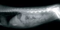
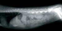
Image 1
Multiple secondary bile ducts are dilated with possible calcified walls and acoustic shadowing noted. No masses noted within the liver parenchyma. The gall bladder is mildly distended, and its walls are not thickened or hyperechoic.

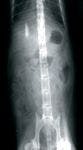
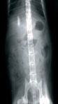
Image 2
The gall bladder does contain a lot of sludge material. The common bile duct is greatly dilated with large amount of accumulated sludge material in its lumen and possible calcified walls with some acoustic shadowing noted.

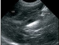
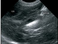
Image 3
The spleen shows an inhomogeneous texture in its parenchyma, and no masses are noted. The left and right kidneys are similar in size, shape and echotexture. Each kidney shows an inhomogeneous texture in the renal cortex.

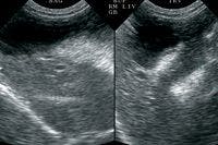
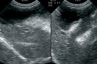
Image 4
No masses or calculi were noted in either kidney. The urinary bladder is distended with urine and contains some urine sediment material - no masses or calculi noted.

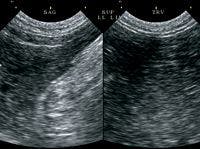
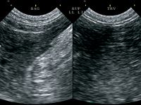
Image 5
The left and right adrenal glands are similar in size and shape. The stomach, small intestine and colon are normal. The pancreas is prominent and shows an inhomogeneous texture in its parenchyma.

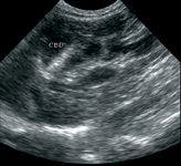

Image 6
Case management:
The tentative diagnosis is chronic cholangitis and biliary tract occlusion. There was no obvious evidence of cancer noted during this abdominal ultrasound study.
Paragraphs taken from my recent geriatric textbook (Geriatrics & Gerontology of the Dogs and Cats , Second Edition. WB Saunders Co., Philadelphia, 2004): "On ultrasonography, severe ascending cholangitis associated with thickening of the extrahepatic biliary system and inflammation within the lumen of the intrahepatic bile ducts may be observed. Ultrasonography also may show coexisting extrahepatic bile duct obstruction (enlarged gall bladder, distended and tortuous common bile duct, and obvious intrahepatic bile ducts), cholecystitis (thickened, laminar appearance to gall bladder wall, adjacent fluid accumulation), and pancreatitis (prominent, easily visualized enlarged pancreas with adjacent hyperechoic fat). Cytologic evaluation of liver aspirates or imprints may reveal suppurative inflammation.
"Cats with extrahepatic bile duct obstruction should have their biliary occlusion decompressed, if possible. If biliary tract decompression cannot be accomplished, the biliary pathway may be rerouted by a cholecystoenterostomy. Biliary diversion is a vital early therapeutic intervention in the prevention or control of sepsis in obstructive suppurative cholangitis."
This cat would benefit most from having biliary tract decompression done by exploratory laparotomy. Otherwise, medical management for biliary tract disease can include the following:
- Fluid therapy according to the cat's needs;
- Prednisone (2-4 mg/kg PO SID or divided BID with titration to the lowest effective dose during the next several months) - an important part of the general treatment plan;
- Metronidazole (5.0-7.5 mg/kg PO BID) or daily enrofloxacin;
- Vitamin E at oral dose of 200 IU per day or daily Denosyl SD4;
- 1 mg injectable vitamin ±2 every 2-4 weeks;
- Diet that the cat will eat well.