Testicular cancer remains easily preventable disease
Signalment: Canine, Labrador Retriever, 12 years old, male, 73 pounds.
Signalment:
Canine, Labrador Retriever, 12 years old, male, 73 pounds.
Clinical history:
The dog presented for ear infection. Therapy has included ketoconazole and Otomax.
Physical examination:
The findings include rectal temperature 101.2° F, heart rate 110/min, respiratory rate 20/min, pink mucous membranes, normal capillary refill time, and normal heart and lung sounds. The physical examination showed BAR, asymmetrical testicles, tense abdomen and ear infection.


Table 1: Results of laboratory tests
Laboratory results:
A complete blood count, serum chemistry profile and urinalysis were performed and are outlined in Table 1.
Thorough abdominal ultrasonography was performed with the dog positioned in dorsal recumbency.
My comments:
The liver shows an inhomogeneous to homogeneous texture in its parenchyma. No masses noted within the liver parenchyma. The gall bladder is mildly distended, and its walls are not thickened or hyperechoic. The gall bladder does contain some sludge material. The spleen shows an inhomogeneous texture in its parenchyma - no masses noted. The left and right kidneys are similar in size, shape and echotexture. Each kidney shows an inhomogeneous texture in the renal cortex. No masses or calculi were noted in either kidney. The urinary bladder is distended with urine and contains some urine sediment material - no masses or calculi noted.

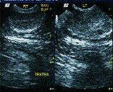
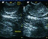
Image 1.
The prostate gland shows a symmetrical shape and a mixed echogenicity in its parenchyma. The left testicle is enlarged and some cavitations noted in the parenchyma. A mixed echogenic irregular-shaped mass-like structure is seen attached to the cranial aspect of the left testicle. The right testicle is normal in shape and echotexture. The left and right adrenal glands are similar in size and shape. The stomach, small intestine and pancreas are normal. The pancreas is prominent and shows a decreased inhomogeneous texture in its parenchyma.
Case management:
In this case, most likely neoplasia of the left testicle is present. There was no obvious evidence of cancer noted during this abdominal ultrasound study. Castration is recommended and histopathologic examination done.

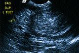

Image 2.
Review on testicular tumors
Testicular tumors are common tumors in older intact male dogs. The incidence in dogs is not very high because of the large number of dogs that are castrated.
However, in intact male dogs these tumors are considered fairly common. Testicular tumors are most common in intact older male dogs; however, they can occur in intact males of any age. There does not appear to be any breed predilection for these tumors. The current cause of testicular tumors is unknown. Dogs that have one or both testicles that are not descended (cryptorchid) are 13 times more likely to develop a tumor in the undescended testicle than dogs with normal testicles. Except for the increased risk of these tumors in cryptorchid dogs, no other risk factors are readily apparent.

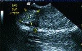

Image 3.
There are three common types of testicular tumors: Sertoli cell tumors, seminomas and interstitial cell tumors. While there are differences in the types of tumors, they are often treated similarly and are, therefore, commonly lumped together as testicular tumors. Sertoli cell tumors show signs of swelling of the testicular and scrotal area.
If the dog is cryptorchid, the swelling will occur in the inguinal or abdominal area depending on the location of the testicle. Up to 50 percent of the Sertoli cell tumors will produce estrogen, and the dog will suffer signs of hyperestrogenism.
These include an enlarged prostate gland, enlarged mammary glands and nipples, symmetrical hair loss, anemia, and the tendency to attract other male dogs. Sertoli cell tumors may metastasize to the abdomen, lung, thymus and brain; however, this occurs in less than 15 percent of the cases. Seminomas will also appear as swellings of the testicle, scrotum and inguinal or abdominal area.

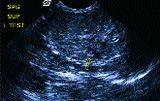
Image 4.
Seminomas produce estrogen or metastasize in less than 5 percent of the cases. Interstitial cell tumors show very few signs and do not produce estrogen or metastasize. They are usually incidental findings and not considered to be much of a problem.
Diagnosis is based on history, clinical presentation and pathological identification through a biopsy or microscopic examination of the removed tumor. Dogs suspected of a testicular tumor should also have abdominal and thoracic radiography to check for metastasis as well as a serum chemistry profile and a CBC.
Treatment
Treatment usually consists of surgical neutering. Because of the success of testicular removal and the low rate of metastasis, neutering is often the only treatment needed. Some dogs have been treated successfully with chemotherapy and in dogs that have metastasis chemotherapy is sometimes recommended.
The prognosis for dogs with treated testicular cancer is usually very good. The low rate of metastasis makes surgical neutering very successful and curative in most dogs. Dogs that develop hyperestrogenism from Sertoli cell tumors will often have a regression of signs, once the tumor has been removed. In severe hyperestrogenism that results in anemia, some animals may require transfusions and more aggressive treatment with erythropoietin therapy. The prognosis for testicular tumors that have metastasized is more guarded and the outcome varies widely depending on location, type and treatment.
Easy prevention
Testicular cancer is easily prevented, and with good neutering policies could be virtually eliminated from the dog population. Neutering in young dogs prevents aggression, roaming, urine marking and a variety of other unwanted male behaviors. The surgery is safe and relatively inexpensive, and in the long run saves the owner money. Dogs that are used for breeding can be castrated when they are no longer used for breeding. Dogs that are cryptorchid should always be castrated, and the owner should insist that both testicles be removed.
Since cryptorchidism is considered to be an inherited trait, cryptorchid dogs should never be used for breeding purposes. Because the retained testicle is 13 times more likely to develop a tumor, it should always be removed.
Dr. Hoskins is owner of DocuTech Services. He can be reached at (225) 955-3252, fax: (214) 242-2200, or e-mail: johnny@docu-techservices.com.


