Surgery STAT: Traumatic fragmented medial coronoid process: Diagnosis
Signs, diagnostic testing of this painful condition that can affect any dog.
This article is the first of a three-part series covering elbow disease in dogs. This first part reviews a new subset of elbow dysplasia, traumatic fragmented medial coronoid process (TFMCP). Part 2 will cover rehabilitation therapy for the elbow after surgical treatment. The final article will discuss intra-articular medical treatment options for dogs.
TFMCP is a condition of the elbow joint that commonly occurs in active dogs. It appears that affected dogs can be any size or age, in contrast to the classic fragmented medial coronoid process (FMCP) that affects the elbows of skeletally immature large- or giant-breed dogs.
Clinical signs
Dogs with TFMCP have a history ranging from intermittent offloading of the affected forelimb to marked lameness, and they often show little response to rest and nonsteroidal anti-inflammatory drugs (NSAIDs). Onset of clinical signs is insidious and exacerbated by exercise. Also, as the lameness persists, its severity may increase. An affected dog often places its carpus in a slightly exaggerated valgus position when it sits or stands, and it may circumduct its antebrachium and abduct its elbow during the swing phase of its stride. Because pain is elicited when the shoulder is extended, many affected dogs are mistakenly treated for shoulder pathology. But this pain is more likely caused by simultaneous elbow extension. Extending the shoulder and elbow causes tension in the biceps-brachialis muscle complex, which exerts pressure on the medial coronoid and overlying inflamed joint capsule and results in pain.
Pathogenesis
TFMCP's cause and pathogenesis are not well understood. Abnormal repetitive loading (e.g., landing from a jump, hitting contacts or a flyball box) may cause subchondral microfractures to develop. Increased repetitive loading can also occur from contracting the biceps-brachialis muscle complex, generating a force that rotates the medial coronoid into the radius. The microcracks that develop disturb the bone's mechanical properties, and if the microcracks are not repaired properly through normal body mechanisms, fatigue fractures may arise. Additionally, osteocyte loss, indicated by decreased osteocyte densities, is associated with microdamage after fatigue loading. Excess load can lead to fatigue microdamage of the subchondral trabecular bone and subsequent fracture, which may play an important role in the pathogenesis of TFMCP. Dogs with elbow dysplasia may be further predisposed because of asymmetric growth of the radius and ulna during development, resulting in elbow joint incongruity. This joint incongruity causes abnormal contact patterns in the elbow, specifically at the coronoid trochlear articulation, which is theorized to increase the load on the medial coronoid process. Regardless of the cause of the condition, if TFMCP is left untreated, it will likely lead to progression of secondary osteoarthritis.
Diagnostic tests
A complete diagnostic evaluation involves a history taking, gait analysis, a physical examination and orthopedic and neurologic examinations. In patients with TFMCP, physical examination reveals discomfort when the medial compartment of the elbow joint, specifically the medial coronoid process, is directly palpated and with elbow hyperflexion. The patient may be reluctant to allow full end-range flexion, and crepitus may be noted when the elbow is put through its range of motion. Joint effusion may be detected as a fluctuant swelling beneath the lateral or medial epicondyle of the humerus, and, depending on how long the condition has existed, muscle atrophy may be noted in the affected forelimb.

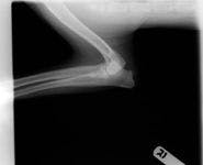

Photo 1: A "clean" lateral elbow radiograph of a dog with TFMCP.
Further diagnostic tests to help differentiate causes of elbow pathology include arthrocentesis, imaging and arthroscopy. Radiographs are of little value in identifying the fragment or line of separation seen with classic FMCP or TFMCP (Photo 1). More advanced imaging techniques such as computed tomography, magnetic resonance imaging and arthroscopy may help confirm the condition. Arthroscopic evaluation of the elbow joint has the advantages of direct observation and magnification of all principal intra-articular structures, dynamic evaluation of tissues during range-of-motion exercises and palpation of intra-articular tissues with arthroscopic instruments. Consequently, arthroscopic exploration can help definitively diagnose TFMCP when a fragment or cartilage fissure is observed (Photo 2). In a small percentage of cases, advanced imaging (computed tomography, magnetic resonance imaging) indicates fragmentation of the coronoid not seen with arthroscopy. In such cases, the microcracks are thought to be within the coronoid bone beneath the cartilage surface.

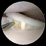

Photo 2: An arthroscopic image of a dog with TFMCP.
Treatment basics
Treatment of TFMCP is multimodal, including medical, surgical and rehabilitation therapy. The goals of such treatment are to relieve the patient's pain, maintain its limb function and return the patient to a normal level of activity and competition.
Depending on the severity of the disease, arthroscopic treatment may include fragment removal (Photo 3), débridement of diseased tissues, creation of vascular access by abrasion arthroplasty, forage, microfracture and subtotal coronoid ostectomy. Compared with a traditional surgical arthrotomy, arthroscopy results in better visualization of structures within the joint, less soft tissue trauma, shorter surgery and hospitalization times, decreased risk of infection and shorter recovery times.


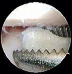
Photo 3: Arthroscopic removal of a fragment in a dog with TFMCP.
After arthroscopic treatment, rehabilitation therapy and medical management are initiated. These topics will be covered in Parts 2 and 3 of this series.
Dr. Canapp is an ACVS board-certified surgeon who practices orthopedic surgery and sports medicine at the Veterinary Orthopedic & Sports Medicine Group in Annapolis Junction, Md.