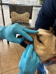
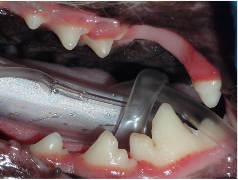
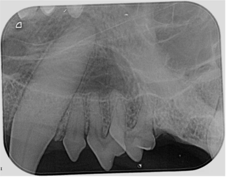
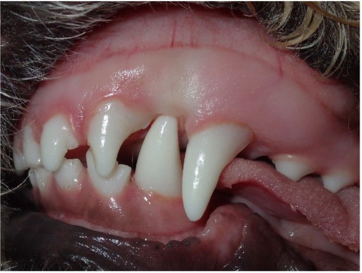
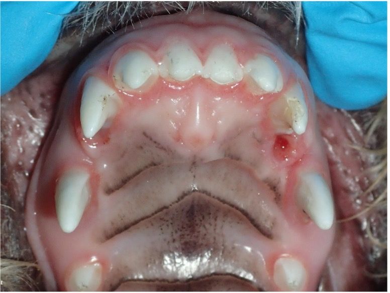
Periodontal disease is the most common disease affecting companion animals today and studies show that by 2 years of age 70% of cats and 80% of dogs have some form of periodontal disease.1,2This staggering statistic implies that a significant portion of puppies and kittens seen for wellness exams will suffer from periodontal disease within 16 months of completing their vaccine appointments. (Figures 1 and 2)
What can we do to mitigate these staggering figures? Imagine averting dental cleanings with 10-15 extractions for small breed or feline patients at age 4 years by initiating proactive dental disease prevention in their youth.
By the time clients detect bad breath or tartar and calculus build up, periodontal disease has already entrenched itself, possibly irreversibly. Only stage 1 periodontal disease (PD) can be reversed to a stage 0 through professional dental cleaning (PRO) and proactive home care. Once PD progresses to stages 2-3, treatment involves a PRO, addressing periodontal pocketing, oral surgery, and client education on home care routines that can be committed to.
Stages of Periodontal Disease
The degree of severity of PD relates to a single tooth; a patient may have teeth that have different stages of periodontal disease.3
To gain insight into their pet’s oral health, we can ask clients specific questions to better understand their pet's oral health:
- Is your pet drooling more?
- Does your pet drop toys or treats while chewing?
- Does your pet carefully hold toys or treats in their mouth?
- Does your dog resist playing tug of war when they used to love it?
- Is your pet acting head shy?
- Has your pet changed its eating habits?
Normal (PD0)
Clinically normal; gingival inflammation or periodontitis is not clinically evident.
Stage 1 (PD1)
Gingivitis only without attachment loss; the height and architecture of the alveolar margin are normal.
Stage 2 (PD2)
Early periodontitis: Less than 25% of attachment loss or, at most, there is a stage 1 furcation involvement in multirooted teeth. There are early radiologic signs of periodontitis. The loss of periodontal attachment is less than 25% as measured either by probing of the clinical attachment level, or radiographic determination of the distance of the alveolar margin from the cementoenamel junction relative to the length of the root.
Stage 3 (PD3)
Moderate periodontitis: 25-50% of attachment loss as measured either by probing of the clinical attachment level, radiographic determination of the distance of the alveolar margin from the cementoenamel junction relative to the length of the root, or there is a stage 2 furcation involvement in multirooted teeth.
Stage 4 (PD4)
Advanced periodontitis: More than 50% of attachment loss as measured either by probing of the clinical attachment level, or radiographic determination of the distance of the alveolar margin from the cementoenamel junction relative to the length of the root, or there is a stage 3 furcation involvement in multirooted teeth.
What you can do to advocate for your patient’s dental health
As veterinary professionals, we must advocate for dental health during every wellness exam and educate clients on preventative measures. Oral health assessments can be integrated into various appointment types including wellness exams for puppies and kittens during the vaccine series, adult wellness exams, new patient exams and presurgical exams for routine procedures such as spays and neuters.
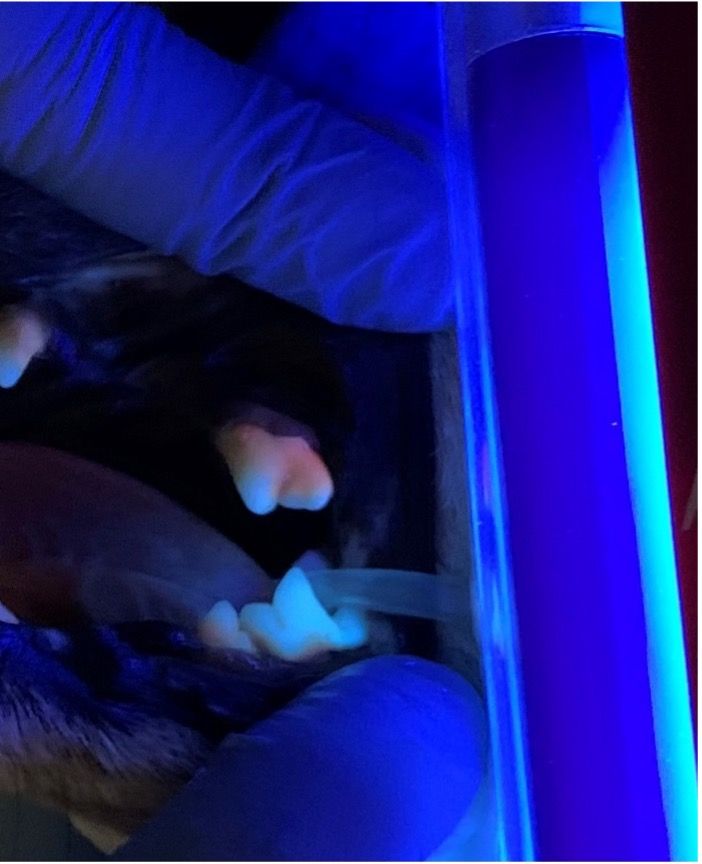
There are visualization tools we can use to show clients the current state of their pet’s teeth and gum line such as ultraviolet light to illuminate tartar build up. (Figure 3)
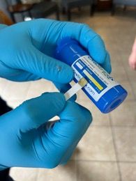
When visual exams are challenging, thiol test strips (Figure 4) allow detection of infection in the mouth. Negative results can be celebrated as a success, and positive results can be used to educate the owner as to why a PRO is recommended.
At puppy and kitten exams, the team can offer client education on breed-specific dental health issues. Malocclusions, eruption of adult teeth and missing teeth can be watched closely at each appointment. Abnormalities such as cleft palates and displaced teeth can also be assessed during these exams.
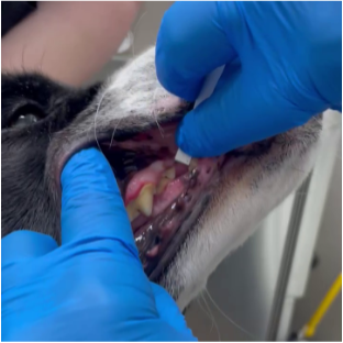
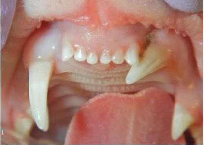
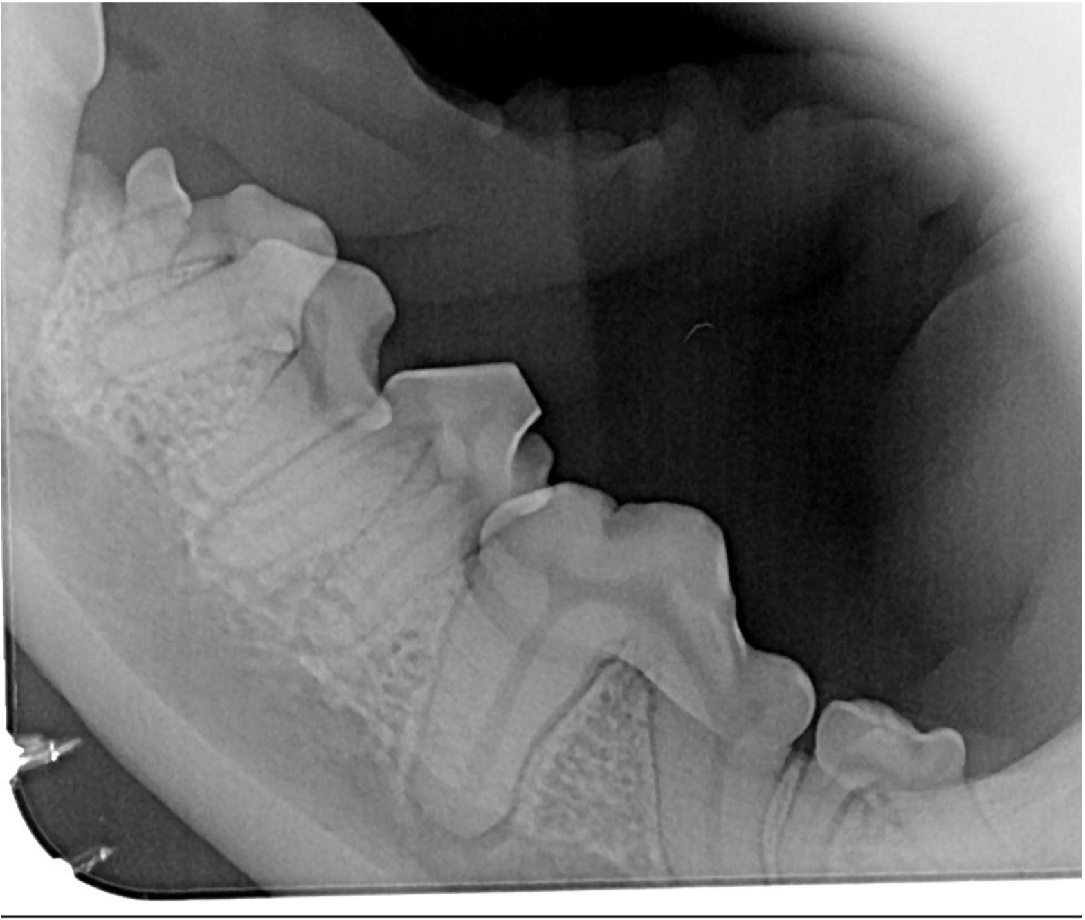
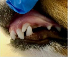
Routine spay or neuter exams offer a prime opportunity for oral health assessments, allowing early intervention for dental abnormalities. The team will assess crowding of teeth (Figure 5) that may allow PD to occur at a faster rate and visualize missing, fractured, displaced teeth causing trauma, and persistent deciduous teeth. This is a perfect time to advocate for baseline dental radiographs (Figure 6) and oral thiol tests to see what is happening under the gum line and decide when their first PRO may be needed.
For small breed dogs or mini/micro dogs, it’s possible to recommend a PRO before they reach 1 year of age. Early identification of unerupted or displaced teeth causing pain when chewing or closing the mouth allows for timely intervention. The urgency remains the same for persistent deciduous teeth; if a deciduous tooth coexists with an adult tooth, prompt attention is necessary to address the issue.
Now that we have established the timing for evaluating our patients for dental disease and other dental abnormalities, let’s explore additional services that can be provided alongside preventative PROs.
Creating a preventative dental culture
A team with preventative dental culture allows us to see how prevention of PD really works. For team buy-in:
- Educate everyone about dental disease and how it progresses.
- Meet with staff to create questions the front staff can use when fielding calls about dental cleaning appointments on the phone.
- Use/create dental health report cards or dental questionnaires the technicians can start in the room on intake.
- Have doctors and technicians use thiol test strips to detect the presence of periodontal disease.
The Veterinary Oral Health Council (VOHC) is a great resource to provide information on home care products as a team and proactive approaches to make oral health important for the staff and clients.
Home care is one of the biggest tools we have in our toolkit for PD prevention. Exploring products and techniques available on the VOHC website can help identify suitable options to address tartar and plaque buildup across various patient profiles. The VOHC seal ensures the product will help control plaque and tartar. With many products on the market, relying on a dependable source is key to knowing which recommendations will prove effective.
Clients must assess their ability to engage in teeth brushing with their pet. While brushing remains the gold standard for prevention, not all pets readily accept it. In such cases, alternate options become necessary to ensure compliance such as rawhide chews, edible chews, water additives, oral gels, teeth wipes, and sprays, all of which, when used as directed, effectively target tartar and calculus build-up.
In an ideal scenario, every veterinary hospital would have proactive dental technicians and doctors, significantly impacting the trajectory of our clinics. Technicians, who often initiate client education, particularly regarding dental evaluations, play a crucial role. By asking the right questions and openly discussing our patients’ dental needs, we can garner strong support from both staff and clients.
Developing questionnaires for staff to use during check-in or intake can prompt clients to consider their pet's oral health. Additionally, dental health report cards, containing details about the pet’s oral health status, recommended home care products, and any conducted tests, can be distributed post-appointment. These measures contribute to a comprehensive approach to dental care and enhance overall clinic performance.
When our patients exhibit a positive thiol test, show signs of PD, or report oral discomfort, we recommend a comprehensive approach. This includes a thorough anesthetized oral exam and probing, full mouth dental radiographs, and ultrasonic scaling. Preventative measures yield shorter anesthetic dental procedures, reduced oral surgeries, less systemic disease, and lower financial costs for clients.
Understanding breed-specific abnormalities
Educating staff about the unique dental abnormalities present in many dog and cat breeds can significantly improve awareness and care for these pets. Early identification and intervention are key to effectively addressing these dental issues. In our hospitals, we frequently see these breeds with early onset periodontal disease or other dental issues.2,3
- Brachiocephalic felines: Persians, British shorthairs, and exotic shorthairs often exhibit abnormally positioned teeth due to crowding associated with the brachiocephalic skull shape. (Figure 7)
- Brachiocephalic canines: Dogs with brachiocephalic skull shapes experience crowding and rotated teeth, leading to advanced PD. Additionally, unerupted teeth (Figure 8) in these breeds may result in dentigerous cysts if left untreated.
- Poodles and doodles: These breeds can have linguoversion of mandibular canine teeth (Figure 9), also known as base narrow, which often causes occlusal trauma on the roof of the mouth (Figure 10). Early intervention during deciduous and adult tooth stages is crucial.
- Shetland sheepdogs are prone to rostroversion of maxillary canine teeth (Figure 11), sometimes requiring orthodontic treatment if causing oral trauma.
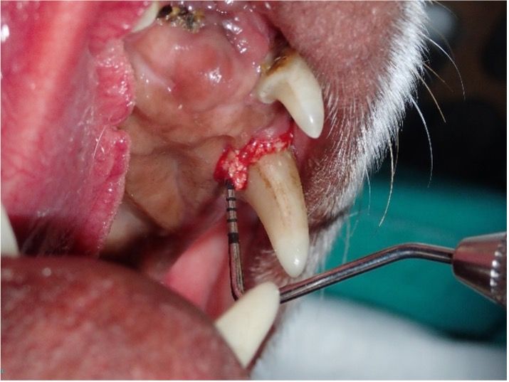
- Dolichocephalic canines:Known for advanced pocketing on the palatal aspect of their maxillary canines, untreated cases may lead to oronasal fistulas. Dachshunds are overrepresented for this condition. (Figure 12)
- Shar-Pei: This breed can suffer from a congenital defect called tight lip syndrome, where the lower lip curls over the mandibular incisors, causing lingual displacement and interfering with normal mandibular growth. Early intervention is essential to ensure oral comfort and health. If the owner notices any of the above changes, a complete oral health exam should be considered. Using thiol strip tests, along with a complete oral exam, PRO, and full mouth dental radiographs, can effectively identify the underlying cause of discomfort and address it promptly.
Early prevention significantly impacts our patients' oral and overall health, fostering a longer and healthier quality of life. By championing oral health initiatives now, we envision a future where many patients enjoy healthy mouths well into their senior years.
Winckler embarked on her veterinary journey in general practice. With a Certified Veterinary Technician license, she honed her skills in general practice until 2016, when she transitioned to the dental specialty field, joining Arizona Veterinary Dental Specialists. In 2023, Carrie achieved additional certification as a Veterinary Technician Specialist in Dentistry. An advocate for dental wellness, Carrie enjoys educating others on the importance of oral care for all animals. She is currently an instructor for Thrive Pet Healthcare’s Dental Growth Program, which empowers general practices nationwide to set higher standards in dentistry.
References
- Wiggs RB, Lobprise HB. Veterinary Dentistry : Principles and Practice. Lippincott-Raven Publishers; 1997.
- Lobprise HB. Blackwell’s Five-Minute Veterinary Consult Clinical Companion : Small Animal Dentistry. Wiley-Blackwell, A John Wiley And Sons, Inc., Publication; 2012.
- Wolf HF, Hassell TM, Rateitschak-PlüssEM, Rateitschak KH. Color Atlas of Dental Medicine: Periodontology. Georg Thieme Verlag; 2005.
- Holmstrom SE. Veterinary Dentistry: A Team Approach E-Book. Elsevier Health Sciences; 2018.

")