Staging and management of chronic kidney disease (Proceedings)
Chronic kidney disease (CKD) is a common problem that affects an estimated 0.5 to 7% of dogs and 1.6 to 20% of cats.
Chronic kidney disease (CKD) is a common problem that affects an estimated 0.5 to 7% of dogs and 1.6 to 20% of cats. Nephron damage associated with CKD is usually irreversible and can be progressive. Renal failure results when three-quarters or more of the nephrons of both kidneys are not functioning. Whether the underlying CKD primarily affects glomeruli, tubules, interstitial tissue, or renal vasculature, irreversible damage to any portion of the nephron renders the entire nephron nonfunctional. Healing of irreversibly damaged nephrons occurs by replacement fibrosis and therefore a specific etiology is often not determined. Chronic kidney disease occurs over a period of weeks, months, or years and is an important cause of death in dogs and cats. It is often not possible to improve renal function in CKD and therefore treatment is aimed at stabilizing renal function. Importantly, there is increasing evidence that dietary and anti-hypertensive treatments can decrease the progressive nature of CKD.
Etiology
The cause of canine and feline CKD is usually difficult to determine. Due to the interdependence of the vascular and tubular components of the nephron, the end point of irreversible glomerular or tubular damage is the same. Morphologic heterogeneity between nephrons exists in the chronically diseased kidney with changes ranging from severe atrophy to marked hypertrophy. These histologic changes are not process specific and, therefore, an etiologic diagnosis is frequently not possible. The more common renal diseases that have been associated with the development of CKD in dogs and cats include glomerulonephritis, amyloidosis, tubulointerstitial disease, pyelonephritis, nephrolithiasis, polycystic kidney disease, feline infectious peritonitis, leptospirosis and neoplasia. Progressive diseases that destroy nephrons at a slow rate allow intact nephrons to undergo compensatory hypertrophy which can delay the onset of renal failure. In these cases, when renal failure finally occurs, the nephron hypertrophy can no longer maintain adequate renal function and < 25% of the original nephrons are functional. This points out the need for early diagnosis and intervention.
Staging of Canine and Feline CKD
The International Renal Interest Society (IRIS) was created to advance the scientific understanding of kidney disease in small animals at the 8th Annual Congress of the European Society of Veterinary Internal Medicine in Vienna, Austria in 1998. Seventeen independent veterinary nephrologists from eight countries serve on the IRIS Board with the mission of helping practitioners better diagnose, understand, and treat canine and feline renal disease. The following table was developed by the IRIS Board as guide to staging feline CKD.


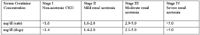
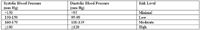
Serum creatinine concentrations must always be interpreted in light of the patient's urine specific gravity and examination findings in order to rule out pre- and post-renal causes of azotemia. The above stages are further classified by the presence or absence of proteinuria and systemic hypertension as follows:


Prevalence of Feline CKD
The distribution of staging of 786 cases of CKD in cats was reported by Dr. Jonathan Elliott from the Royal Veterinary college in London at the 2004 ACVIM Forum as follows: Stage I = 33.3%, Stage II = 37.2%, Stage III = 15.4%, and Stage IV = 14.1%. Similar data has not yet been determined for dogs.
Pathophysiology
The pathophysiology of CKD can be considered at both the organ and systemic level. At the level of the kidney, the fundamental pathology of CKD is loss of nephrons and decreased glomerular filtration. Reduced glomerular filtration results in increased plasma concentrations of substances that are normally eliminated from the body by renal excretion. In addition to excretion of metabolic wastes and maintenance of fluid and electrolyte balance, the kidneys also function as endocrine organs and catabolize several peptide hormones. Therefore hormonal disturbances also play a role in the pathogenesis of CKD. For example, decreased production of erythropoietin contributes to the nonregenerative anemia of CKD and decreased metabolism and excretion of parathyroid hormone and gastrin contribute to osteodystrophy and gastritis, respectively. Finally, part of the pathophysiology of CKD is brought about by compensatory mechanisms. For example, the osteodystrophy of CKD occurs secondary to hyperparathyroidism which develops in an attempt to maintain normal plasma calcium and phosphorus concentrations. Similarly, the individual glomerular filtration rate of intact nephrons increases in CKD in an attempt to maintain adequate renal function; however, proteinuria and glomerulosclerosis may be consequences or "trade-offs" of this hyperfiltration.
Clinical Signs and Diagnosis
Clinical signs of CKD may not be present in early stages and when present in later stages, are usually nonspecific (lethargy, depression, anorexia, gastroenteritis, and dehydration). Occasionally uremic breath and/or oral ulcers may be observed. Unique signs of CKD (vs. acute renal disease) include a history of weight loss and polydipsia-polyuria, poor body condition, nonregenerative anemia, small and irregular kidneys, and renal secondary hyperparathyroidism. The classic diagnosis of renal failure based on renal azotemia (persistent azotemia superimposed on the inability to concentrate urine) pertains to CKD stages II-IV. Stage I CKD (non-azotemic CKD) could be diagnosed in cats and dogs with persistent proteinuria, urine concentrating deficits, increases in serum creatinine over time, even if the values remain in the normal range (e.g., serum creatinine that increases form 0.6 to 1.2 mg/dl could indicate a 50% reduction in GFR), or abnormal renal palpation or renal ultrasound findings.
In general, the diagnostic approach to patient once CKD has been identified and staged is focused on three areas: 1) characterization of the renal disease, 2) characterization of the stability of the renal disease and function, and 3) characterization of the patient's problems associated with the decreased renal function. Further definition of the renal disease (beyond a standard minimum data base) could include for example, quantitation of proteinuria, measurement of blood pressure, urine culture, kidney imaging, and possibly kidney biopsy. The stability of the renal function would be assessed by serial monitoring of abnormalities identified during the initial characterization of the renal disease. This monitoring should always include serum biochemistry profiles, urinalyses, quantitation of proteinuria, and measurement of blood pressure but may also include follow-up urine cultures and ultrasound examinations. Characterization of the renal disease and its stability is most important in the earlier stages of CKD when appropriate treatment has the greatest potential to improve or stabilize renal function. Characterization of patient problems becomes more important in the later stages of CKD when clinical signs tend to be more severe. In the later stages of CKD, diagnostic (and subsequent therapeutic) efforts should directed at assessing anorexia, vomiting, acidosis, potassium depletion, hypertension, anemia, etc.
Management
Similar to the diagnostic approach to CKD, the therapeutic approach should also be tailored to fit the patient's stage of disease. For example, disease specific treatments for nephroliths and bacterial pyelonephritis as well as treatments designed to slow the progression of renal disease (so called renoprotective treatments) will be of most value in the earlier stages of CKD. Renoprotective treatments include dietary change designed to reduce serum phosphorus concentrations and angiotensin-converting enzyme inhibitors designed to normalize systemic and intraglomerular blood pressures and reduce proteinuria. In the later stages of CKD, treatment tends to be focused on decreasing the patient's clinical signs associated with the decreased renal function.
Hypertension
Systemic hypertension is relatively common in cats and dogs with CKD. Although the exact mechanism of the hypertension is not known, a combination of glomerular capillary and arteriolar scarring, decreased production of renal vasodilatory prostaglandins, increased responsiveness to normal pressor mechanisms, and activation of the renin-angiotensin system may be involved. Gradual reduction of dietary salt intake is often recommended as the first line of treatment; however, there are no studies that document the efficacy of dietary salt reduction in lowering blood pressure. In many cases vasodilators (angiotensin converting enzyme inhibitors [ACEI] and calcium channel antagonists [CCA]) may be necessary to control hypertension. Systemic hypertension may contribute to progressive nephron loss by causing irreversible glomerular damage via increased intraglomerular pressures and glomerulosclerosis. Although ACEI will normalize intraglomerular pressures via efferent arteriole vasodilatation, CCA (e.g., amlodipine) may be necessary in addition to control systemic hypertension, especially in cats. In cats with reduced renal mass and concurrent hypertension, amlodipine had an antihypertensive effect and reduced the risk of ocular injury and neurologic complications induced by hypertension. In another study of cats with reduced renal mass, treatment with benazepril sustained single nephron GFR and increased whole kidney GFR, while reducing the degree of systemic hypertension. Similar results have been observed in dogs with naturally-occurring CKD treated with enalapril. Based on these studies, angiotensin-converting enzyme inhibition may be an effective treatment to slow the rate of progression of renal failure in dogs and cats with renal disease.
Dietary Management
Reduction of dietary phosphorus and protein intake is the cornerstone of management of CKD. Dietary management may not only allow the animal to live more comfortably with decreased renal function but may also significantly prolong survival. Ideally, dietary protein reduction allows all essential amino acid requirements to be met without excesses. This is accomplished by feeding lowered quantities of high biological value protein and results in decreased need for renal clearance of urea and other nitrogenous metabolites. It is important to keep in mind when feeding reduced protein diets, that the energy requirements of the body have a higher priority than does protein anabolism, and therefore, if the available carbohydrates and fats are insufficient to meet caloric requirements, endogenous proteins will often be broken down as a source of energy. Catabolism of endogenous proteins for energy increases the nitrogenous waste the kidney must excrete and exacerbates the clinical signs of renal failure.
Researchers postulate that protein requirements for patients with CKD are higher than those of normal animals. Ideally, dogs and cats with CKD should receive a minimum of 2.0 - 2.0 and 3.3 - 3.5g protein/kg/day, respectively. Twenty percent of the 70 to 80 Kcal/kg/day in cats should be high quality protein. A good recommendation for dietary protein reduction is to feed the maximum amount of high biological value, highly digestible protein that the animal can tolerate at his/her level of renal function. (Dietary protein reduction refers to decreased protein intake compared to normal protein intake. Most commercial pet foods contain relatively high levels of protein. Dietary protein should never be restricted, that is fed at a level that is less than the patient's dietary requirements.) A favorable response to therapy is a stable body weight and serum creatinine and albumin concentrations with decreasing serum urea nitrogen and phosphorus concentrations. Moderate dietary protein reduction should be employed early in the course of renal failure and use of more severely reduced protein diets should be reserved for patients that are refractory to moderate dietary protein reduction.
Management of the hyperphosphatemia that occurs in CKD is closely related to dietary protein reduction inasmuch as protein reduced diets are also phosphorus reduced. An increase in plasma phosphorus concentration occurs in CKD as a result of decreased renal excretion. Concurrently, decreased renal production of the active form of vitamin D3 decreases intestinal absorption of calcium which, in conjunction with impaired renal reabsorption of calcium, decreases plasma ionized calcium concentrations. Decreased vitamin D3 and serum calcium concentrations stimulate parathyroid hormone (PTH) secretion which facilitates renal excretion of phosphorus and increases serum calcium concentrations by increasing renal calcium reabsorption and calcium absorption from bones and the gastrointestinal tract. The "trade-offs" for this hyperparathyroidism, however, can be severe and include osteodystrophy, bone marrow suppression, and soft tissue mineralization. Soft tissue mineralization occurs predominately in damaged tissue and if mineralization occurs in renal tissue the result may be a progressive decline in renal function. If the product of the serum calcium and phosphorus concentrations is greater than 50-70 mg/dl, the patient is at risk for soft tissue mineralization. Studies in cats with remnant kidney CKD have shown that normal dietary phosphorus intake is associated with microscopic renal mineralization and fibrosis and these changes were prevented by reducing dietary phosphorus. Similarly in both cats and dogs with naturally occurring CKD, feeding a diet specifically formulated to meet the needs of cats with CKD, together with phosphate binding drugs if required, controls hyperphosphatemia and secondary renal hyperparathyroidism, and is associated with an increased survival time.
In addition to feeding a phosphorus-restricted diet, administration of enteric phosphate binders will help combat hyperphosphatemia. Enteric phosphate binders do not directly lower plasma phosphorus but bind phosphorus in the intestinal tract and prevent absorption. Enteric phosphate binders are generally ineffective if dietary phosphorus intake is not restricted. In many cases of canine CKD, low dosages of 1, 25-dihdroxycholecalciferol (calcitriol) have been associated with decreased PTH concentrations and decreased renal mortality. Conversely, preliminary studies in cats with CKD have not demonstrated a benefit to calcitriol treatment.
Calorie Malnutrition
Vomiting and anorexia are common in dogs and cats with CKD and can often result in decreased caloric intake. Causes of vomiting and anorexia include: 1) stimulation of chemoreceptor trigger zone by uremic toxins, 2) decreased excretion of gastrin and increased gastric acid secretion (plasma gastrin concentrations in cats with chronic renal failure may be as high as 20 times the normal concentrations), and 3) gastrointestinal irritation secondary to uremia. Vomiting may be treated with metoclopramide, which blocks the chemoreceptor trigger zone. Metoclopramide also increases gastric motility and emptying without causing gastric acid secretion and is the drug of choice for vomiting associated with renal failure. H2 receptor blockers (famotidine and ranitidine) have been shown to effectively decrease gastric acid secretion, which may attenuate vomiting in cats with CKD. Oral ulcers, stomatitis, and glossitis may occur as a result of gastritis and vomiting or the effect of uremic toxins on mucosal membranes and will often also result in anorexia. If vomiting has been controlled but anorexia persists, placement of a gastrostomy tube (especially in cats) will often facilitate the maintenance of caloric intake and hydration status.

Urinalysis offers a noninasive, rapid screening for canine cancer detection
February 9th 2024This is the first rapid test using urine developed by the Virginia Tech College of Engineering, College of Agriculture and Life Sciences, and the Virginia-Maryland College of Veterinary Medicine
Read More


Sweet pee new remedy in feline diabetes
November 9th 2023A novel class of drugs normalizes blood glucose in type 2 diabetic cats by dumping sugar into urine rather than modulating glucose uptake in the tissues but patient selection and close monitoring are crucial to using them safely
Read More

