Small herbivore dental disease (Proceedings)
Rabbits, guinea pigs, chinchillas are all monogastric, hindgut-fermenting herbivores adapted to a course, high-fiber diet.
Rabbits, guinea pigs, chinchillas are all monogastric, hindgut-fermenting herbivores adapted to a course, high-fiber diet. They graze and browse almost continuously, chewing on plant material, gradually wearing their teeth down as a result. To compensate, the teeth (incisors and cheek teeth in these species) continue to erupt throughout life, at up to 2 mm per week. The rates of dental wear and tooth eruption are in equilibrium, and if they fall out of synch, problems occur. The dynamic process of mastication, tooth wear, and tooth growth can be thrown out of balance by improper husbandry, which is why dental disease is among the most common presenting complaints in exotic companion mammal practice.
Pathophysiology
The possession of "open-rooted" teeth that continue to grow throughout life is the most important peculiarity of rabbits and rodents. This single factor underlies many of the diseases typical of these species, and is the primary reason why dental disease is so frequent as well. Lagomorphs and rodents are herbivorous, with highly specialized nutritional requirements and digestive physiology. This is the primary reason why dental disease is frequently followed by other secondary disease processes, which often actually produce the first clinical signs and symptoms noted by owners and veterinarians alike. As prey species, the small herbivores frequently minimize clinical symptoms, making early detection of dental disease a challenge. Small herbivores are highly dependent on normal dental function. As such, dental disease in rabbits, guinea pigs, and chinchillas often has a tremendous impact on health in general.
Etiology
The three main causes proposed for dental disease are congenital, nutritional, and metabolic bone disease. The primary congenital abnormality of teeth is incisor malocclusion, and this occurs mostly in dwarf rabbit breeds. Nutritional causes include insufficient dietary fiber and dental wear (all species), and vitamin C deficiency (guinea pigs). Metabolic bone disease can be due to nutritional deficiency of calcium or vitamin D, insufficient sunlight, or renal secondary hyperthyroidism (rabbits). Of course, dental disease can with any process that disrupts the teeth or bones of the skull (e.g. trauma, infection, or neoplasia).
Terminology
That portion of a tooth extending above the gum line is the clinical crown; extending below the gum line is the reserve crown. Teeth that continue to erupt throughout life are termed elodont or aradicular. The "root" of an elodont tooth is more accurately termed the apex. Teeth that normally exhibit long clinical crowns are referred to as hypsodont (e.g. incisors and cheek teeth of rabbits, guinea pigs, and chinchillas); those with short crowns are termed brachyodont (e.g. cheek teeth of rat- and squirrel-like rodents). Visually indistinguishable aradicular hypsodont premolar and molar teeth are simply referred to as cheek teeth. The long space separating the incisors from the cheek teeth in rabbits and rodents is the diastema. Dental pathology other than congenital disease is called acquired dental disease (ADD).
Presenting signs
Malocclusion of the incisor teeth is the most common clinical presentation of dental disease. While dental disease affecting the cheek teeth is a more frequent clinical diagnosis, diseases affecting the incisors are typically more obvious and apparent to owners.
Animals with dental disease tend to hypersalivate or drool. In mild cases, the animal may still be eating, but only selects certain food items from the previous menu. The animal may chew differently, hold its head at an odd angle, or otherwise indicate problems eating or drinking. It may go to food and show initial interest, but refrain from eating due to difficulty or pain. With advanced disease, dysphagia leads to anorexia. Odor from the mouth may be detected, and pus or blood may be evident in severe cases. Lumps or swellings may be evident on the jaw or skull. Exophthalmos, epiphora, or blepharospasm can also be signs. If asked, owners may report that there are fewer droppings than normal, that droppings are smaller than normal, or softer, etc. Weight loss, lethargy, and unkempt appearance are also common. There may also be vocalization or erratic behavior due to discomfort.
Diagnosis
Dental disease can usually be confirmed during physical examination. A bivalve nasal speculum (illuminated attachment for Welch Allyn otoscope/ophthalmoscope handle) makes oral examination much easier and provides a far better field of view than a penlight or otoscope. Malocclusion or malposition of incisors and/or cheek teeth will indicate disease. Sharp edges and spurs that form along the edges of teeth can cause lesions on adjacent lips, buccal mucosa, gingival, or the tongue. The buccal aspect of upper cheek teeth and the lingual aspect of lower cheek teeth are trouble spots to examine closely.
Examination under general anesthesia provides a more thorough examination. Loose teeth, pus, blood, and oral ulcers all indicate dental disease. A light source with magnification (i.e. illuminated loupe, rigid endoscope) is extremely helpful for identifying subtle lesions. Skull radiographs (V/D, lateral, and oblique views) are important for assessing tooth apexes and reserve crowns, and for characterizing odontogenic abscesses. CT and MRI imaging may be necessary in more complex cases
Equipment
To treat the gambit of dental lesions encountered with small herbivores, a number of specialized instruments should be considered. The basic setup for removing dental spurs will include a Lempert rongeur, diamond file, rabbit/rodent mouth gag, cheek dilator, and tongue spatula. For elevating and removing teeth there is the Crossley incisor luxator, the Crossley cheek tooth luxator, feline notched elevators (concave- and convex-cutting), and cheek tooth extraction forceps. To probe the distal aspect of the clinical crown on the last cheek tooth of each quadrant, the author uses Billeau flexible wire ear loop curette. A straight dental hand piece or Dremel tool is also useful for reducing crowns or sharp edges. A rodent/rabbit dental table that serves as work station, patient positioner, and mouth gag is also available.
Anesthesia
Most small herbivores do not regurgitate or vomit. They frequently hold food material in their oral cavities (especially guinea pigs), which could result in aspiration. Fasting for an hour prior to dental procedures will allow the oral cavity to empty. Atropine or glycopyrrolate will reduce salivation. Isoflurane and sevoflurane are the anesthetics of choice. Premedication with midazolam (0.25-0.50 mg/kg IM) and buprenorphine (0.02-0.05 mg/kg SQ or IM) will reduce the level of anesthetic gas required. Induce by mask or chamber, and usually maintain by mask. These species are "obligate nasal-breathers", therefore the mask only needs to cover the nose.
For certain dental procedures it will be necessary to intubate the patient. This can be a challenge in rabbits and rodents because in these species the glottis is small, the tongue is so shaped that it hides the larynx, and the larynx is smaller than the trachea. Especially in the rabbit, the vocal cords are strong and can easily deflect the tip of the endotracheal tube into the esophagus. Visualization of the larynx is aided by hyperextension of the head and neck. A small-bladed laryngoscope (e.g. Miller 0 neonatal blade) is used to depress the tongue and elevate the soft palate. A canine otoscope can be used instead of a laryngoscope in smaller patients. Direct visualization of the trachea can also be achieved using an endoscope. The endoscope is positioned so the larynx is in view, and an endotracheal tube is passed parallel to the scope and into the trachea. Further, with some scopes it is possible to put endoscope directly inside the tube like a stylet, and to visually guide the scope/tube assembly into the trachea.
Use a circulating water heat pad intraoperatively to prevent hypothermia; provide a warm area for recovery as well. For prolonged procedures, hospitalization and post-operative support is strongly advised. Observation is continued until the animal is once again eating on its own.
Treatment
Incisors
Overgrown incisors can be trimmed with wire cutters, however this method runs the risk of fracturing the tooth. A better option is to cut the tooth down to the desired level with a Dremel cutting wheel or dental burr, being mindful of the heat generated and careful not to overheat the tooth. The author usually attempts to restore a beveled cutting edge to the occlusal surface of the incisor after reduction.
Removal usually involves extraction of all incisors. The incisor is first elevated on its mesial and distal aspects using a Crossley incisor luxator, then on the labial and lingual surfaces using the curved feline notched elevators. While the periodontal ligament is being luxated, the tooth is loosened using only the fingers so as not to accidentally break the tooth. The tooth is alternately rotated clockwise and counterclockwise while alternately placing traction and apical pressure on the tooth. Once the operator feels as though the incisor could be pulled out, the tooth is firmly twisted and driven apically so as to cause permanent damage the apical germinal tissue. Finally, the incisor is extracted using only the fingers. The defect can be flushed with sterile saline and closed or left open to heal, but packed with hemostatic sponge to prevent food from entering the cavity.
Cheek teeth
Premolar/molar points and spurs are rongeured, and then filed smooth with a diamond hand file, dental hand piece, or Dremel rotary bit. The operator must used caution with a rotary tool, however, that excessive trauma does not occur at either the rotating bit or along the spinning shaft. A safer alternative for filing cheek teeth is the Dremel oscillating engraver, the kind of engraver used to personalize valuables. The sharp engraving tip is removable, and has the same shaft diameter as the rotary bits. We replaced the engraving tip with a ball-shaped diamond bit. The result is very effective vibrating bit that will grind teeth but not hurt soft tissue. Clinical crowns may be reduced in similar fashion.
Extraction of cheek teeth is usually easier than incisor removal because cheek teeth are usually not removed unless they are already diseased and loose. The periodontal ligament of the mesial and distal tooth surfaces are luxated with the transverse blade of the Crossley cheek tooth luxator, and the buccal and lingual aspects are luxated with the saggital blade. The process is continued until the tooth is sufficiently loose to be extracted with cheek tooth forceps. The remaining cavity is packed with hemostatic sponge to prevent food packing, and left to heal by secondary intention.
Comments on particular species
Rabbit
Domestic rabbits are descended from the European rabbit, Oryctolagus cuniculus, and belong to the order Lagomorpha. The lagomorphs (rabbits, hares, and pikas) are characterized by the presence of a second small pair of upper incisors or "peg teeth". The relationship of rabbits to rodents is controversial; lagomorphs were once considered to be a suborder of the Rodentia. Wild European rabbits eat a diet of graze and browse which is high in fiber and low in energy. A large quantity of poorly digestible fiber is essential for normal peristalsis and dental wear.
The nasolacrimal duct of the rabbit courses from a single ventral lachrymal punctum, through the maxilla, into the nasal cavity. It courses very near the apexes of the first cheek tooth and the incisor, and may be occluded by dental disease of either. The dental formula of the rabbit is: 2(I 2/1, C 0/0, P 3/2, M 3/3). Because rabbits are obligate nasal-breathers, and mouth-breathing is a poor prognostic sign. Rabbits are notoriously prone to laryngospasm. Insertion of an endotracheal tube therefore has to occur during inspiration, when the vocal cords are open. Endotracheal intubation is easiest in large breeds.
Dental malocclusion
Rabbits have elodont, hypsodont incisors and cheek teeth. Without the regular wear that a course, fibrous diet provides, cheek teeth overgrow and develop sharp edges and points. These may cause buccal ulcerations along the upper arcades, and lingual ulcers along the lower arcades. Apical elongation into the mandible and maxillary bone also occurs, as a result of increased occlusal contact. Treatment is by trimming or filing molar points, and/or shortening overgrown cheek teeth to allow the mouth to close and reduce occlusal contact. Recurrence is likely, and routine follow-ups every 4-6 weeks may be necessary. Providing timothy grass hay ad lib helps to prevent this problem.
Mandibular prognathism (lower incisors projecting in front of the upper incisors) is analogous to brachycephalic syndrome in dogs, and is most common in dwarf rabbits. Incisors normally grow 2-2.5mm per week. Unopposed, the upper incisors curve inward and grow up toward the roof of the mouth. The lower incisors tend to grow outward and upward, occasionally through the upper lips or nose. These teeth can be trimmed periodically (once every 4-6 weeks) or surgically extracted. Extracted incisors occasionally regrow, requiring a second attempt at removal.
Odontogenic abscesses
Skull and jaw abscesses typically stem from apical tooth infections. Prognosis has historically been poor, however new approaches show great promise. In most cases, the affected tooth must be removed. One method involves weekly surgical debridement, followed by wound packing with antibiotic soaked synthetic gauze, and then temporary wound closure. The process is repeated until the abscess shrinks and dead space is eliminated. After three "cleanup" cycles, the fourth treatment involves implanting antibiotic impregnated PMMA beads and permanent wound closure. An alternative method is to marsupialize the abscess and aggressively flush, debride, and pack the abscess with an antimicrobial agent (e.g. sugar, honey, povidone iodine) daily for up to several weeks. The patient is kept on systemic antibiotics throughout abscess treatment, and antibiotic selection is based upon culture and sensitivity testing. Anaerobic coverage is important.
Metabolic bone disease
There is strong evidence for metabolic bone disease as a cause of ADD in rabbits. Harcourt-Brown has demonstrated that affected rabbits exhibit osteopenic bone in comparison with unaffected rabbits, and that progressive osteodystrophic disease affects the teeth and bones of the skull. The visual and radiographic features are typical of nutritional secondary hyperparathyroidism. The dietary calcium intake of rabbits offered an inappropriate diet mixture of pellets, seeds, and cereal grains has been show to fall short of the minimum required for mineralization of bone. Vitamin D, parathyroid hormone, and calcium derangements have been documented in rabbits housed indoors versus those housed outside. If dietary calcium levels are corrected in the early stages of dental disease, horizontal ridges in the incisor enamel will grow out, and normal dental appearance will be restored. ADD is uncommon in laboratory rabbits (usually fed a balanced pelleted ration), wild rabbits, and pet rabbits housed outdoors.
Guinea pig
Guinea pigs have elodont incisors and cheek teeth. They have large tongues and small, narrow oral cavities. The dental formula is: 1/1 incisors, 0/0 canines, 1/1 premolars, 3/3 molars. The incisors are generally white in color.
Scurvy
Guinea pigs cannot metabolize endogenous vitamin C. They require 15-25 mg per day in their diet. Ascorbic acid is necessary for collagen synthesis. A lack of vitamin C results in defective collagen, including that which is necessary to anchor teeth tightly in their sockets. Without it, teeth loosen and malocclusion results.
Dental malocclusion
Over time, sharp edges and points may develop on cheek teeth. Ulcerations, and in severe cases, entrapment of the tongue commonly occur. With insufficient wear, the molar crowns may elongate and, if the mouth cannot fully close, the incisors may elongate as a result. Treatment is by trimming or filing molar points, and/or shortening cheek teeth to allow the mouth to close. Primary incisor malocclusion is rare, and incisor trimming alone is rarely indicated. Particularly with guinea pigs, incisor malocclusion suggests cheek tooth abnormalities. Providing timothy grass hay ad lib and adequate vitamin C helps to prevent this problem. Recurrence is likely, but prognosis is good with regular recheck and tooth trims in most cases.
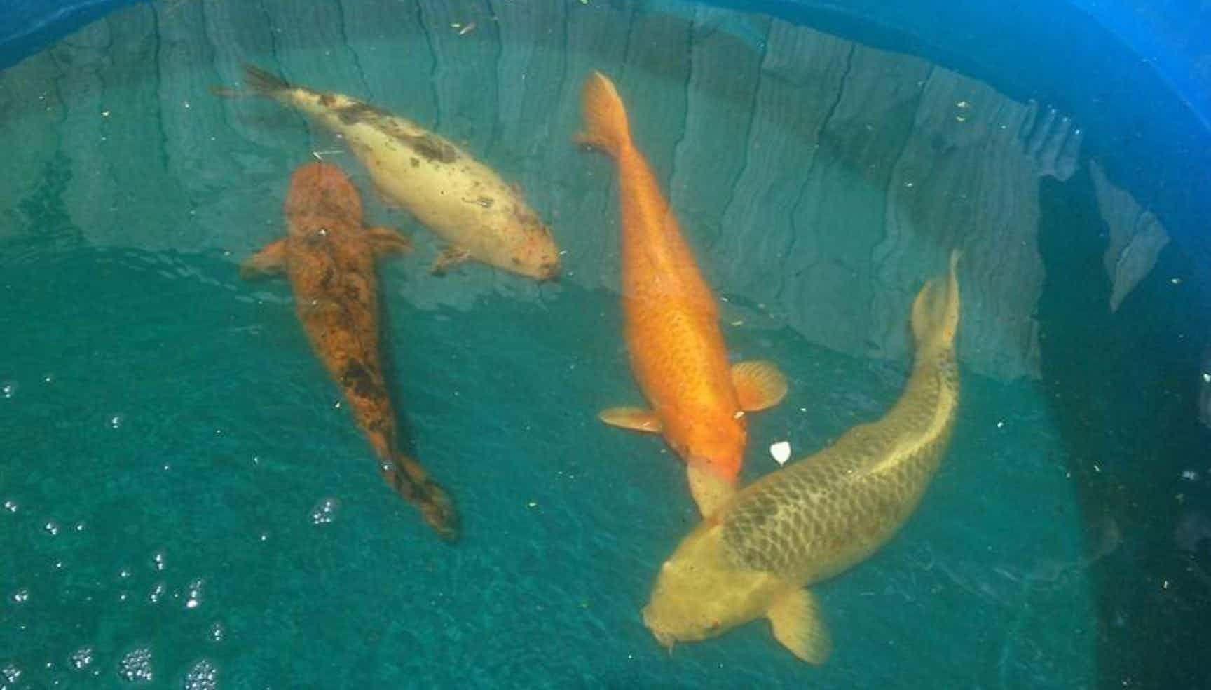
Chinchilla
Chinchillas have elodont incisors and cheek teeth. The dental formula is the same as for the guinea pig. The incisors are usually orange-colored. Chinchillas have vestigial cheek pouches.
Dental malocclusion
Also called "slobbers", as chinchillas are particularly prone to developing a wet chin, face, chest, and forepaws. Molar and incisor malocclusion is common: Crossley detected dental abnormalities in 35% of apparently healthy chinchillas presenting for clinical examination, and incisor abnormalities in 55% of those presenting with signs of illness. Uneven wear can cause sharp edges and points on cheek teeth, resulting in oral ulcerations. The buccal aspect of upper molars and the lingual aspect of lower molars are the trouble spots.
Tooth elongation
The natural diet of chinchillas is tough, fibrous, and low in energy content. As a result, wild chinchillas must chew a lot of food to meet their energy requirements and provide adequate tooth wear. Low roughage diets prevent natural tooth wear from occurring. As cheek teeth elongate, increased occlusal contact occurs. Affected teeth are forced apically and begin to curve; remodeling of the surrounding tissues results. The tooth apex begins to intrude through the jaw bone. Lesions can be palpated along the ventrolateral surface of the mandible. Radiographically, they can be seen extending dorsally into the orbits, nasal cavity, and sinuses as well. Exophthalmos and damage to the lachrymal ducts may result. Winking and squinting can indicate dental disease beneath the affected orbit. Tooth root dysplasia leads to increased curvature of teeth, inability to chew properly, spike and point formation, and secondary infection. Unfortunately, there is no cure. Severely diseased teeth can be removed. Opposing teeth may also need to be pulled. Frequent rechecks and repeated cheek tooth trimming are indicated. Long term pain relief and syringe feeding may be necessary. The long term prognosis is poor, but a good quality of life can be offered with regular tooth trims and monitoring.
Supportive care
Supportive care measures are often the key to a good outcome following dental procedures. The clinician should consider hospitalization so that the patient can be observed, his progress can be monitored, and additional nursing can be provided if necessary. In spite of their thick fur, small herbivores can lose body heat quickly. When sick or anesthetized, thermal support should be provided through the use of incubators and thermal pads. In order to avoid causing heat stress, do not let ambient temperatures exceed 80°F. Provide nutritional support to by syringe-feeding gruel such as Oxbow Critical Care Formula. Additional options include vegetable baby foods and soaked, ground pellets. Feed approximately 20-30 ml/kg q 8 hrs. This is continued and then gradually reduced until fecal production and appetite return to normal.
Fluid rate is 75-150 ml/kg/day, depending on severity. Fluid support can be provided by both oral and injectable routes. Provide fluid support orally with oral electrolyte solutions such as Rebound OES (Virbac) or Pedialyte (Abbott) at 15-20 ml/kg q 8 hrs; parental fluids are typically given SQ at a rate of 25-35 ml/kg q 8 hrs. Warm all fluids to body temperature prior to administration. Fluids are continued and then gradually reduced until urine output and drinking return to normal. Gradually reduce fluid therapy as urine output and drinking return to normal.
Motility modifiers are indicated in cases of GI stasis, bloat, and reduced fecal output, whenever intestinal obstruction or perforation has been ruled out. Metoclopromide 0.5 mg/kg PO, SC, IM, and cisapride 0.5 mg/kg PO are administered q 8-12 hrs for 3-5 days, until appetite and fecal production return to normal. Prokinetics may be used in alone or in combination.
Avoid the temptation to give antibiotics "just in case". Especially in small herbivores, antibiotics can have negative effects on appetite and digestion. Routine trims (even those with ulcerations) do not usually require antibiotics. Antibiotics are indicated for those cases in which infection is present or contamination has occurred. Antibiotics are selected based upon the most likely pathogen and the safety with regard to affecting cecal flora. Until the results of culture and sensitivity testing are known, an antibiotic combination that provides broad-spectrum coverage against both aerobic and anaerobic bacteria should be chosen. Penicillin G benzathine/penicillin G procaine combination has been widely used in the rabbit (at 80,000 IU/kg q48h SQ), but should NOT be used in guinea pigs or chinchillas. Chloramphenicol is safe in rabbits and rodents at 50 mg/kg q12h PO. Azithromycin is also safe and effective orally in rabbits and guinea pigs at 15-30 mg/kg q24h PO, however owners must be advised to discontinue the drug if anorexia or diarrhea occurs; the author has no experience with its use in chinchillas. Enrofloxacin 5-10 mg/kg PO 12 hr, and trimethoprim/sulfa 30 mg/kg PO q 12 hr are poor choices for infected teeth and odontogenic abscesses alone, but either antibiotic may be useful in conjunction with metronidazole 20 mg/kg PO q 24 hr. Systemic antibiotic therapy may be necessary for up to 4 weeks post-operatively in with severe infections.
Powdered bark of the Slippery Elm tree is an effective demulcent, forming a soothing film over mucous membranes, and relieving minor pain. It can be used to help alleviate oral ulceration, such as occur in small herbivores from dental malocclusion, molar spurs, and iatrogenic causes. The powder is sold in health food stores. A dilute paste is made using tap water (one 250 mg capsule into 15 cc water) and administered after hand feeding. Treatment may be applied directly to lesions (i.e. 0.50cc into each buccal cavity).
Small herbivores are more stoic and less communicative than their larger counterparts with regards to showing pain. At the same time, most are prey species, hard-wired for "flight", and are therefore more susceptible to the negative effects of pain. In our practice, buprenorphine and meloxicam have proven to be universally safe and effective in small exotic mammals. Buprenorphine is usually administered as preemptive analgesic 30 min prior to surgery, 0.02-0.05mg/kg SQ or IM, depending on severity and species. The dose may be repeated in 6-12 hrs. Meloxicam is typically dosed at 0.2-0.3mg/kg q 24 hrs for 5-7 days for post-operative pain.
Prognosis
The prognosis for patients with dental disease varies by the severity of the lesion. Incisors malocclusion frequently requires repeat trimming, however removal of the incisors is practical in the species presented here. ADD of the cheek teeth is generally considered progressive and non-reversible; the prognosis for return to normal is poor. Mild cases can be managed by intermittent trimming and filing of the teeth. With advanced ADD, removal of loose or infected teeth, and treatment of odontogenic abscesses are frequently indicated. The goal of treatment is to provide the animal with a good quality of life by minimizing discomfort, maintaining body condition, and enabling the pet to eat on its own if possible.
Prevention
Parents who produce prognathic young should be removed from the breeding program (particularly rabbits). High-carbohydrate, high-sugar diets meet the caloric needs of herbivores before adequate fiber intake and dental wear has occurred. Therefore a course diet, high in fiber and low in simple-carbohydrates, is recommended. Long-stem grass hay intake should be offered ad lib, and a moderate amount of pellet and greens should be offered daily. Treats such as dried fruit and yogurt drops should be minimized or eliminated. A complete physical exam and laboratory database are recommended every 6-12 months. Any suspicion of dental disease should indicate a closer oral examination while sedated. In addition, skull radiographs will aid in early detection of dental disease.
References
Capello V, Gracis M. Rabbit and Rodent Dentistry Handbook, Lake Worth, Fl: Zoological Education Network, 2005.
Harcourt-Brown F. Textbook of Rabbit Medicine, Oxford: Butterworth-Heineman, 2002.
Quesenberry K, Carpenter J. Ferrets, Rabbits, and Rodents: Clinical Medicine and Surgery, 2nd Ed. St. Louis: Saunders, 2004.
Taylor M. A wound packing technique for rabbit dental abscesses. Exotic DVM 2003; 5(3):28-31.
Tyrrell KL, Citron DM, Jenkins JR, et al. Periodontal bacteria in rabbit mandibular and maxillary abscesses. J Clin Micro 2002; 40(3):1044-1047.
Crossly DA. Dental disease in chinchillas in the UK. J Small Anim Pract 2001; 42:12-19.


UN, WHO address public health concern over avian flu transmission to humans
April 18th 2024Veterinary professionals working with certain animals are advised to take precautionary steps to minimize risk of infection, while researchers in Texas study potential H5N1 vaccines, antivirals, and antibody therapies for humans
Read More


