Should circulatory shock heighten suspicion of hypoadrenocorticism?
Signalment: Canine, Maltese, 2 years old, male, neutered, 8.4 lbs.
Signalment
Canine, Maltese, 2 years old, male, neutered, 8.4 lbs.
Clinical history
The dog has been shaking for three days and currently is being treated with amoxicillin. The dog is anorexic and lethargic and is walking acceptably.


Johnny Hoskins, DVM, Ph.D., Dipl. ACVIM
Physical examination
The findings include rectal temperature 101.2° F, heart rate 165/min, respiratory rate 20/min, pink mucous membranes, normal capillary refill time, and normal heart and lung sounds. The dog has a non-painful abdomen but constipated stool palpated. The neurological examination is normal.
Laboratory results
A complete blood count, serum chemistry profile and urinalysis were performed and are outlined in Table 1.


Table : Results of laboratory tests
Ultrasound examination:
Thorough abdominal ultrasonography was performed with the dog positioned in dorsal recumbency.
My comments:
The liver shows an inhomogeneous texture in its parenchyma. No masses noted within the liver parenchyma. The gall bladder is mildly distended, and its walls are not thickened or hyperechoic. The gall bladder does contain some sludge material. The common bile duct is slightly dilated. The spleen shows a homogeneous to inhomogeneous texture in its parenchyma - no masses noted.


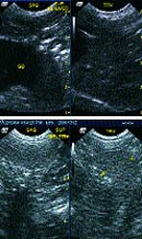
Image 1 and 2.
The left and right kidneys are similar in size, shape and echotexture. Each kidney shows an inhomogeneous texture in the renal cortex. The renal pelvis is slightly dilated in the right kidney. No masses or calculi were noted in either kidney. The urinary bladder is distended with urine and contains some urine sediment material. No masses or calculi noted. The left and right adrenal glands are similar in size and shape. The stomach, small intestines and colon are normal. The pancreas shows an inhomogeneous texture in its parenchyma.
Case management:
In this case, most likely hypoadrenocorticism is present. There was no obvious evidence of cancer noted during this abdominal ultrasound study. At this point, an ACTH stimulation test is warranted now, and then therapy for hypoadrenocorticism is started with fluids and prednisone. After the results of the ACTH stimulation test are known, then Florinef or injectable mineralocorticoid is started. The following information may be helpful.
Review on hypoadrenocorticism
Primary hypoadrenocorticism is most often diagnosed in young dogs. Secondary hypoadrenocorticism resulting from ACTH deficiency is relatively common in dogs afflicted with deficiencies of the pituitary gland.
The clinical signs of hypoadrenocorticism are intermittent vomiting, diarrhea, weight loss, lethargy, anorexia and weakness. These signs often resolve with fluid therapy and/or corticosteroid treatment. Physical examination of dogs in an acute hypoadrenal crisis shows weak pulse, bradycardia, prolonged capillary refill time, depression and severe muscle weakness. Clinical findings of hypoadrenocorticism that heighten the index of suspicion include a normal or slow heart rate in the face of circulatory shock and the waxing and waning course of disease prior to collapse.


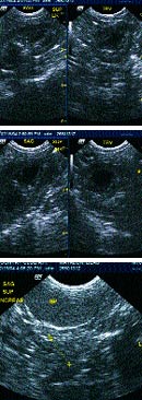
Image 3, 4, and 5.
Severe hyponatremia and hypochloremia associated with hyperkalemia are the hallmarks of hypoadrenocorticism. Although a serum Na:K ratio of less than 27:1 is considered suggestive of hypoadrenocorticism, it is never pathognomonic. Gastrointestinal disease, acute renal failure, and postrenal azotemia may also cause a low Na:K ratio. Some dogs with hypoadrenocorticism, especially those dogs with only a glucocorticoid deficiency, will not show the typical electrolyte disturbances. Azotemia and hyperphosphatemia are also associated with primary hypoadrenocorticism and make it difficult to differentiate from the azotemia of acute renal failure. The hematologic abnormalities are eosinophilia and lymphocytosis or normal eosinophil and lymphocyte counts in the face of any metabolic stress. The anemia of hypoadrenocorticism usually results from ongoing hemorrhagic gastroenteritis and not caused by the endocrine problem itself. Although hypoglycemia is more commonly associated with secondary or atypical hypoadrenocorticism, it is infrequently seen with primary hypoadrenocorticism. Urine specific gravity is frequently low and is attributed to an inadequate medullary gradient due to sodium depletion and decreased medullary blood flow. Dilute urine along with azotemia and hyperkalemia may easily be mistaken for acute renal failure.
Diagnosis
Diagnosis of primary hypoadrenocorticism is based on clinical signs, expected electrolyte disturbances, and confirmation with an ACTH stimulation test. The technique used for performing the ACTH stimulation test includes:
1) baseline cortisol sample should be collected with the initial blood work and synthetic ACTH should be administered intravenously during the initial fluid therapy, and
2) one hour after ACTH administration, a second cortisol sample is collected and a glucocorticoid is then administered in the suspected dog with hypoadrenocorticism. Intramuscular injection of synthetic or gel ACTH may not be absorbed in dogs in circulatory shock; thus, intravenous administration of synthetic ACTH is preferred. If glucocorticoids must be administered before cortisol is measured, dexamethasone sodium phosphate is used, because dexamethasone will not interfere with the cortisol assay used in the diagnostic laboratory. Endogenous plasma ACTH may be measured to determine if the hypoadrenocorticism is primary or secondary. This specimen must be collected in an EDTA tube, spun within one hour of sampling, and stored in a plastic container before corticosteroids are administered.

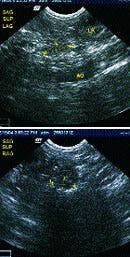
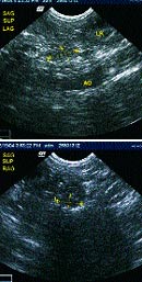
Image 6 and 7.
Dogs with primary hypoadrenocorticism will exhibit a subnormal response to ACTH administration. The baseline cortisol concentration is usually low or undetectable and the post-ACTH cortisol concentration is also low or undetectable. Endogenous plasma ACTH concentrations are dramatically increased in animals with primary hypoadrenocorticism as a result of loss of negative feedback to the pituitary gland caused by decreased serum cortisol concentrations. In the case of secondary hypoadrenocorticism, which is caused by a pituitary deficiency of ACTH, the endogenous ACTH concentrations are usually decreased (<20 pg/ml). The response to exogenous ACTH is diminished, but not as dramatically as for primary hypoadrenocorticism. Baseline cortisol and post-ACTH cortisol concentrations may be in the normal range.
Treatment
Treatment of the Addisonian crisis includes: 1) fluid therapy for electrolyte stabilization, 2) glucocorticoid replacement therapy, 3) treatment of gastrointestinal disease, and 4) mineralocorticoid replacement therapy.
Normal saline solution without potassium supplementation is the preferred fluid solution for the hypoadrenal crises. Treatment of hyperkalemia can be achieved using fluid therapy alone. If hyperkalemia is life-threatening, intravenous administration of calcium chloride or calcium gluconate may be used to counteract the adverse effects of potassium on the heart. Glucocorticoid and mineralocorticoid therapy must be initiated after diagnostic tests for hypoadrenocorticism have been performed. Glucocorticoid therapy, using ultra-short acting corticosteroids such as dexamethasone sodium phosphate and prednisolone sodium succinate, is indicated. Dexamethasone may be preferred in some dogs that require immediate glucocorticoid administration, as it will not interfere with the cortisol assay; in addition, a single dose of short-acting corticosteroid will not suppress the hypothalamic pituitary adrenal axis.
Long-term therapy of primary hypoadrenocorticism involves the use of mineralocorticoid supplementation as oral fludrocortisone (0.1 mg per 10 lb orally every 24 hours) or injectable deoxycorticosterone pivalate (2 mg/kg every 21-30 days). Electrolytes should be monitored once weekly until the dog is stable on replacement therapy. Most dogs with secondary hypoadrenocorticism and those supplemented with deoxycorticosterone pivalate require a low dose of glucocorticoids (0.2 mg/kg orally every 24 hours). About 50 percent of Addisonian cases supplemented with fludrocortisone require glucocorticoid supplementation.


")


