The respiratory system: From the nose to the diaphragm (Proceedings)
The respiratory system extends from the tip of the nose to the diaphragm.
General Comments
• The respiratory system extends from the tip of the nose to the diaphragm.
• Take good quality radiographs.
• Take as many radiographs as needed to make the diagnosis or to determine that other diagnostic methods are needed.
• Take sequential radiographs to monitor a disease process and response to therapy.
• Intervention may be required to obtain a definitive diagnosis.
• Some animals never get completely better and others go on to die despite your best efforts.
• Have the radiographs reviewed by the radiologist if you are not sure what is happening.
Anatomical Structures
The x-rays pass through many structures when the thorax is radiographed and all of these structures can have different presentations depending on their configuration. These structures are:


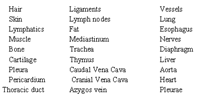
Lung Lobes:


Common Conditions of The Nasal Cavity and Sinuses
Infection (Bacterial, viral, fungal)
Foreign body
Trauma
Neoplasia
Parasitism
Congenital
Common Conditions Of The Trachea
Congenital Malformation (hypoplasia)
Trauma
Neoplasia
Foreign body
Parasitism
Chondromalacia
Infection
Common Conditions Of The Bronchi
Bronchitis (the dog has been used as an animal model of this disease in humans). Bronchiectasis
Pathology of the Lung
Alveolar air spaces
Interstitial tissues
Airways
Vessels
a) Radiographic findings when disease processes involve the alveolar:
Air bronchograms
Air Alveolograms
Ill-defined infiltrates
Lobar border visualization
b) Radiographic findings when disease processes involve the interstitial tissues:
Interstitial Pattern-
Short linear opacities criss-cross randomly
Nodular densities round or irregular in contour with well-defined borders,
admixture linear and nodular densities.
c) Radiographic findings when disease processes involve the bronchi and vessels
Bronchovascular Pattern:
Vessel or conducting airways change in appearance,
Increased prominence
Decreased prominence
Ill-defined borders
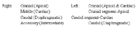
Vessels - Cranial lung lobe vessels (artery or vein) at edge of cardiac silhouette should be approximately ¾ diameter of the 3rd or 4th rib at approximately the level of the trachea and the transverse diameter of the vessels should be equal to each other.


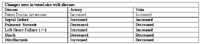
Changes seen in vessel size with disease:
Changes Normally Seen as Animal Ages ("Normal For Age")
Pleural thickening
Increased linear markings - interstitial fibrosis
Nodular densities - occasionally calcified - metaplastic mineralization-alveolar microlithiasis, osteomata
Increased density of tracheal and bronchial walls - mineralized cartilages
Hyperlucency -emphysema., air trapping
The Lung - Radiographic Assessment
Like any other organ, the lung can respond to noxious stimuli in a finite number of ways. One can try to emorize a distinctive radiographic appearance for each type of lung lesion; however, the final result will probably be utter frustration as the lung changes all begin to look alike.
As such, a systematic approach based on morphoanatomic structures is suggested. The basis for this system is pattern recognition; once the pattern is recognized, then a list of differential diagnoses can be made. Subsequent tests can be performed to exclude or confirm a diagnosis.
When Evaluating The Lungs, You Should Know
Pattern of involvement
Lung lobes/regions involved
History
The Three Patterns Recognized Stem from Lesions Involving
Alveolar Pattern
The alveolar pattern develops as the air within the alveolar ducts and alveoli are replaced by a substance of soft tissue opacity. Often, we cannot tell whether it is blood, inflammatory or non-inflammatory exudate, aspirated material, neoplastic cells or a collapsed lung that has caused the change in lung density; however, we can say that the alveoli no longer contain air (no longer are radiolucent but are of a soft tissue opacity). Radiographically, the alveolar pattern may show one or more of the following signs:
• Lobar involvement: as one lobe becomes opacified and the surrounding lobes remain aerated, the borders of the involved lung lobe become visible.
• Air bronchograms: if the bronchi and bronchioles contain air while the surrounding alveoli are of a soft tissue density (radiopaque); then the air filled bronchi and bronchioles appear as branching radiolucencies surrounded by radiopaque alveoli.
• Air alveolograms: occur when there is a patchy intermixing of opacified groups of alveoli and normal aerated radiolucent alveoli. The lung will then appear as a mottled admixture of radiolucencies and radiopacities.
• Ill-defined fluffy or wispy margins of infiltrate: representing opacified alveoli that may either have an indistinct border with normal aerated lung or become confluent and coalesce with other regions of diseased lung.
Recognition of any or all of the above alveolar changes indicates alveolar air spaces are involved.
Interstitial Pattern
Interstitial tissues of the lung consist of the supporting collagenous and elastic tissues, smaller vessels, capillaries, lymphatics and an admixture of cell types located permanently in the supporting network or migrating into and out of it, basement membranes and body fluids. It is the tissues on which the alveolar lining cells reside and through which blood and gasses move. Although barely perceptible as an entity in a normal thoracic radiograph, their presence is attested to by the fact that normal aerated lung is still more radiopaque than just air (i.e. free gas in the pleural space/pneumothorax, emphysema, bulla, blebs—all contain air that is more radiolucent than normal lung).
In a disease process, the interstitial tissues may thicken because of an increase in the number of cell types and fluid normally present or because of proliferation of altered cells or migration of new cell types into this tissue.
Bronchovascular Pattern
The bronchi and blood vascular supply are usually seen to a variable extent in the normal thoracic radiograph. When their presence becomes abnormally obvious or their size changes, then one can say there is a lesion occurring in the Bronchovascular component of the lung. If the vessels are altered, then the vascular component is abnormal and if the bronchi are altered, then the bronchial component is said to be abnormal.
Normally, the walls of the larger bronchi can be seen as fine-lined soft tissue opacities within the lung that taper as they extend peripherally. An artery and vein are located next to each bronchus. In the lateral radiograph, the artery is dorsal and the vein is ventral to the bronchus. In the dorsoventral radiograph, the artery is located lateral and the vein medial to the bronchus.
If the bronchus is seen in cross section, it will appear as a circular density with a lucent center. The density occurs because of the soft tissue components of the bronchial wall; whereas, the radiolucent center represents the air within the bronchial lumen.
The bronchial tree can become prominent if the alveoli are filled with soft tissue opacity material, if their walls thicken or if the cartilaginous components ossify or calcify. Any or all of these changes may occur with ronchitis, bronchiectasis, old age degeneration and calcification, bronchial neoplasm or any disease process affecting the air passages.
Normal bronchi can appear prominent if the surrounding alveoli become opacified, the ability to see the bronchi in this situation is known as "air bronchograms".
Pneumothorax vs. Hyperinflated Lung!
You must make sure you can distinguish hyperinflated lung from pneumothorax. When a pneumothorax is resent, the lung is collapsed and it is therefore more opaque, free gas is present in the parietal pleural space so the edge of the collapsed lung can be seen, the vessels in the collapsed lung do not extend to the parietal pleural and the heart may be displaced away from the sternum; When hyperinflated lung is present, the lung is less opaque, free gas is not seen in the pleural space, vessels can be seen extending to the pleura and the heart may be lifted away from the sternum. If you insert a transthoracic needle into a chest that you believe is a pneumothorax and it is truly a hyperinflated lung, you may kill the animal!!!