Perform exploratory laparotomy with large intra-abdominal soft tissue mass
Signalment. Feline, domestic shorthair, 13 years old, female spayed, 13 lbs. Clinical history. The owner presents the cat for recent hair loss on its back.
Signalment
Feline, domestic shorthair, 13 years old, female spayed, 13 lbs.


Clinical history
The owner presents the cat for recent hair loss on its back.
Physical examination
The findings show rectal temperature 101.8°F, pink mucous membranes, mild to moderate dental disease, and very firm abdominal mass in the ventral abdomen. Normal thoracic auscultation is noted.


Laboratory results
The cat tested negative for FeLV and FIV infection. A complete blood count, serum chemistry profile, and urinalysis were performed and are outlined in Table 1.
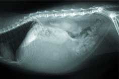
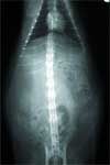
Radiographic review:
Survey thoracic and abdominal radiographs are done. The thoracic radiographic views are unremarkable. The abdominal radiographs follow:


My comments
The abdominal radiographs show a large mass effect located in the ventral region of the cranial to mid-abdomen that is displacing surrounding abdominal organs.
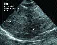
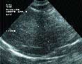
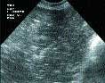
Ultrasound examination
Thorough abdominal ultrasound is performed with the cat positioned in dorsal recumbency.
My comments
The liver has an abnormal shape and shows a mixed echogenicity. There is increased echogenicity in the right lateral and caudate liver lobes. No masses noted within the liver parenchyma. The gall bladder is mildly distended, and its walls are not thickened or hyperechoic. The gall bladder does contain some sludge material. The spleen shows an inhomogeneous texture - no masses noted. A nodular lesion is present in the body of the spleen – this is an insignificant benign lesion.

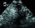

The left and right kidneys are similar in size and shape. The left kidney shows increased echogenicity. The right kidney shows an inhomogeneous texture. No masses or calculi were noted in either kidney. The urinary bladder is distended with urine and contains some urine sediment material; no masses or calculi noted. The left and right adrenal glands are similar in size and shape. The stomach, small intestines and colon are normal. The pancreas is not visualized. A large echogenic mass with no visible capsule is located caudal to the liver, dorsal to the spleen and cranial to the left kidney. Fine needle aspirations of this echogenic mass indicate a fatty appearance.
Case management
The tentative diagnosis is an intra-abdominal neoplasia. I suspect that this abdominal mass is an intra-abdominal lipoma. The general radiographic appearance of this soft-tissue mass is most compatible with a lipoma. At this point, an exploratory laparotomy should be performed for possible removal (or debulking) of this large intra-abdominal soft tissue mass.

One should to be careful and make sure all surface vessels are observed during the dissection of this soft tissue mass. Also, do cut through the soft tissue mass once excised from the abdomen for firm tissue and submit firm tissue samples for histopathologic examination to rule out a liposarcoma or lipomatosis.
Feline lipoma review
Typical dialogue from a cat owner about lipoma might include: "My cat developed a lump of fat under his ear, and it needs to be removed. It has grown during the past year since I had it checked, and the veterinarian said it was fat and not to worry. Now I'm worried. Is it a difficult operation? I'm just kind of mad that it wasn't removed when I first discovered it. Is it normal procedure to wait?"
Potential answer to the question: Fatty tumors (lipomas) are not as common in cats as they are in other species, but they do occur. Usually it is possible to identify a lipoma quite easily by aspiration of the lump since only fat cells are recovered when this is done. Subcutaneous lipomas do have a very characteristic feel as well. These are very easy to remove in most cases, and it is standard practice in veterinary medicine to leave them alone unless they begin to interfere with normal activity or function of some organ. The only real problem with waiting would be misidentification of the lump originally.

Fortunately, most of the things that feel like lipomas are also benign, so the odds are good that your cat will have a routine, uneventful surgery. You should have any excised lump examined by a veterinary pathologist to ensure the removed lump represents a benign lipoma.
Benign lipomas most commonly occur in the subcutaneous tissues of the thorax and abdomen. See the Web site www.vet.uga.edu/vpp/CLERK/Nation for additional details.
Lipomas are typically soft masses that are easily movable in the subcutis. Benign lipomas can also occur infrequently within the thoracic and/or abdominal cavities. Lipomas are on cross section white and oily and may have a thin capsule. Large lipomas may show central necrosis.
Cytologic examination of aspirate smears shows primarily mature adipocytes. Surgical excision or monitoring for changes in size and shape of the subcutaneous mass(es) are the preferred clinical management in most affected animals.


Benign lipomas may also occur as invasive and nonencapsulated. Although a benign tumor, they may infiltrate soft tissues, especially muscles, fasciae, tendons, blood vessels, lymph nodes, joint capsules and nerves. Muscle infiltration is typically so extensive that surgical cures are nearly impossible. Owners should be informed that at least 50 percent of animals affected with infiltrative lipoma have recurrence after seemingly complete surgical excision, except for those that have limb amputation for appendicular tumor.
Amputation of an affected limb should be recommended only when the tumor affects the animal's quality of life; this tumor causes little problem unless it interferes with limb movement, causes pressure-related pain or develops in a vitally important anatomic site. Infiltrative lipoma has not been reported to metastasize. However, there is one report of bone and joint invasion.


")


