Negative palmar angle syndrome in racing horses
Early recognition and correcton can prevent this potentially career-ending pathology.
A client calls to say his performance horse is slightly off. You arrive at the barn and begin your examination by pulling out your hoof testers, expecting to find an abscess or perhaps a close nail. The hoof testers indicate pain over the heels and navicular area. You mentally go down the differential list: bruising, corns, sheared heels, thrush, foreign body, navicular lesions, bursitis, tendonitis, desmitis, synovitis, arthritis, fracture, etc.
But one thing missing from that list, a common cause of lameness that's often overlooked, is negative palmar angle syndrome (NPAS). NPAS refers to the condition of progressive heel collapse and its consequences on gait and performance. Treatment and outcome are facilitated by grading the severity according to physical and radiographic features. While Grades I (mild) and II (moderate) can be corrected with trimming and routine shoeing, Grades III (severe) and IV (complicated by flexor contracture) require more intensive mechanical intervention and patience.
Physical characteristics
The typical foot with NPAS is often labeled as a long toe, low heel foot or the specific heel abnormality as an under-run or under-slung heel. While these terms ably describe some of the common external abnormalities of the hoof capsule, they do little to describe what's going on inside. The damage to the tissues within the collapsing or collapsed heel area, including the various coria (e.g., wall, sole, bar, frog), and the bony, synovial and connective tissue structures is, as the saying goes, out of sight, out of mind.
In addition to the classic long toe, low heel appearance of the hoof, signs of NPAS include landing toe first, painful or awkward breakover medially or laterally when asked to turn, anxiety when shoeing, resistance during training, inability to take canter leads and lameness that ranges from mild to severe depending on the type and severity of the associated pathology.
Unfortunately, it usually takes persistent lameness for us to realize that something is profoundly wrong with these feet. And even then, we search for specific abnormalities to which we can pin a medical label, such as navicular bursitis or deep flexor tendonitis. We continue to miss the point that these medical conditions often are an end result of structural failure of the hoof capsule and, thus, damage to its contents. In my opinion, recognizing this problem early and working to correct it can prevent many of these potentially career-ending pathologies.

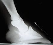

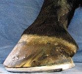
Photo 1: A horse with collapsed heels often has 50 percent to 60 percent of the solar surface area in front of the frog.
Identifying this problem is easy. Horses with weak or collapsed heels often have an elongated foot instead of a nicely rounded one. A well-trimmed healthy hoof has strong heels that are set back to the widest part of the frog. And when looking at the solar surface of the hoof, two-thirds of the surface area is behind the apex of the frog. In contrast, a horse with collapsed heels often has 50 percent to 60 percent of the solar surface area in front of the frog (Photo 1).
Radiographic characteristics
The presence of collapsed heels and a hoof capsule that is "running forward," as evidenced by examination of the solar surface as described above, should be a red flag. Regardless of whether the horse is lame at the time, it is important to obtain lateral radiographs of the feet. Use a radiographic technique that yields good soft tissue detail, and center the beam at the solar margin of the third phalanx (P3) to ensure a true lateral. These radiographs are important both to confirm the presence of a negative palmar or plantar angle (PA) and to develop an appropriate treatment plan (see "All about PA").

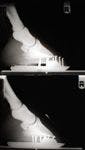
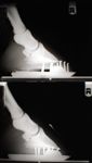
Photo 2: Relative sole depth is reversed in a foot with NPAS, so there is more depth under the tip of the third phalanx than under the wings.
Although several subtle abnormalities may be evident on a good soft tissue radiograph in feet with NPAS, the two most obvious and most important are a negative PA and abnormal digital alignment (DA).
- PA. When looking at the area beneath P3 on a true lateral radiograph of a healthy foot, there is more sole depth under the wings of P3 than under the tip, thereby creating a positive PA. Relative sole depth at these two locations is reversed in a foot with NPAS, so there is more sole depth under the tip of P3 than under the wings (Photo 2).
- DA. On a lateral radiograph that includes the pastern, the long axes of the three phalanges (and their dorsal surfaces) can be represented by a straight line in a well-trimmed horse with healthy feet. This normal alignment is broken in a horse with NPAS by the displacement of P3 as a consequence of distortion and ultimate collapse of the hoof capsule in the heel region. In fact, it is not uncommon to find osteophytes in the region of the extensor process of P3 in these feet, as the distal displacement of the wings of P3 tips the extensor process closer to the dorsal surface of P2.
Possible mechanisms
As professionals, we have long recognized that this hoof shape and these radiographic changes are abnormal. What we perhaps have failed to fully appreciate is the range of consequences and the long-term impact of this abnormality. Weakening and collapse of the horn in the heel region allows P3 to displace (in short, for the wings to drop, and, thus, for the entire bone to tip up). In the process, it alters the loads on various structures within the hoof capsule, rendering them more vulnerable to overload even under conditions of normal use. Whether a horse is at rest or in motion, repetitive and cumulative damage occurs in these feet because the reserve capacity of this marvelous structure (the equine foot) is lost when the heels collapse.


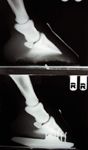
Photo 3: In the Grade I form of NPAS, sufficient sole depth under the tip of the third phalanx is present, so trimming alone can restore a normal plantar angle and digital alignment.
In fact, many of the specific digital pathologies we can now identify with sophisticated imaging techniques (e.g., subtle abnormalities in the navicular bone and related structures, deep digital flexor tenopathies, distal interphalangeal synovitis or arthritis) may have their foundation in this rather ordinary phenomenon of heel collapse. For example, calcification of the navicular portion of the deep flexor tendon is common in horses with NPAS, as is remodeling of the navicular bone and P3.


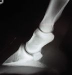
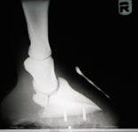
Photo 4: In the Grade II form of NPAS, there is insufficient sole depth under the tip of the third phalanx to correct the problem with trimming alone.
As for what causes NPAS, it is likely a multifactorial problem. Research has shown that when Thoroughbreds start galloping (i.e., race training), their hoof angles decrease unrelated to farriery changes. It has also been observed that foals born and raised in sandy, humid soil conditions do not develop good heels. There may be a genetic component to this problem as well. And improper trimming and shoeing must be on the list of contributing factors. Furthermore, farriers who do not have a good understanding of NPAS and its management may have difficulty correcting these feet and may actually worsen the condition. For completeness, it's also worth noting that NPAS can be a consequence of deep flexor tenotomy, particularly if DA is not properly restored at the time of surgery.


Photos 5: In the Grade III form of NPAS as shown in this photograph and radiograph, the hoof is trimmed to create two planes on the solar surface. A plantar angle of zero is created in the forward portion of the foot, and the heels are "floated" behind the quarters.
Grading system
The diagnosis and treatment of NPAS is discussed at length in the November 2010 issue of The Journal of Equine Veterinary Science.1 That paper focuses on a simple four-tier grading system for categorizing and managing these feet, as the severity of the problem dictates the finer points of the treatment approach. Briefly, the grading system can be summarized as follows:
- Grade I: mild. There is sufficient sole depth under the tip of P3 that trimming alone can restore a normal PA and DA (Photo 3).
- Grade II: moderate. There is insufficient sole depth under the tip of P3 to correct the problem with trimming alone; the hoof is trimmed to achieve a PA of at least zero (and into the positive range, if possible), and then a full rocker shoe is used to correct the PA and DA (Photo 4).
- Grade III: severe. The hoof is trimmed to create two planes on the solar surface. A PA of zero is created in the forward portion of the foot (from the quarters to the toe), and the heels are "floated" behind the quarters. The heels are lightly rasped to remove the defective horn and bring the bearing surface back to the widest part of the frog. A full rocker shoe is nailed to the forward portion of the hoof, and a soft composite material (e.g., Equi-Pak—Vettec) is used to fill the gap between shoe and heel while the heels regenerate (Photos 5 & 6).
- Grade IV: contracted. These cases are complicated by flexor contracture, causing a "post-legged" appearance in which the pastern is more vertical than normal and the fetlock angle is reduced. These horses may even knuckle forward at the fetlock. Correction is involved and protracted but can be rewarding (Photo 7).

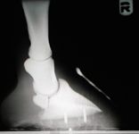
Photos 6: In the Grade III form of NPAS as shown in this photograph and radiograph, the hoof is trimmed to create two planes on the solar surface. A plantar angle of zero is created in the forward portion of the foot, and the heels are "floated" behind the quarters.
Treatment
In my experience, success depends on obtaining good lateral radiographs, gauging the severity of NPAS and applying an appropriate trimming and shoeing strategy according to the severity of the disorder and the needs of the individual horse.


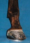
Photo 7: Grade IV forms of NPAS are complicated by flexor contracture, causing a "post-legged" appearance in which the pastern is more vertical than normal and the fetlock angle is reduced.
If this condition is not treated appropriately, severe contracture of the heels can develop, followed by quarter cracks or heel fractures, either of which makes the management of NPAS more complicated and delays the return to soundness. Suppurating corns may also develop and can lead to proximal migration of the infection into the collateral cartilages and even the distal interphalangeal joint.
Conclusion
NPAS is a treatable condition. Unfortunately, many affected horses that are not treated properly are retired, some after having undergone neurectomies because nothing else has made them comfortable. But with a little know-how and some patience, you can treat these cases and be rewarded for your efforts with a satisfied client and a happy horse.
Andrea E. Floyd, DVM, has specialized in equine podiatry for more than 25 years. She is the owner of Serenity Equine, Evington, Va., and the author of Equine Podiatry. Dr. Floyd is a member of the American Veterinary Medical Association, American Association of Equine Practitioners and the American Farriers Association.
REFERENCE
1. Floyd AE. Use of a grading system to facilitate treatment and prognosis in horses with negative palmar angle syndrome (heel collapse): 107 cases. J Equine Vet Sci 2010;30(11):[in press].


