MRI propels lameness diagnosis
Though many experts believe magnetic resonance imaging (MRI) is underused in equine veterinary medicine, greater availability of the technology and access to interpretation experts are facilitating greater adoption, especially when diagnosing lameness.
Though many experts believe magnetic resonance imaging (MRI) is underused in equine veterinary medicine, greater availability of the technology and access to interpretation experts are facilitating greater adoption, especially when diagnosing lameness.


The integrated table at Alamo Pintado Equine Medical Center is able to hold 550 pounds, so they are able to place a portion of the horse on the table within the magnet, allowing them to image a larger portion of the horse.
"I think the message to equine practitioners right now is that they tend to think that they don't have to worry about MRI because they can't afford it and that it is not available," says Bob Schneider, DVM, Dipl. ACVS, equine orthopedic surgeon at the Washington State University Veterinary Teaching Hospital. "But it is available. We're not talking about something that's in the near future. It's pretty much in use widely right now."


MRI has exceptional sensitivity and specificity, provides physiologic and anatomic information about bone and soft tissue and is useful to identify occult subchondral bone and soft-tissue injuries. MRI has made it possible to identify injury to the bone, tendons and ligaments that could not previously be identified in horses.


MRI provides several sequences composed of hundreds of images in each sequence.


Sagittal view of a cushingoid horse with no macroscopic evidence of an adenoma present.
"We have been able to diagnose lameness problems we didn't even realize existed before MRI," Schneider says.
Ultrasound works for some problems but has limitations in imaging all the structures of the foot. Radiographs can detect major bone injuries clearly, but MRI is more sensitive, provides an even clearer picture and can identify accompanying soft-tissue injury. MRI produces a clear display of anatomy on any plane and allows for visualization of different tissue structures. The image along with a good physical can cue clinicians into what might be significant about an animal's presentation.
Helping make a firm diagnosis
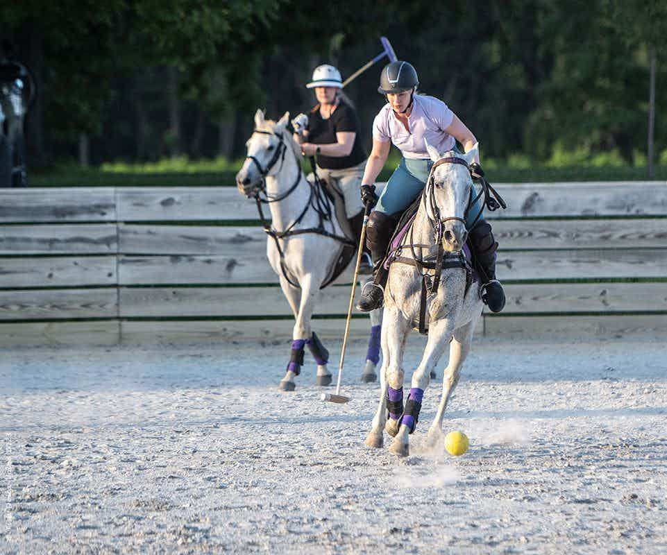
When lameness develops in performance horses, a definitive diagnosis is necessary to provide effective treatment and an accurate prognosis. Lameness evaluations, radiographs and ultrasound are typically used to identify the cause of lameness. MRI should be considered in cases when the lameness has been localized to the distal portion of the limb, but a definitive diagnosis can not be made from radiographs or ultrasound, says Chad Zubrod, DVM, MS, Dipl. ACVS, of Oakridge Equine Hospital in Edmond, Okla. The results of MRI evaluation may change the type of treatment prescribed in many cases. New and novel treatments may be beneficial and improve the horse's prognosis for return to athletic performance.
Fractured navicular bone at the central eminence with mild palmar displacement. Increased fluid accumulation at the central eminence is indicative of inflammation.
"There is a whole realm of diagnosis I would never consider making before — potentially painful injuries, which were essentially next to impossible to diagnose without MRI," says Jake Hersman, DVM, of Animal Imaging in Irving, Texas. "With MRI, it has been rewarding noting various soft-tissue injuries, such as deep flexor tendon or collateral ligament injuries, that have changed the way we handle some of these horses. We could get some very good indications that a horse had a collateral ligament (tear) or possibly other injuries, but we never were certain. There are also a whole lot of changes in the navicular bone itself that we see with MRI that we couldn't with regular films."


Canine with a likely acquired portosystemic shunt.
Availability
Animal Imaging uses a 1.0-Tesla high-field magnet, which requires general anesthesia. The actual imaging time for a unilateral sequence is about 45 minutes, and a bilateral sequence requires about twice as long. With the system used at Animal Imaging, images can be sent to radiologists — typically Washington State University clinician Pat Gavin, DVM, PhD, Dipl. ACVR — for review, as well as other consulting colleagues, as soon as they are acquired. Generally, MRI fees run between $1,000-1,600 and are considered reasonable compared to exploratory surgery that used to be required to diagnose some causes of equine lameness, sources say.


This proton density transverse section through the right-hind foot just proximal to the navicular bone shows a dorsal- to plantar-directed tear in the medial lobe of the deep digital flexor tendon, shown by a high signal intensity defect (arrow) within the normally low signal intensity deep digital flexor tendon (black).
"When a horse is referred to our facility for imaging, the procedure is performed, and the horse is sent back to the referring veterinarian for treatment as long as the referring veterinarian has the means necessary to treat the horse," Zubrod explains. If not, the patient is treated in-house after consulting with the referring veterinarian. Following imaging, they will send a report and images to the owner and referring veterinarian that describes the abnormalities found on the MRI and outlines the recommended treatment or treatments. This eliminates the need for a veterinarian to know how to read an MRI in order to refer a patient for this imaging modality.


This proton density transverse section through the left-front pastern shows desmitis of the lateral branch of the oblique distal sesamoidean ligament, shown by increased signal intensity centrally (white) in the enlarged ligament (arrows).
"I think that referring veterinarians need to know which cases would benefit from an MRI and recommend MRI to the client," Zubrod says. "Getting a definitive diagnosis is the best thing for the patient, and we should not make the decision for the client as to whether an MRI is economically feasible for them. As veterinarians, we should be offering the client the best diagnostics and treatment available, and let them make the decisions whether or not to utilize these techniques."
Technique, use
MRI is unique from other imaging techniques in that there are different sequences to look at, and each sequence contains hundreds of images. In the same tissue, fluid, for example, can look different on different sequences. In certain sequences fluid is white, while in other sequences, fluid is black. Depending on the imaging sequence, the same tissue will have different signal intensities.


This gradient echo coronal section (left) through the right-hind fetlock joint shows subchondral bone damage to the medial condyle of the third metacarpal bone, shown by increased signal intensity (white) indicative of inflammatory fluid just above the joint (arrow).
Consequently, learning to use MRI effectively requires a crash course in the process, the technology and of course, practice in interpreting the images.
"It is a matter of first learning the physics of MRI, understanding how MRI works," notes Natasha Werpy, DVM, who specializes in equine imaging and runs the MRI center at the Equine Orthopaedic Research Center at Colorado State University.
Her colleagues concur.
"There is a learning curve as far as interpreting MRI images, though there are some veterinarians who have got quite a lot of expertise at interpreting them, it's really a role for specialists," says Wayne McIlwraith, BVSc, PhD, DSc, FRCVS, Dipl. ACVS, director of the Equine Orthopaedic Research Center at Colorado State University.
This proton density sagittal section through the right front fetlock joint shows subchondral bone and cartilage damage to the distal aspect of the third metacarpal bone, shown by increased signal intensity (white) indicating inflammatory fluid surrounded by decreased signal intensity (black) indicating sclerosis (arrows).
Werpy works at a reading center that helps interpret the images the center produces. She says she can log onto a permissions-based server to view images as quickly as she needs to.
This interpretive expertise enables practitioners of all skill levels to use the technology.
Veterinary MRI units are available to equine practitioners in many locations around the country, including many universities and larger equine clinics.
This proton density transverse section through the left front proximal metacarpus shows desmitis of the dorsomedial aspect of the proximal suspensory ligament, shown by increased signal intensity (grey/white instead of black) diffusely in this region (arrows).
During the next few years, there will be more training courses offered to learn how to interpret the images, as well as a driving force for more equine MRI radiologists, as they realize the benefits of the technology.
More units will come on line, too, making MRI a viable option in almost any part of the country.
"The thing about MRI that is so important to realize is that it really allows you to differentiate different types of lesions at a level that we were never able to do before," Werpy says.


This proton density transverse section through the left front coffin joint of a horse with subchondral bone and cartilage damage of the distal aspect of the second phalanx, as shown by an area of decreased signal intensity (arrows) in the medial condyle.
The many stages of injuries, from acute lesions with fluid to scar tissue and adhesions, will have better opportunity for diagnosis with MRI, and those horses likely will get treated differently as equine practitioners will be developing treatments for different stages of injuries that they were unable to do before. Thus, MRI is on the cutting edge of targeted treatment, especially in performance horses.
"Over time we will be able to separate lesions by stages and types. They might be better treated with specific treatments for a specific stage of injury," Werpy says. "We'll be able to hopefully develop treatments more specifically to give a better prognosis. That is the way I look at it when I read these studies."