Malocclusion and tooth-on-tooth trauma
While an uncommon option, odontoplasty can resolve some cases.
The two main indications for therapeutic intervention when dealing with malocclusions in veterinary dentistry are tooth-on-tooth trauma and tooth-on-tissue trauma. This article focuses on tooth-on-tooth trauma, but tooth-on-tissue trauma may also be occurring; both of these conditions must be addressed.
Treatment options for tooth-on-tooth trauma
The result of malocclusions involving tooth-on-tooth trauma is abnormal tooth wear that often results in dentin or pulp exposure in one or both teeth. Subsequent loss of tooth vitality is a likely end result of any tooth-on-tooth malocclusion. Intervention is indicated whenever this relationship occurs. Orthodontics, endodontics, extraction and, occasionally, odontoplasty are all possible treatment modalities. The decision on which treatment (or which combination of treatments) to use depends on the often complex arrangement present before and after attempts at resolution.
When evaluating malocclusions, the top consideration should always be the least-invasive treatment for the patient. The question then becomes: What is the most feasible line of therapy?
If, for instance, extractions are the obvious choice, it may not be what the pet guardian has in mind. If the condition dictates that orthodontic movement is also an option, and referral is available, orthodontics must be discussed and offered as an alternative.
Not only are careful evaluation and planned therapy critical in these cases, post-therapeutic anatomy must be evaluated to assess the new occlusion once adjustments have been made: Is there new tooth-on-tooth or tooth-on-tissue trauma present? If I alter the crown height of this tooth
2 mm, how will that affect the rest of the dentition?
Case study: Odontoplasty
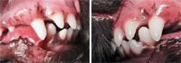
A 10-month-old spayed female mixed-breed dog had a persistent deciduous right mandibular canine tooth (804) (Photo 1). Radiographs demonstrated near total root resorption, and the tooth was easily extracted.


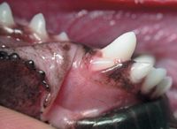
Photo 1: A persistent deciduous right mandibular canine tooth (804) in a 10-month-old dog. This tooth was extracted.
More important, the left mandibular canine tooth (304) was contacting the distal aspect of the left maxillary third incisor (203) (Photo 2). Sole contact of this type does not disperse occlusal forces evenly, but rather concentrates them on two teeth. Under normal conditions, occlusal contact is shared by multiple teeth, including the incisors and molars in dogs. Left alone, wear will likely progress to damage the incisor, exposing dentin and compromising tooth vitality. Mandibular canine contact with the palatal mucosa is also likely if wear continues.

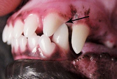

Photo 2: The left mandibular canine tooth (304) is contacting the distal aspect of the left maxillary third incisor (203).
Selective extractions, crown amputations and vital pulp therapy or movement are all options, but a simple, minimally invasive procedure called odontoplasty is the best option in this particular case.
Odontoplasty is not commonly used because its effectiveness is limited to select cases in which minor crown reduction is needed. Odontoplasty requires using a sterile, water-cooled, fine diamond bur to remove dentin and enamel, while not entering the pulp chamber. A minimum of 1 mm of dentin should be left coronal to the pulp chamber. Dental radiography is used to estimate this landmark.
If dentin is exposed, dentinal bonding, followed by a very thin layer of composite, seals the open dentinal tubules. Polishing disks prepare the composite for two additional layers of unfilled resin. This procedure allows the crown height to be reduced to eliminate all destructive occlusal forces. Consider using radiography to determine the tooth's further eruptive potential (Photo 3). In this case, the apices of the mandibular canines were radiographically closed, and further eruption was not anticipated.


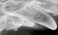
Photo 3: A radiograph of another patient showing an open apex of the right mandibular canine tooth (104).
A closer look at the dog's dentition showed a close relationship between the right mandibular canine tooth (404) and the right maxillary third incisor (103) (Photo 4). If odontoplasty is performed as described, 404 will then likely contact 103. So consideration must be given to performing odontoplasty on tooth 404 as well (Photos 5A and 5B).


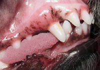
Photo 4: A close relationship between the right mandibular canine tooth (404) and the right maxillary third incisor (103).


Photos 5A and 5B: Odontoplasty is followed by dentinal bonding, a thin composite, polishing and two additional layers of bonding agent on both mandibular canines. The result of odontoplasty and the composite restoration is seen here for both mandibular canines.
Next, you must determine if any other malocclusions will result from the new arrangement. Extubating the patient momentarily to check the occlusion and confirm that there are no additional problems is generally required. In this case, the molars came to rest in proper occlusion, and the mandibular incisors came to rest on the cingula of the maxillary incisors, recreating the normal occlusion as seen at the two-week recheck (Photo 6).


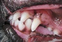
Photo 6: No additional problems were noted at the two-week recheck.
This concludes a three-part series on malocclusions. In the next series, I will demonstrate multiple examples of what appear to be normal mouths that radiographically are very abnormal.
Dr. Beckman is associated with Affiliated Veterinary Specialists, Orlando, Fla.; Florida Veterinary Dentistry and Oral Surgery, Punta Gorda, Fla.; Animal Emergency Center of Sandy Springs, Atlanta; and Dallas Veterinary Dentistry & Oral Surgery, Dallas. www.veterinarydentistry.net


