Making sense of respiratory patterns (Proceedings)
It is suspected that mammals generally adopt a respiratory pattern that meets their metabolic needs with the least metabolic energy cost.
It is suspected that mammals generally adopt a respiratory pattern that meets their metabolic needs with the least metabolic energy cost. Because of the phenotypic diversity in healthy dogs and cats, how this is achieved varies dramatically between patients - what works best for a Borzoi may not work at all for a Pug. In addition to these species and breed differences, respiratory diseases cause characteristic changes in respiratory frequency, tidal volume, airflow velocity, and patterns of muscle activation that can provide clues for the clinician to help localize the cause of abnormal respiratory function. This lecture will present the basic elements of how respiratory patterns change with disease states and emphasize important physical examination findings.
Dyspnea vs increased respiratory effort
The term dyspnea refers to the experience of distress secondary to respiratory disease. In man it is most often associated with increased resistance to airflow but may also be caused by anything leading to clinically significant hypoxemia or hypercapnia (especially if acute). Like pain, its presence is inferred from facial expressions and behavioral signs of distress. Dyspnea is a true medical emergency; our goal is to relieve it within minutes of presentation. In contrast, the terms labored breathing or increased respiratory effort refer to the physical manifestations of increased work of breathing. This may be objectively measured, but most of the time is inferred from subjective appraisal of physical signs. Whereas an animal with dyspnea is having a crisis, an animal with labored breathing may be very well adapted to its condition and be free from distress. Where a given patient lies on this spectrum must be inferred from their behavior. In general, as an animal approaches dyspnea it must focus more and more conscious effort on the act of breathing. Animals that are interactive and engaged with their environment usually still have significant physiological reserves; those that appear withdrawn and focused on the act of breathing may be close to the edge of respiratory arrest. A notable exception to this generalization is the puppy. Juvenile dogs will often remain active, alert, and hungry even with advanced respiratory failure and may go straight to the brink of death before showing compelling behavioral signs of distress.
Fixed airway obstructions
Neoplasia, constrictions, compression from abscesses or trauma: A 'fixed' obstruction refers to one that does not change appreciably with the phase of respiration. As the airway lumen narrows with advancing disease, the animal will adopt a prolonged inspiration and exhalation that is characteristic of fixed obstructions. Respiratory frequency falls as the inspiratory phase becomes noticeably prolonged. As the problem becomes more severe, more energy is expended on both inspiration and exhalation. The intercostal muscles 'suck in' from markedly negative pleural pressures created by vigorous diaphragm contraction, and the animal may actively exhale, first by contracting the intercostal muscles alone, later by a combination of intercostals and abdominal muscles. The term 'abdominal breathing', referring to active contraction of the abdominal wall, can ONLY assist with exhalation and is a response to air trapping that slows exhalation to unacceptable rates.
Neoplasia and constrictions from scarring are typically insidious in onset, initially causing clinical signs only with exercise and usually allowing plenty of time for adaptive strengthening of the respiratory muscles. If acute, as for example from cervical swelling from trauma, the animal may have the same physical features as aspiration of a foreign body, including dyspnea and cyanosis.
Foreign body: This disorder is characterized by an acute onset of respiratory difficulty, often accompanied by coughing/retching and behavioral signs of dyspnea. Marked cyanosis may be present as hypoxemia occurs before there is any adaptive shift in hemoglobin oxygen affinity and cardiovascular responses are vigorous, with good perfusion of skin and mucus membranes. In the author's experience, inspiration is more severely affected than expiration but this would be expected to vary with location of the obstruction. Most obstructions are at the level of the larynx or tracheal bifurcation.
Brachycephalic syndrome: This syndrome is classically characterized as restriction of the upper airway secondary to excessive or extreme phenotypic expression of the abnormalities selectively bred for in some chondrodysplastic breeds. Although some anatomic features of the syndrome produce a dynamic obstruction, most individuals have at least some component of fixed obstruction. Any combination of stenotic nares, stenotic nasal cavity, elongated soft palate, myopathy of the pharyngeal opening musculature, laryngeal dysplasia, everted laryngeal saccules, and trachea hypoplasia are possible.1-3 Collapse of the cervical and/or intrathoracic portion of the trachea is sometimes seen. Some dogs have concurrent abnormalities of respiratory drive4, and some acquire rib fractures and/or hiatal hernia.5 The breathing pattern is characterized by prolonged inspiration, and (usually) comparatively easy exhalation.6 Affected dogs routinely develop stertorous sounds with the slightest provocation. In spite of evidently high resistance to air flow, affected animals are generally not focused on breathing and relatively free from distress, even if their arterial pO2 is very low. This finding fits with the observation in many species that chronic hypercapnia and hypoxemia invoke adaptive responses that resets chemoreceptor response thresholds to relatively high values of pCO2 and low values of pO2.
Dynamic obstructions
Laryngeal paralysis: Although some dogs with idiopathic laryngeal paralysis have features of a fixed airway obstruction, this is probably the best example of a dynamic extrathoracic obstruction.7 In this disorder, deterioration of the recurrent laryngeal nerves results in a loss of innervation of the abductor muscles of the larynx. It may occur as an isolated neuromuscular abnormality or be part of a polyneuropathy that may include swallowing disorders and/or megaesophagus. Most often, it is insidious in onset and many owners may recognize that clinical signs have been present for months only in retrospect after a crisis. During inspiration, the larynx is prone to collapsing into the lumen secondary to the subatmospheric pressure that creates a Venturi effect as described in Bernoulli's principle. Vigorous inspiration becomes prolonged and either stertorous (if the laryngeal structures vibrate) or attended by a high pitched squeak (if obstruction is nearly complete). Many dogs seem to be able to prevent this for long periods by adopting an inspiratory pattern that is slow in frequency with a relatively prolonged inspiration and large tidal volume. They can thus avoid symptomatic obstruction unless forced to increase ventilation by the demands of activity or environment (by inducing panting from heat exposure). When signs occur in these dogs they often come on dramatically as the dog worsens the obstruction with rapidly escalating inspiratory efforts that in turn worsen the obstruction. Many present with cyanosis (from acute worsening of the obstruction) and hyperthermia (from the heavy muscular effort).
Tracheal collapse, bronchial collapse: These terms describe the obstruction of a large airway due to chondromalacia, compression, or in the case of the trachea, a redundant dorsal ligament. Clinical signs will depend upon which portion of the airway system is affected. Extra-thoracic tracheal collapse will tend to occur during inspiration when intraluminal pressure is subatmospheric and the dorsal ligament is sucked into the tracheal lumen. Intrathoracic collapse is more likely to occur during exhalation when pleural pressures approach (or exceed) intraluminal airway pressure. Either event may stimulate coughing via stimulation of submucosal nociceptors, but intrathoracic collapse usually causes more severe problems than cervical tracheal collapse. Animals with cervical tracheal collapse are often asymptomatic except with activity; once they begin collapsing the airway they may quickly transition to a pattern characterized by vigorous attempts at inspiration with loud stertor or sounds of 'choking off'. Animals with intrathoracic airway collapse may have no signs other than persistent cough. If severe, they may begin trapping air at relatively high lung volumes and be forced to recruit accessory abdominal muscles of expiration in an effort to forcibly 'burp' air past obstructed airways.
Feline asthma: If bronchospasm is severe, this syndrome has all the features of a fixed airway obstruction. In most patients, however, it is likely that restriction to airflow is worse on exhalation as small airway diameter falls with declining lung volume. Wheezes are commonly auscultated, but auscultatory evidence of large airway collapse is typically lacking.
Restrictive lung disease
Restrictive diseases are characterized by pulmonary changes that reduce lung compliance and make the lungs more difficult to inflate. At the same time, the underlying disorder typically compromises the oxygenating capacity of the lung. Arterial hypoxemia and lung inflammation or edema are direct respiratory stimulants and provoke an increase in desired minute alveolar ventilation. Thus, when severe, the animal is faced with the need to increase ventilation with lungs that are increasingly stiff and difficult to inflate. The archetypical pattern that results from this combination is breathing at a relatively fast rate and small tidal volume.
Clinically, the animal develops an increased frequency with rapid shallow inspirations. As inspiration becomes more difficult (or fatigue develops with acute illness), one of the first behavioral changes is a reluctance to lie in lateral recumbency. As difficulties progress, the dog may have trouble remaining in sternal recumbency for prolonged periods and position changes become more frequent. Finally it may be difficult to lie down for any significant period and the dog will prefer to sit or stand, even in the face of exhaustion. During this progression, the dog begins to extend the head and neck and recruit accessory muscles of inspiration, including the scalenes and the sternomastoids8;9. The forelimbs are abducted as breathing difficulty becomes greater. The head may bob up and down with respiratory effort. If the elastic recoil of the lung is sufficient, exhalation may be completely passive. If there is excessive air trapping from airway collapse, or if passive recoil of the lung does not provide sufficient emptying the dog may exhale actively (contraction of intercostal muscles) and may recruit accessory muscles of expiration (abdominal muscles).
In contrast to what is seen with airway obstruction or pleural space disease, hypoxemia from lung disease typically causes more pallor than cyanosis, yielding a pale-to-grey color of the mucus membranes. If the underlying cause is rapidly progressing the animal may develop fatigue of the respiratory muscles quickly and go on to develop hypoventilation (respiratory failure).
Pulmonary edema/pneumonia: Any increase in the water content of the lung tissue will reduce its compliance by increasing the opening pressure required to inflate pulmonary exchange units. As edema develops, regions of the lung will lose ventilation and venous admixture increases. Respiratory frequency increases, in some cases associated with stimulation of pulmonary C fibers (juxtacapillary receptors) from the increased fluid pressure of the interstitium, and in some from hypoxic stimulation of chemoreceptors. Because the lungs are relatively stiff, the prototypical pattern will be relatively shallow breaths at a relatively high frequency. As these disorders tend to develop rapidly, respiratory fatigue may develop quickly and hasten death in the absence of ventilatory support.
Idiopathic pulmonary fibrosis: This progressive disorder of cats and dogs results in loss of function (hypoxemia) and compliance, and is may be accompanied by cough. Compared to pulmonary edema, this disorder progresses relatively slowly and there is ample time for training of the respiratory muscles and development of fatigue resistance. Affected animals are often not in any respiratory distress until functional loss is severe.
Pleural space disease
The thoracic girth is normally maintained by the opposing forces of lung (which wants to collapse) and rib cage (which wants to spring outward). Separation of the two with a layer of air or fluid in the pleural space produces a reduction in lung volume and an increase in thoracic girth. The effects of pneumothorax on breathing has been studied more than the effects of pleural effusion, but it appears that dynamic lung compliance, inspiratory duration and tidal volume decrease significantly with either disorder.10;11 As lung volume falls exchange units are lost as the reduction in volume becomes sufficient to favor small airway collapse, leading to hypoxemia due to venous admixture and if severe, hypoventilation. At the same time this is occurring the total intrathoracic volume is growing, flattening the diaphragm (placing it at a mechanical disadvantage) and shifting external intercostal muscle function towards exhalation.
As the disorder progresses, inspiratory efforts increase and accessory muscles of inspiration may be recruited. The diaphragm will contract maximally, maintaining good excursion in spite of its flattened profile in exhalation. Pneumothorax directly stimulates phrenic nerve activity in a manner independent of the effects on gas exchange. 10
Diaphragmatic paralysis
Diaphragm paralysis may be caused by injury to the phrenic nerve(s), degeneration of the phrenic nerves, or severe motor unit diseases such as botulism. If the condition is isolated to phrenic nerve involvement, dogs compensate by increasing the activity of the intercostal, levator costae and parasternal muscles. Although this may be a conscious effort in response to chemical stimulation, contraction of the normal diaphragm stimulates receptors whose output inhibits efferent activity to the intercostal and levator costae muscles. Clinically, the dog has an increased frequency, marked inspiratory excursion of the anterior ½ of the thorax, and sometimes paradoxical movement of the abdomen. The latter is characterized by a failure of the abdomen to maintain its girth during inspiration or a frank reduction of girth if the diaphragm and abdominal viscera are sucked into the thorax by the vigorous inspiratory efforts happening up front.
Intercostal paralysis
Weakness or paralysis of the intercostal muscles may be seen as part of a generalized myopathy, motor unit disease, and spinal cord injury due to trauma or neoplasia. In this patient, the thoracic wall may fail to maintain its normal position during tidal breathing and move paradoxically during diaphragm contraction. Because these dogs are typically paralyzed, if their lungs are otherwise normal they usually have adequate respiratory function with diaphragm movement alone.
Disorders of the bony structures forming the thoracic bellows

Several disorders affect the bellows mechanism of respiration, including rib fractures and breakdown of the chondral arch. Rib fractures are painful and may limit tidal volume, prompting a compensatory increase in respiratory frequency. In addition, fractured ribs disrupt the action of the intercostal muscles and may produce paradoxical motions of regions of chest wall that further compromise ventilation.
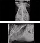
Some animals with stiff lungs or chronic airway disease will eventually suffer collapse of the costachondral arch (as depicted in a cat below), possibly secondary to excessive forces generated by a hypertrophied diaphragm. When this happens, the diaphragm is shortened, losing its dome shape and most of its mechanical advantage. When viewed with fluoroscopy, contraction of the diaphragm results in pulling the caudal aspect of the sternum closer to the spine, instead of forcing the caudal ribs to move in a cranial direction and increasing thoracic volume. Because these animals typically have chronic lung or airway disease and already suffer significant functional loss, their condition is often made acutely worse when the contribution of the diaphragm to inspiration is compromised.
Summary of signs to look for
1. Working hard to breath, behavioral and facial features of distress: This represent dyspnea – a true medical emergency that must be relieved within minutes!!
2. Working hard to breath, no behavioral signs of distress, interested in environment: This usually represents a successful adaptation to respiratory disease, not as severe of an emergency. Note that puppies may die of respiratory failure without showing evidence of dyspnea, but that would be rare in any alert cat or adult dog.
3. Cyanosis (very blue + normal CRT): Hypoxemia from airway obstruction or pleural space disease; cardiovascular function usually normal.
4. Pale grey, slow CRT: Hypoxemia from heart failure
5. Audible long-duration noise at mouth: Large airway obstruction
6. Long inspirations and exhalation: Constant large airway obstructions
7. Long inspiration, normal exhalation: Dynamic obstructions such as laryngeal paralysis
8. Long/forceful exhalation +/- cough: Intrathoracic airway collapse or obstruction
9. Contraction of abdomen: Occurs only in exhalation, represents recruitment of those accessory muscles of exhalation, and is called abdominal breathing. Seen when there is resistance to exhalation (e.g., small airway collapse), especially when the frequency (RR) is high.
10. Vigorous, large excursions of chest and abdomen: Rule out pleural space disease (especially if cyanosis is present!)
11. Fast, shallow breathing: Think stiff lungs: edema, fibrosis. Inflammation, disseminated neoplasia.
12. Refusal to lie lateral, head extended, forelimbs abducted, head bobbing: Seen with stiff lungs and large effort.
Reference List
Schotland HM, Insko EK, Panckeri KA, et al. Quantitative magnetic resonance imaging of upper airways musculature in an animal model of sleep apnea J Appl Physiol 1996;81(3):1339-1346.
Petrof BJ, Pack AI, Kelly AM, et al. Pharyngeal myopathy of loaded upper airway in dogs with sleep apnea. J Appl Physiol 1994;76(4):1746-1752.
Hendricks JC, Kovalski RJ, Kline LR. Phasic respiratory muscle patterns and sleep-disordered breathing during rapid eye movement sleep in the English bulldog. Am Rev Respir Dis 1991;144(5):1112-1120.
Hendricks JC, Kline LR, Kovalski RJ, et al. The English bulldog: a natural model of sleep-disordered breathing. J Appl Physiol 1987;63(4):1344-1350.
Hardie EM, Ramirez O, III, Clary EM, et al. Abnormalities of the thoracic bellows: stress fractures of the ribs and hiatal hernia. J Vet Intern Med 1998;12(4):279-287.
Amis TC, Kurpershoek C. Pattern of breathing in brachycephalic dogs. Am J Vet Res 1986;47(10):2200-2204.
Amis TC, Smith MM, Gaber CE, et al. Upper airway obstruction in canine laryngeal paralysis. Am J Vet Res 1986;47(5):1007-1010.
Farkas GA, Rochester DF. Contractile characteristics and operating lengths of canine neck inspiratory muscles. J Appl Physiol 1986;61(1):220-226.
Muza SR, Criner GJ, Kelsen SG. Effect of lung volume on the respiratory action of the canine sternomastoid. J Appl Physiol 1996;80(3):852-856.
Lee BP, Chiang ST, Hwang JC. Effects of intrapleural pressure on phrenic nerve activity 4. Chin J Physiol 1986;29(2):79-90.
Krell WS, Rodarte JR. Effects of acute pleural effusion on respiratory system mechanics in dogs. J Appl Physiol 1985;59(5):1458-1463.