Localization of azotemia: Test your skill
Azotemia is defined as an abnormal concentration of urea, creatinine, and other nonprotein nitrogenous substances in blood, plasma or serum.
Azotemia is defined as an abnormal concentration of urea, creatinine, and other nonprotein nitrogenous substances in blood, plasma or serum.
Azotemia is a laboratory finding with several fundamentally different causes. Since nonprotein nitrogenous compounds (including urea and creatinine) are endogenous substances, abnormal concentrations in serum may be caused by
- an altered rate of production (by the liver for urea; by muscles for creatinine), or by
- an altered rate of loss (primarily by the kidneys). Assuming that the rates of production are constant, determination of serum urea nitrogen (SUN) and serum creatinine (SC) concentrations is commonly used to assess kidney function (specifically glomerular filtration rate).
However, azotemia may be caused by factors that are not directly related to the urinary system and by abnormalities of the lower urinary tract not directly related to the kidney. Therefore, azotemia should not be used as a synonym for primary renal failure or uremia.
Although the concentrations of serum urea nitrogen and creatinine commonly are used as crude indices of glomerular filtration rate, meaningful interpretation of these endogenous markers depends on recognition and evaluation of prerenal, primary renal, and post-renal factors that influence their rate of production and their rate of loss. In addition, knowledge of urine specific gravity values is of great significance in localizing different types of azotemia. If sufficient clinical evidence is present to warrant examination of a patient's renal function by determining the serum concentration of creatinine or urea nitrogen, then the pretreatment specific gravity of urine should be routinely evaluated at the same time.
Test your skill
Table 1 provides a summary of findings observed in azotemic dogs and cats with a variety of different diseases. Based on the information provided, would you localize the type of azotemia in each patient as prerenal, primary renal and/or post renal?


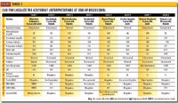
Table 1: Can you localize the azotemia? (Interpretations at end of discussion)
My interpretations are summarized at the end of this Diagnote. The following discussion summarizes basic mechanisms associated with different types and combinations of azotemia associated with impaired excretion of urea and creatinine. It does not include non-urinary factors that may result in mild increases in the rate of production of these metabolites (consumption of high-protein diets, gastrointestinal hemorrhage).
Normal values
See Table 2 for urine specific gravity reference ranges. The normal reference range for serum urea nitrogen concentration in cats is approximately 14 to 34 mg/dl; it is approximately 10 to 30 mg/dl in dogs. The normal reference range for serum creatinine concentration in dogs and cats is approximately 0.6 to 1.5 mg/dl. (Note: In practice, it is usually best to use normal reference ranges that have been derived for the laboratory providing your hospital with test results.)


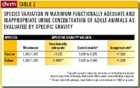
Table 2: Species variation in maximum functionally adequate and inappropriate urine concentration of adult animals as evaluated by specific gravity
Prerenal azotemia
Causes and pathogenesis:
Non-urinary factors (consumption of high-protein diets, gastrointestinal hemorrhage) may result in mild increases in the rate of production of urea (SUN = <60mg/dl; SC = normal). In this instance, both renal function and renal structure are normal. Consult reference texts for further details. Non-urinary diseases may also reduce glomerular filtration as a result of reduction or renal blood flow. Inadequate perfusion of normal glomeruli with blood, regardless of cause (dehydration, cardiac disease, shock, hypoadrenocorticism, decreased plasma colloidal osmotic pressure) may cause prerenal azotemia (Table 1, case No. 2 and 4). Prerenal azotemia is initially associated with structurally normal kidneys capable of quantitatively normal renal function, provided compromised renal perfusion is corrected prior to the onset of ischemic nephron damage. However, if poor perfusion of the kidneys persists, prerenal azotemia may progress to intrarenal (primary) renal failure.
Diagnosis (Table 3) — A diagnosis of prerenal azotemia should be considered if abnormal elevation in the serum concentration of urea nitrogen and creatinine is associated with adequately concentrated urine (specific gravity >1.030 in dogs; specific gravity >1.035 in cats). Detection of adequately concentrated urine in association with azotemia indicates that a sufficient quantity of functional nephrons (>1/3 in dogs) are present to prevent primary renal azotemia.
Significant elevations in the serum or plasma concentration of urea nitrogen or creatinine due to primary renal failure usually is not recognized in dogs or cats until approximately 70 to 75 percent of the nephron population is nonfunctional. Elevation in urine specific gravity associated with prerenal azotemia reflects a compensatory response by the body to combat low perfusion pressure and blood volume by secreting antidiuretic hormone (and other substances) to conserve water filtered through glomeruli.

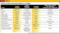
Table 3: Localization of different forms of azotemia
Following restoration of renal perfusion by appropriate volume replacement therapy, restoration of normal concentrations of serum urea and creatinine typically occur in approximately one to three days.
Prognosis: The prognosis of prerenal azotemia is dependent on reversibility of primary cause. The prognosis is favorable for renal function if perfusion is rapidly restored. However, complete loss of renal perfusion in excess of two to four hours may result in generalized ischemic renal disease. With the exception of shock, this degree of reduced renal perfusion is uncommon. Thus the onset of generalized renal disease would be expected to require a longer period of altered renal perfusion.
Postrenal azotemia
Causes and pathogenesis:
Diseases that impair or prevent excretion or urine through the urinary tract may cause postrenal azotemia. In patients with postrenal azotemia, the kidneys are structurally normal initially, and capable of quantitatively normal function provided the underlying cause is corrected. However, if the underlying cause is allowed to persist, death from alterations in water, electrolyte and acid-base and endocrine balance, in addition to accumulation of metabolic waste products, will occur within a few days. If partial obstruction to urine outflow allows the patient to survive for a longer time, varying degrees of hydronephrosis may subsequently occur.


Answers
Complete obstruction of urine outflow (i.e. obstruction in urethra, bladder or both ureters) that persists for more than 24 hours usually results in postrenal azotemia. Unilateral ureteral occlusion (an example of renal disease) is not associated with azotemia unless generalized disease of the non-obstructed kidney is also present. Azotemia that occurs as a sequela to rupture of the excretory pathway (most commonly the bladder) is primarily related to absorption of urine from the peritoneal cavity. Unless damaged as a result of hypovolemic shock or trauma secondary to the underlying cause of rupture of the excretory pathway, the kidneys are structurally and functionally normal.
Diagnosis: Because of variability, the urine specific gravity of patients with post-renal azotemia is not relied on to the same degree for assessment of renal function as it is in patients with primary renal and prerenal azotemia. A diagnosis of post-renal azotemia is based on the integration of clinical findings. Lesions causing urine outflow obstruction are commonly associated with:
1) elevation in serum urea nitrogen and creatinine concentration,
2) oliguria or anuria, dysuria and tenesmus,
3) detection of obstructive lesion(s) by physical examination (urethral plug, herniated bladder), radiography, ultrasonography,
4) variable urine specific gravity values (Table 1, Case 1). Rupture of the excretory pathway is commonly associated with:
- progressive elevation in serum urea nitrogen or creatinine concentration,
- progressive depression, painful abdomen, ascites;
- a history of trauma and associated physical examination findings,
- inability to palpate the urinary bladder,
- detection of a modified transudate or exudate by abdominocentesis;
- abnormalities detected by ultrasonography, or retrograde contrast (positive or negative) cystography or urethrocystography.
Prognosis of obstructive lesions: If the patient has total obstruction to urine outflow for a period of three to six days, then death from uremia will occur. Death usually occurs before significant hydronephrosis has time to develop. Alteration of fluid, acid-base, electrolyte, nutrient and endocrine balance, and accumulation of metabolic waste products cause death. The prognosis is favorable for renal function if adequate urine outflow is rapidly restored. The long-term prognosis is dependent on the reversibility of the underlying cause.
Prognosis of rupture of the excretory pathway: If a persistent rent in the excretory pathway is of sufficient magnitude to result in progressive azotemia, then it is likely that the patient will die if it is not repaired. The prognosis for renal function is favorable if the rent is repaired or heals. The long-term prognosis is dependent on the reversibility of the underlying cause.
Primary intrarenal azotemia
Causes and pathogenesis: Intrarenal azotemic renal failure may be caused by a large number of disease processes (glomerular, tubular, interstitial and/or vascular) which have in common damage of approximately three-fourths or more of the parenchyma of both kidneys.
Diagnosis: In dogs, functional impairment of at least two-thirds of the nephrons is indicated if a dehydrated patient (that has not received fluid, diuretic or glucocorticoid therapy) has impaired ability to concentrate urine. Total loss of ability to concentrate and dilute urine does not always occur as a sudden event, but often develops gradually. For this reason, a urine specific gravity between approximately 1.007 to 1.029 for dogs, or 1.007 to 1.034 for cats, associated with clinical dehydration or azotemia is indicative of intrarenal azotemia (Table 1, Case 5 and 8, Table 3,). Total inability of the nephrons to concentrate or dilute urine (so-called fixation of specific gravity or isosthenuria) results in the formation of urine that is similar to that of glomerular filtrate (approximately 1.008 to 1.012).
If a hydrated patient has an elevation in the serum concentration of urea nitrogen and creatinine, and impaired ability to concentrate or dilute urine, it is likely that impairment of at least three-fourths of the functional capacity of the nephron mass has occurred. (Table 1, Case 3 and 6, p. 18S). Additional studies (ultrasonography, radiography, biopsy, exploratory surgery) are required to establish the underlying cause of primary azotemic renal failure.
Prognosis: Depending on the biological behavior of the disease in question, primary renal failure associated with intrarenal azotemia may be reversible or irreversible, acute or chronic, oliguric or polyuric. Chronic irreversible azotemic renal failure is usually slowly progressive. When formulating a prognosis and therapy, recall that renal lesions do not directly cause uremic signs. They are related to varying degrees of fluid, acid-base, electrolyte and nutrient imbalances, vitamin and endocrine alterations, and retention of waste products of protein catabolism which develop as a result of nephron dysfunction caused by an underlying disease.
Caveat: In dogs with progressive disease resulting in primary renal failure, intra-renal azotemia usually occurs after loss of the ability to concentrate urine to at least a SG of 1.030. Likewise, most cats with naturally occurring primary renal diseases associated with moderate to severe intrarenal azotemia (serum creatinine >3.5 mg/dl) cannot concentrate urine to specific gravity values >1.035. However, we have encountered some cats with naturally occurring primary renal failure and mild azotemia (serum creatinine < 3.5 mg/dl) that could concentrate urine to values substantially in excess of 1.040. Therefore, in cats mild azotemia (serum creatinine = approximately 1.6 to 3.5 mg/dl) associated with urine specific gravity values >1.035 to 1.040 does not conclusively rule out intrarenal azotemia. Further studies of cats are needed to determine the urine SG value that best indicates an adequate population of functional nephrons to prevent signs associated with primary renal failure.
Azotemia associated with glomerulotubular imbalance
Causes and pathogenesis:
In some patients with primary renal failure caused by generalized glomerular disease, azotemia may be detected prior to marked impairment in urine concentrating capacity (Table 3 ). The renal lesion in such patients must be characterized by glomerular damage which is sufficiently severe to impair renal clearance of urea and creatinine, but which has not yet induced a sufficient degree of ischemic atrophy and necrosis of renal tubular cells to prevent varying degrees of urine concentration.
Diagnosis: This group of patients may be differentiated from patients with pre-renal azotemia by failure of a search for one of the extrarenal causes of poor perfusion, by persistent proteinuria and by persistent azotemia despite restoration of vascular volume and perfusion with appropriate therapy (Table 1, Case 6; Table 3).
Caveat: Caution should be used when interpreting urine specific gravity values in patients with substantial proteinuria since specific gravity may be slightly elevated by the effect of protein. Addition of 40 mg of protein per 100 ml of urine will increase the urine specific gravity by approximately 0.001.
Combinations
Combinations of primary intrerenal azotemia, prerenal azotemia and/or postrenal azotemia.
Pathogenesis: Many symptomatic azotemic patients with primary renal failure are clinically dehydrated. Dehydration associated with azotemia and impaired urine concentration is evidence that a combination of intrarenal azotemia and prerenal azotemia are present. Likewise, in recent years, we have encountered increasing numbers of cats with primary chronic azotemic renal failure that have developed uremic crises as a result of postrenal azotemia caused by obstruction of one or both ureters with calcium oxalate uroliths.
Diagnosis: Combinations of causes of azotemia should be considered on the basis of:
- a previous history of compensated primary renal failure,
- detection of primary extrarenal disease processes in addition to generalized renal disease,
- detection of clinical dehydration,
- the patient's response to therapy (Table 1, Case 5 and 8 ). Patients with uremic crises precipitated by reversible extra-renal factors (pancreatitis, dietary indiscretion) may rapidly respond to supportive and symptomatic therapy, as evidenced by a rapid and significant reduction in the magnitude of azotemia. The therapeutic response of patients with uremic crises caused by progressive irreversible destruction of nephrons is usually slower, as evidenced by only a marginal reduction in the magnitude of azotemia.
Prognosis: The influence of the severity of azotemia on the prognosis should be withheld until the magnitude of azotemia is reassessed after correction of the prerenal or post-renal components.