How to identify the pruritic dog
One of the most frustrating and time-consuming problems in everyday veterinary medicine is the presentation of the dog with pruritus.
One of the most frustrating and time-consuming problems in everyday veterinary medicine is the presentation of the dog with pruritus. It is essential as the first step in solving this problem to obtain a good history from the owner. That includes age, breed of dog, duration of pruritus, seasonal vs. nonseasonal occurrence, areas of the body involved, and response to any medications either topical or systemic that have been administered. The next step is a good physical examination and several quick in-house tests that can rule in or out many of the various differentials of pruritic skin disease. This two-part article will focus on the most common differential diagnoses of pruritus in the dog.
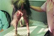
Flea allergy dermatitis - alopecia of the caudal thighs, ventral tail and perineum.
Scratching the surface
The first differential to rule out in working up any case of pruritus should be ectoparasites - either fleas, Cheyletiella, scabies, Demodex, lice or poultry mites (which can affect dogs). This is probably the quickest differential to rule out since combings and scrapings can be performed in the office with the results known in a matter of minutes.
Of course in the case of fleas or Cheyletiella, if the dog has just been bathed, a false negative may occur.
Fleas should be suspected, even if none are found, if the areas of the body involved include the dorsal tail head and medial thighs ("naked butt"). If the owner has several pets, particularly indoor/outdoor cats, this should add to the suspicion.
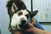
Atypical presentation of a patient with Cheyletiella - periocular and nasal erythema and crusting.
In my area of the country, the Midwest, flea season is commonly late summer through fall, but in warm areas this differential needs to be considered year round. Always question the owner if they have seen fleas on any of the other pets, as some owners consider a few fleas per pet to be normal. It must be stressed to the owner that in a non-flea allergic pet, fleas may not be a problem, but in a flea sensitive dog, it may be the entire problem.
Surprisingly some clients have never seen a flea or flea dirt, so I make a point of showing them. Some dermatologists will perform intradermal skin testing for fleas as proof to the owner of the pet's flea sensitivity. Many flea allergic animals will also develop a secondary bacterial pyoderma particularly in the groin.

More typical presentation of a patient with Cheyletiella or "walking dandruff."
Bacterial pyoderma in some patients can be pruritic along with the concurrent flea allergy. Thankfully, in the past few years, we have seen the emergence of effective flea treatments. My suggestion for flea allergic patients is to treat all the cats and dogs in the household with an adulticide such as Advantage or Frontline Topspot and use an insect growth regulator either systemically or as a house treatment. In the case of Advantage, I usually have the owner apply one tube today, then repeat in l5 days, then once monthly (off label). Treat the concurrent bacterial pyoderma until one week past clearing with an appropriate antibiotic for the skin such as cephalexin, lincomycin, Clavamox or Baytril.
Overlooked culprits
Cheyletiella or "walking dandruff" has reached almost epidemic proportions in our area of the country. If you do not think Cheyletiella exists in your area, maybe you are not checking for it. It should be suspected in dogs of all ages, particularly those that come in contact with other pets via grooming or kenneling.
Probably the most common breed of dog I see affected is the American Cocker Spaniel. Also suspect this mite in an elderly patient that has never had a skin problem previously (that is true for any of the following contagious mites). Often the owner reports that the pruritus "comes and goes" as opposed to the constant pruritus associated with scabies.
There are many techniques to detect Cheyletiella mites including combings, scotch tape technique and skin scrapings. I feel the easiest is to just use a flea comb and place the combings in oil on a microscope slide, cover with a glass cover slip and observe under low power.
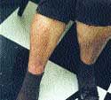
Owner with bites from Cheyletiella mites on her legs.
The mite is large, so when present, it is easily visible. Sometimes the mite is not seen but the elliptically shaped eggs, which resemble hookworm eggs, are present. Be suspicious of Cheyletiella in a patient with "chronic hookworm" disease as you may be mistaking Cheyletiella eggs for hookworm eggs. The typical presentation of a patient with Cheyletiella is one with "dandruff" but I have had patients present with periocular crusting, acral lick granulomas, chronic sneezing and just plain pruritus with very little if any dandruff.
Many patients with Cheyletiella will "ripple" along the back when combed. Other pets in the household may be asymptomatic carriers particularly those with underlying internal medicine diseases such as cancer or Cushing's disease. Some pet owners will develop lesions as this has been reported to affect approximately 30 percent of owners. Treatments for the pets include lime sulfur dips weekly for four weeks, or pyrethrin shampoos weekly for four weeks, or topical Revolution, or injectable or orally administered ivermectin at 200mcg/kg/wk x four weeks (not for use in herding breeds and use with caution in geriatric dogs of any breed), or milbemycin at l mg/kg every other day for 16 days (off label). The patient must be heartworm negative before using ivermectin, Revolution or milbemycin.

Scabies mites as viewed under the microscope from a skin scraping. Note the group of elliptically shaped eggs in the center.
Whichever treatment is chosen, all dogs and cats in the household need to be treated as well as the environment. Environmental treatment can be any premise spray that is used to eradicate fleas. Re-exposure to the mite seems to be a problem for some patients depending upon their environment. Our office tends to see dogs that visit horse barns where barn cats reside, homes that foster pets, and dogs that are professionally groomed as having the most problems with this ectoparasite.
Difficult diagnosis
Even more difficult to find is the canine scabies mite as reportedly it can be missed 50-70 percent of the time. Suspect scabies in a dog with ear edge, hock, ventral abdomen or elbow involvement where antipruritic doses of steroid are not effective. Any age dog may be affected and there is usually the history of exposure to another dog, fox, wolf or coyote (members of the canis species) via kenneling, grooming, or free roaming. Many dogs with scabies will be scratching nonstop in the exam room rather than be curious about their new surroundings. The diagnosis of the disease is made by performing deep skin scrapings of any of the affected areas and observing the scrapings in oil under low power. Mites, fecal pellets or eggs may be seen.
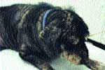
Hypothyroid patient with scabies. Hypothyroid patients with scabies can have extremely large numbers of mites.
Treatment options include lime sulfur dips weekly for four weeks, or topical Revolution used at 15-day intervals for a total of three doses (off label), or ivermectin 200mcg/kg weekly for four weeks (avoid in herding breeds and geriatrics of any breed), or milbemycin l mg/kg every other day for 16 days (must be heartworm negative before using ivermectin, milbemycin or Revolution), or paramite dip weekly for four weeks (although there seem to be some pockets of paramite resistance in some regions of the country). The environment should also be treated using a premise spray or professional exterminator. Any dog in contact with the affected patient needs to be examined and/or treated. As with Cheyletiella, the mite will bite humans but prefers not to live on us, so itchy owners can be an extra clue when looking for the diagnosis of canine scabies. "Scabies incognito" has been reported where mites are never found yet the animal responds to treatment. It emphasizes the fact that if you suspect scabies, treat for it. This mite normally does not affect cats, but one report found that until the cat was treated, the dog did not improve. Since many of these patients have a secondary bacterial pyoderma, be sure to treat that with appropriate antibiotics. It has been reported that hypothyroid patients can have extremely large numbers of mites, so you may want to consider thyroid testing in patients with a large mite burden that may be showing other characteristics of hypothyroidism.
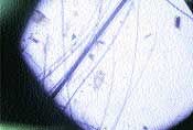
Poultry mite as viewed in oil under the microscope.
Other considerations
Other less common but troublesome ectoparasites affecting dogs includes the very large poultry mite and lice. We have diagnosed patients with poultry mites when checking combings for Cheyletiella. It is a very large mite, orange-to-red if it has been feeding on the patient, and almost resembles a "spider" under the microscope. This mite is not necessarily confined to rural dogs, as we have detected it in city dwelling animals. Reportedly, this mite can also affect humans via a bird's nest near an open window. Treatments are as for scabies or Cheyletiella and if the nest is located, move it away from any entrance to the home. Lice is most often seen with the naked eye due to its size and moving around on the exam table. Treatment is the same as for Cheyletiella, including treatment of the environment.
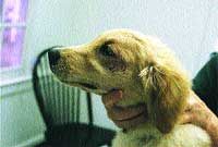
Focal area of alopecia under the right eye. Skin scrapings found Demodex mites in this young Golden Retriever.
Demodex mites in some patients can be quite pruritic and most often are accompanied by a bacterial pyoderma which adds to the pruritus. Again, skin scrapings are necessary to make the diagnosis. It helps first to squeeze the area you will be scraping to help dislodge the mites making them easier to find. Demodicosis can be focal or generalized so it is always important to perform deep scrapings on any area of hair loss. Demodicosis can affect any age of dog but young dogs and elderly dogs seem to be more affected. There appears to be a genetic tendency in some breeds such as Collies, Boston Terriers, Shih Tzu, German Shepherds, Old English Sheep Dogs, Boxers, English Bulldogs, Shar Peis, Chihuahuas, Pugs, Dobermans and Staffordshire Terriers. When a focal lesion in a young dog is diagnosed, therapy includes benzoyl peroxide topically with continued monitoring of the patient for the emergence of other lesions. Steroids are contraindicated in any form. In a young patient with generalized demodicosis treatment options include: Amitraz dips performed at weekly or every two week intervals until negative scrapings, then an additional two dips (must be >16 weeks of age), or ivermectin 200-800mcg/kg/day orally until negative scrapings then an additional 60 days (must be heartworm negative and a nonherding breed) or milbemycin 0.5-lmg/kg bid until negative scrapings then an additional 60 days (must be heartworm negative). Benzoyl peroxide shampoos and antibiotics to treat the secondary bacterial pyoderma are also suggested as ancillary treatments. Demodex otitis is usually accompanied by a yeast or bacterial component, but avoid using any products with topical steroids. It cannot be emphasized enough that steroids in any form are contraindicated. When generalized demodicosis is diagnosed in an older dog where there was no prior steroid use to "encourage" the mites, the patient needs to be checked for underlying systemic disease. Cushing's disease, hypothyroidism, allergy or underlying neoplasia may accompany generalized demodicosis in the older patient. These patients are treated with one of the above-mentioned therapies while also addressing treatment of the underlying internal medicine disease.
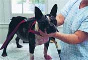
Generalized demodicosis in juvenile Boston Terrier unable to resolve the disease because of concurrent steroid use.
With all of the above mentioned pruritic diseases, most often diagnosis is possible right in the office via performing combings or scrapings. Don't miss a differential because you are in too much of a hurry to check scrapings! Veterinary technicians can perform the combings or scrapings prior to your seeing the patient. Nothing makes an owner or yourself more angry than to "miss" a mite and proceed onto the next differential of pruritus which may be costly and more time intensive. Now that we have ruled out ectoparasites by performing our scrapings and combings, we are ready to proceed onto the remaining differentials of pruritus including bacterial pyoderma, food allergy, or atopy which will be discussed in the next article.
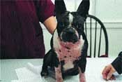
Same patient after one month of milbemycin and weaning off steroids.