

|Articles|May 1, 2010
How to handle common foaling complications and injuries
Author(s)Ed Kane, PhD
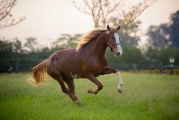
Foaling, like human delivery, should be uneventful. But the process can be fast and violent, and it can present serious health problems to the mare and foal.
Advertisement
Foaling, like human delivery, should be uneventful. But the process can be fast and violent, and it can present serious health problems to the mare and foal.
"When mares deliver foals in the standing position, the foaling process is extremely explosive compared even to calving in the cow," says Pete Sheerin, DVM, Dipl. ACT, Rood & Riddle Equine Clinic, Lexington, Ky. "Over such a short period of time, there's just so much abdominal push. When you stimulate a mare's cervix, she starts to push. And many times it's hard to get malpresentations corrected quickly while she's pushing against you."
Difficulties can arise with recumbent deliveries as well. "When mares lie down on their side, they push with all their might," states Karen Wolfsdorf, DVM, Dipl. ACT, Hagyard Equine Medical Institute, Lexington, Ky. "And these are big animals with a lot of musculature and a very strong abdominal press. That's why if you do not have the foal presented correctly, it definitely has a huge impact — the mare can get into serious trouble since she keeps pushing and there is nowhere for that foal to go."
"Most of the time, foaling is uncomplicated. It's those rare occasions that something goes wrong," notes Jennifer Schleining, DVM, MS, Iowa State University College of Veterinary Medicine. "Then you've got some pretty important decisions that you have to make readily. You may have to decide, if I had the option, do I want to save the mare or do I want to save the foal? Time is of the essence — you have a relatively small window to get the foal out properly to save the foal or the mare or both."
What can go wrong
Several foaling complication sequelae might arise. Dystocia, retained placenta and periparturient hemorrhage are the most common and potentially most life-threatening complications for the mare or the foal. Other less common or less severe complications include uterine and rectal prolapse, perineal and rectovaginal lacerations, cervical and vaginal lacerations and uterine rupture.
Photo 1: Obstetric chains applied to a foal in a breech presentation. Note the mare is still standing before extensive contractions begin.
"I think that there's definitely a difference between the most common complications that you see and the most life-threatening things that you see," Wolfsdorf says. "You might be presented with a mare that has a dystocia that ends up with postpartum metritis or a deeper infection in its uterus that can have a vulvar discharge, which needs to be treated. This can be successfully treated with uterine lavage, intrauterine and systemic antibiotics, anti-inflammatory medications and oxytocin."
Most of the problems are those that affect the mare. "Obviously, if we can get a live foal, that's what we want. But we also want to be able to send home a reproductively normal mare so she can continue to produce foals," Sheerin says.
"The first stage of parturition is the one that the mares themselves can manipulate, and they can put off labor until it's the middle of the night when nobody is watching," says Schleining.
Stage two starts when the water breaks. "After you've seen the water break, you should have a foal on the ground within 30 minutes," Schleining states. "That doesn't give you a whole lot of time to get in and intervene and still have a live foal. If you don't have any progress by 30 minutes, you need to figure out why."
Photo 2: The mare is now in lateral recumbency as contractions become more powerful. Assistants continue to apply tension on the obstetric chains with a veterinarian helping position the foal in the birthing canal.
Dystocia
The most common foaling abnormality is a dystocia, in which the foal is coming out in an abnormal position rather than feet and head first. For example, a leg or the head may be back or the foal may be coming out backward. (See Photos 1 through 4, to better understand a breech position delivery.)
Advertisement
Most of the time, as long as you can get the foal repositioned, the situation will be resolved. Most veterinarians are pretty adept at doing this at the farm. If the mare is given an epidural, similar to the procedure in people, then the foal is much easier to reposition, and it can be assisted out. "But if it is a very large foal or the mare continues to strain and you're trying to reposition the foal, you can end up with ruptures of the uterus or other complications," Schleining says.
Photo 3: The foal is now halfway out. The umbilical cord can be seen in the veterinarian's left hand.
To save the foal if it is coming out backward, speed of delivery is important, Schleining says. Air supply is critical. If the foal doesn't breathe within five minutes of expulsion of its chest, or if it is delivered backward and it doesn't breathe within minutes, permanent brain damage or death will occur. Similarly, if the fetal membranes remain on the foal's nose after its chest has been expelled, they should be taken off so it can breathe.
Photo 4: One final push before the foal is completely out. The rib cage is just now visible.
"With the dystocia," says Sheerin, "if you're working with it on the farm, you have a limited amount of time for your manipulations before you decide to refer it or, if referral is not an option, to do something more aggressive." Dystocia that cannot be corrected on the farm and requires referral can have a more serious prognosis. The outcomes of the mare and fetus are often tied to the length of time of dystocia prior to resolution.
"In central Kentucky, we have the luxury of sending everything into the clinic, so the majority of dystocias are sent to the clinic if we can't get them resolved at the farm," Sheerin notes. "Once at the hospital, we'll anesthetize the mares, raise their hind ends, and then do the manipulations that are necessary. In most cases, we can have a controlled vaginal delivery, and then everything is fine." If a practitioner is faced with a difficult case on the farm and the client is a great distance from a referral center, it's best to send them early.
Retained placenta
"I think one of the most common postpartum complications that is sometimes overlooked and that can have severe consequences is retained placenta," Wolfsdorf explains. Retained placenta — still attached to the endometrium after three hours — is the most common postpartum complication (see Photo 5). The nonpregnant horn is more commonly retained than the edematous pregnant horn. It has an incidence of up to 10 percent. According to Wolfsdorf, "higher incidence has been reported in draft mares, mares of increased age and cases of prolonged gestation, hydrops, abortion, stillbirth, twinning, dystocia, placentitis and cesarean section."
Photo 5: This photo depicts a retained placenta. The placenta is tied in a knot hanging from the vulva to apply tension through gravity on the endometrial attachment.
Retained placenta may not be obvious, such as when the whole placenta is retained and hanging from the vulvar lips. "Occasionally, just the tip of a nonpregnant horn may be retained, and if you don't examine the placenta routinely and completely, you can easily miss a piece of placenta," Wolfsdorf says. "If you miss a piece of placenta, then you set yourself up for metritis, endotoxemia, septicemia and laminitis. Eventually, the mare may end up with serious complications. Make sure you examine each placenta completely. Turn it inside out so that the velvety or chorionic surface can be viewed since this is the surface against the endometrium and is most likely to show pathology. In addition, you will be able to identify the tips of both horns by the avillus portion where the oviductal papillae resides."
"With a normal mare that has retained her placenta, I will start uterine lavage as soon as I examine the mare," says Sheerin. "If a majority of the placenta is retained intact, we perform the Burns technique, in which the placenta is filled with fluids and the fluids are held in the placenta for a period of time. We then remove that fluid and treat the mare with a small dose of oxytocin. Most mares will pass their placenta after this treatment. These mares are also given systemic antibiotics and anti-inflammatory drugs."
For mares that have had a uterine artery bleed as well as retained placenta, the procedure is somewhat different. "I will wait probably 24 hours before I start to do anything with the mare, and then the things I do, I do in slow increments," says Sheerin. "I will examine her and perhaps do a little bit of gentle manipulation to allow the placenta to be passed." If the mare stops pushing and enough of the placenta is sitting in the uterus, there's not enough gravity to pull it out. If you can exteriorize a portion of it, then gravity will kick in and slowly break down the attachment, and the mare will pass it. But don't start just tugging on it; gentle traction will suffice.
"If I do lavage on these mares, which I generally start within 24 to 48 hours after foaling, I use small volumes of fluid so I'm not distending the uterus too much," says Sheerin. "A liter or two, flushing it in and out, is adequate. I may do that many times to try to dilute out any potential bacteria, so we avoid dealing with septicemia or endotoxemia as well."
Periparturient hemorrhage
Obviously life-threatening, most cases of periparturient hemorrhage occur in older, multiparous mares. But, recently, it is increasingly seen in primiparous mares of almost any age, 5 to 24. "Hemorrhage may occur from the middle uterine, external iliac, utero-ovarian or vaginal/vestibule-vaginal arteries in late pregnancy and after parturition, and it accounts for 40 percent of periparturient deaths in mares," states Wolfsdorf.
You can see different levels of hemorrhage in these mares. A mare may hemorrhage prior to foaling and exhibit mild, colic signs, or you may find the mare dead. More commonly, a mare hemorrhages after foaling, which may occur from a couple of hours to a couple of days postpartum. "The majority of mares hemorrhage postfoaling or within 24 hours of foaling," says Sheerin. "But we've had mares that hemorrhage prefoaling as well as mares that hemorrhage as far as a week out from foaling."
Three different types of hemorrhage can occur, according to Wolfsdorf. First, mares can rupture a uterine artery and bleed into their broad ligament. This type is usually self-limiting, contained within the ligament by hematoma formation. Affected mares may be mildly to moderately painful and will usually show signs of colic and discomfort and eventually develop mild signs of shock.
A second, more severe and more life-threatening type occurs when the hemorrhage leaks out of the broad ligament intra-abdominally, and the mares go into full-blown shock.
The third type involves intrauterine hemorrhage in which there may be an artery or a blood vessel within the uterus that leaks or is torn during parturition, and the mares bleed into their uterus. "That is usually a more subtle scenario in which you may just find blood coming out of the mare's vulva when she walks or lies down since the uterus gets full," says Wolfsdorf. "When she moves around, there may be a trickle of blood or clots that come out of her uterus."
Veterinarians have a variety of options to treat these mares. "In my experience, it is important to keep mares quiet in a dark stall and keep them sedated, if needed, as well as comfortable with anti-inflammatory agents such as banamine," says Sheerin. "Once we determine that a mare is indeed bleeding, we'll use naloxone, which is an opiate antagonist useful for treating shock, and sedation. We may start the mare on fluids and use aminocaproic acid, which is used in people with hyperfibrinolysis-induced hemorrhage, plus or minus corticosteroids as well as additional fluids. Using fluids obviously is controversial, in that are you going to possibly increase the blood pressure too much by increasing the volume or replacing the volume. Nobody has come up with an answer because it is something that is hard to do any sort of controlled study on, so the evidence is only anecdotal. Thus, the decision is made on a case-by-case basis."
Sending these mares to a referral clinic may add additional stress, increasing blood pressure and possibly causing the broad ligament to rupture. "If there is a complete rent in the broad ligament because of the bleed, and the mare is bleeding profusely into its abdomen, then there is nothing you can do to save it," Sheerin says.
Other complications
During the violence of foaling, and with dystocia, various tissues may be torn. The anatomy of a malpositioned foal can do damage. "If the foal's feet are not positioned correctly, they can damage the cervix and the vaginal vault, and they even can go up dorsally and penetrate through into the rectum," says Wolfsdorf. A foal's feet may come out of the mare's rectum instead of out of the vulva. This scenario may be more common in maiden mares because they haven't relaxed as well and don't dilate as well. "But these injuries are repairable," Wolfsdorf states.
Other kinds of complications are commonly seen but are less severe, including rectovaginal tears, which can occur when a Caslick's procedure has not been reversed prior to foaling or from a large foal. This kind of tear occurs when the foal tears the mare's vulva lips or perineal body, which can develop into a rectovaginal fistula. Other injuries can also occur like bruising post foaling or vaginal and cervical tears. These may have consequences to a mare's fertility, especially with cervical tears, but they are not generally life-threatening.
Another common foaling complication is colic from colon torsions. "We're not sure why it happens exactly," Wolfsdorf says, "but there's a belief that once the mare has had the foal, there is increased space in her abdomen. So she starts eating more because there's less pressure on her stomach. This may result in an increased buildup of gas that causes the colon to torse." With early diagnosis and prompt surgical correction, successful outcomes are possible. But if diagnosis is delayed, it may have a devastating outcome.
Complications associated with straining are not necessarily as common but are equally severe, such as rectal or uterine prolapse postfoaling. Tears in the vagina can also allow the small colon to protrude through the vulvar lips. It's not common, but it is serious, with serious implications.
Conclusion
Foaling should be uncomplicated, but the force of the act, or the unusual positioning of the foal, may change the outcome dramatically. Being prepared for the range of possible complications or injuries is essential to mare and foal wellbeing.
Ed Kane, PhD, is a researcher and consultant in animal nutrition. He is also an author and editor on nutrition, physiology and veterinary medicine with a background in horses, pets and livestock. Kane is based in Seattle.
Newsletter
From exam room tips to practice management insights, get trusted veterinary news delivered straight to your inbox—subscribe to dvm360.
Advertisement
Related Content
Advertisement


Advertisement
Advertisement
