Fluid therapy: Hold the salt (Proceedings)
In this lecture I will outline how to formulate a plan for fluid therapy for supportive care of sick animals using the three classic goals of fluid therapy.
In this lecture I will outline how to formulate a plan for fluid therapy for supportive care of sick animals using the three classic goals of fluid therapy:
1. Provide for maintenance needs until the animal becomes self-sufficient with oral intake
2. Repair of existing deficits of water and electrolytes
3. Replacement of concurrent losses due to ongoing disorders such as vomiting during the planned fluid therapy period.
These are the goals that need to be assessed & met for most sick animals needing fluid therapy. Therapy should be planned and executed over a period of several hours to 1 full day, depending on the needs of the patient and your available resources. At the end of this planned period the physical and laboratory findings are reassessed and the next treatment period is planned.
Maintenance Therapy
Two-thirds of lean body weight in adult mammals is water. Of this, roughly 2/3 is present as cellular water, and 1/3 is extracellular fluid (ECF). 1/4 of the extracellular water, or about 50 cc per kg of lean body weight, is plasma. The single most important determinant of plasma volume is the ECF compartment volume, as plasma volume remains a relatively constant fraction (1/4) of that compartment. The ECF compartment size is controlled by manipulation of the total body sodium content. As the body's content of sodium is increased or decreased, osmoregulation and thirst adjust water balance to maintain osmolality in the normal range.
Maintenance calculations are based on requirements to maintain homeostasis in a normal animal is resting in a cage at room temperature with no activity and no oral food or water. The requirement for water and electrolytes is based on the animal's mass and is not affected by health problems – therefore it is always the same and may be obtained from a table or chart.
The easiest way to administer water and electrolytes needed for maintaining the animal's physiology is to use a commercial maintenance solution. These products have approximately 30-40 mEq/l of sodium, 13-16 mEq/l of potassium, and variable amounts of bicarbonate precursor, calcium, magnesium, and other electrolytes. Although the sodium content is still higher than necessary, these fluids are reliably too low in potassium and need to be supplemented to around 28 mEq/l. If the solution contains 28 mEq/l of potassium, and is administered at the appropriate maintenance rate for water, this will deliver 0.05 - 0.1 mEq/kg/hour of potassium to the patient, meeting its maintenance needs for that electrolyte.
Veterinarians often use replacement solutions (for example, lactated Ringer's solution [LRS]) to provide maintenance water. This provides the patient with > 20 x maintenance for sodium – in fact, one liter of LRS solution provides the amount of sodium present in 100 pounds of dry dog food formulated to A.A.F.C.O. standards. Inappropriate use of replacement solutions in animals with heart disease or other causes of congestion may be a fatal mistake. Many critically ill patients – often immobile and inflamed - are prone to edema and will readily retain these fluids when used for maintenance water. Always think about whether the patient can tolerate the high sodium load it will receive if you use replacement solutions to provide maintenance water!!!
Repair of Existing Deficits
What is required to restore the animal to homeostasis, and how fast do we need to accomplish this? Typically, we administer fluid therapy to animals that we think are dehydrated or at risk for becoming so. In addition to water deficits, sick dehydrated animals often have abnormalities of sodium (content more than concentration), potassium, magnesium, and acid-base regulation. Other abnormalities may need correction with fluid therapy, such as hypoglycemia or hypercalcemia; however, these problems are encountered less often and will not be considered here.
Water deficit is estimated as % dehydration. Dehydration can be classified as iso-, hypo-, or hypertonic based on the concurrent magnitude of sodium loss, and this determines the basic IV solution you will use. Potassium depletion +/- hypokalemia is common in sick, dehydrated animals and is estimated based on a measurement of serum K, evaluation of the underlying disease, and evaluation of clinical findings suggesting cellular K depletion or hypokalemia. Acid-base evaluation is based on knowledge of the underlying disease process, physical exam, and measurement of serum TCO2 (or HCO3) and the Na-Cl difference. The same disorders that cause potassium depletion tend to magnesium depletion as well, and this electrolyte is often administered empirically to patients judged to be potassium deficient.
Estimating dehydration is notoriously imprecise. It is generally thought that as the water content of the skin decreases, it becomes progressively less pliable (elastic) and its turgor (how "full" of fluid/blood it feels) is reduced. More significant dehydration impairs salivation (but remember that nauseous dogs or cats may hypersalivate in the face of severe dehydration). Severe dehydration (especially if isotonic) causes enough loss of fluid & blood from the orbit to cause a sunken appearance of the eyes.
Isotonic Dehydration
The water lost in this class of dehydration is high in sodium (comparable to the concentration found in ECF). Therefore, it comes almost exclusively from the ECF compartment. Cell volume is not affected, but the ECF contracts almost in direct proportion to the volume lost. Thus, plasma volume sustains a big hit, and many animals develop clinical signs of hypovolemia with only 8 - 10% dehydration. Other causes of hypovolemia such as hemorrhage or extravasation of plasma do not cause dehydration - there is no substantial water loss from the body - but has similar effects because the plasma is lost to the environment or to sequestration. The dramatic impact of isotonic dehydration on ECF and plasma volume often gives rise to obvious clinical and laboratory abnormalities.
Isotonic dehydration is corrected with replacement solutions. Replacement fluids are characterized by high sodium content, between 130 and 154 mEq/l. Following intravenous administration, nearly all of the fluid remains in the ECF. Examples include Ringer's solution, lactated Ringer's solution, 0.9% sodium chloride, and several proprietary solutions. Isotonic dehydration can potentially be corrected rapidly, and if accompanied by clinically significant hypovolemia that may mean in well under 1 hour. If hypovolemia is absent or mild, the rate of correction may be more leisurely, but in general one should strive to complete that goal within 24 hours.
Hypertonic Dehydration
Volume depletion is mild in this form of dehydration because the water loss occurs by evaporation or as dilute urine in diabetes insipidus and contains very little electrolyte. Thus, the loss of pure water is distributed throughout total body water, 2/3 of which is cellular and only 5% (50 ml/kg) is plasma. In its pure form, this type of dehydration will not produce significant hypovolemia, but the ECF concentration of sodium will rise. This increase in osmolality will directly stimulate some renal excretion of sodium which limits the severity of the increase in serum sodium concentration due to water loss. Normal dogs and cats in a thermoneutral environment can survive up to 1-2 months without water (particularly if they have access to dry food). Death is not due to shock (hypovolemia), but rather due to deranged cell function as a consequence of water loss.
Animals with hypertonic dehydration require water to restore normal hydration and osmolality. If pure water loss alone was responsible, use 5% dextrose in water. If a 'mixed' form of dehydration is present and substantial ECF was also lost, begin with a replacement solution (e.g., LRS or 0.9% saline) to expand the ECF compartment first, then follow with the dextrose solution. If the hypernatremia is chronic (>2 days), you must replace water and decrease serum [Na+] slowly, with a maximum reduction of serum [Na+] of about 2 mEq Na+/L/hour for 6 hours, followed by a rate of 1/2 mEq/l/hour. Acute reduction of serum [Na+] by >15 mEq/l will cause significant clinical signs of water intoxication (cerebral edema) including "drowsiness", irritability, inactivity, stupor, exaggerated startle response, hyperventilation, seizures, coma (animals).
Hypotonic Dehydration
This form of dehydration is most often seen in animals with isotonic GI fluid losses that are partially restored by drinking water. Consequently, these animals present with variable dehydration (due to water loss), hypovolemia (due to sodium loss) and hyponatremia (due to hypovolemia-induced release of ADH). With the exception of hypoadrenocorticism, severe hyponatremia is uncommon in animals because most affected individuals usually don't drink enough or are vomiting. Dogs with very low cardiac output – usually due to dilated cardiomyopathy – that require high doses of diuretics may become hyponatremic with or without dehydration, as their low output stimulates thirst and release of ADH at a lower threshold of osmolality.
The severity of dehydration and hyponatremia is variable, depending on the animal's ability to drink and the magnitude of hypovolemia, respectively. The degree of hypovolemia is proportional to severity of sodium loss and contraction of ECF. Neurologic signs are more likely to be seen with rapid development due to diarrhea, and are rare in hyponatremia secondary to circulatory failure. Persistent hyponatremia in a treated heart failure patient is a poor prognostic sign.
Serum (ECF) sodium concentration is determined by the relationship between total body sodium content and total body water content. Hyponatremia may occur in animals that are normally-, under-, or overhydrated. If the hyponatremia is due to water retention secondary to heart failure, the treatment is to improve circulation, NOT to administer sodium!! Administration of sodium to a hyponatremic heart failure patient will cause congestion if the animal's blood volume is already increased. Compared to animals with isotonic dehydration, dehydrated hyponatremic animals without congestive heart failure need proportionally more Na than water. If the animal is capable of excreting free water, this may be supplied by any replacement solution. Chronic (> 1-2 days) hyponatremia should be corrected slowly. Experimentally (in rabbits) it has been shown that it is safe to increase serum [Na+] by 12 mEq/L over the first 24 hours, then half that rate until correction is complete. For acute symptomatic hyponatremia you can treat more rapidly but it is probably not necessary to increase serum [Na+] by more than 6 - 8 mEq/l as this will have major impact on cerebral edema.
Most animals with a combination of isotonic dehydration and some other problem will benefit from rapid treatment of any hypovolemia first. For example, a dog with heat stroke and these abnormalities would benefit from rapid administration of a replacement solution (high in sodium) to rapidly restore ECF volume, even though it also has hypernatremia. The remaining water deficit may be completely restored more slowly afterwards.
Replacement of Ongoing Losses
This goal is necessary if an animal continues to lose fluids during the planned period of fluid therapy. Common examples of ongoing fluid loss in hospitalized animals include drainage of wounds, vomiting, diarrhea, polyuria, hyperventilation, or increased evaporation from damaged skin. Electrolyte losses may be high (e.g., sodium loss to wound drainage, potassium losses to vomiting) or low (e.g., evaporation through the respiratory tract). These losses need to be replaced over the course of the planned treatment period to avoid falling behind.
The rate of ongoing water losses is estimated from history initially, then by observation of the animal once in the hospital. Daily (or even 2-3 times daily) body weights, on a single, accurate scale, are the most important method to follow changes in body water. If you estimate fluid volumes by looking at the puddle on the cage floor, be sure to estimate in terms of kitchen measures:
• 1 tsp. = 5 ml 1 Tbls. = 15 ml 1 cup = 240 ml 1 pint = 480 ml
Ongoing isotonic fluid loss should be replaced with replacement solutions. Patients with effusions associated with hypoproteinemia may also benefit from treatment with plasma, albumin solutions, or synthetic colloids to maintain the colloid oncotic pressure of plasma. Hypotonic fluid loss, resulting in hypertonic dehydration of the patient, is usually seen in dogs with high insensible losses do to hyperventilation +/- hyperthermia. This is best replaced with IV fluids low in sodium and other electrolytes, for example 5% dextrose in water or a commercial maintenance solution. Hypertonic fluid loss is not seen clinically in companion animals (or in any mammal, I think!) To replace ongoing losses, first guesstimate how much water the animal will lose during the planning period (6 - 24 hours). The fluid type (high or low sodium) is determined by the nature of the losses.
Potassium Therapy
The amount of potassium required to maintain potassium homeostasis in a normal animal that is given IV maintenance water and no food is approximately 0.05 – 0.1 mEq/kg/hour. If added to a bag of fluid designed to provide water at a maintenance rate, the final concentration is roughly 24-28 mEq/l. If potassium depletion is present, or if ongoing losses include potassium, extra potassium should be given. Many disorders that cause dehydration also cause concurrent depletion of body potassium. Measurement of serum potassium concentration should always be performed to assess patients at risk. However, since 97% of body potassium is present in the ICF, serum [K+] may not accurately reflect the animal's potassium needs. Unfortunately, there is no simple method to accurately determine potassium requirements in depleted animals, and serum [K+] is usually the only available means of laboratory assessment. Although significant body losses of potassium will usually cause hypokalemia, some animals have normal, low or high serum potassium concentrations in the face of severe total body losses.
Signs of potassium depletion include muscle weakness and electrocardiogram (ECG) changes. Skeletal muscle weakness manifests as locomotor difficulties in dogs and cats, and weakness of the neck (ventroflexion) in cats. Smooth muscle weakness may contribute to intestinal ileus. Although this may not be clinically apparent, animals with diarrhea associated with ileus (e.g. parvoviral enteritis) should be monitored and treated aggressively for hypokalemia to prevent aggravation of ileus. Chronic potassium depletion may cause myopathy (with elevated serum CK concentration) and renal disease.
The ECG may show abnormalities including arrhythmias (atrial and ventricular premature depolarizations) or prolongation of the Q-T interval to values greater than 0.25 second. The ECG is a useful way to evaluate the physiological impact of potassium depletion and hypokalemia on the body. For example, a dog with only moderate hypokalemia (serum [K+] = 3.0 - 3.5 mEq/L) with arrhythmias or a long Q-T interval should usually be treated more aggressively than might be predicted from the serum measurement alone. Remember that acid - base disturbances will also affect serum [K]. Non-gap (hyperchloremic) metabolic acidosis will increase serum [K], and metabolic alkalosis will decrease it relative to total body stores.
There is no easy-to-use accurate technique to determine potassium deficit. However, a relatively simple approach to potassium administration may be used. Consider that the amount of IV potassium required by a normal dog or cat to maintain a positive K balance is approximately 0.05 - 0.1 mEq/kg/hour (use the low end for large animals, the higher end for small animals). The routine safe maximal rate of K administration to a K-depleted animal with normal renal function is about 0.5 mEq/kg/hour. Thus, the total potassium administration rate required for all 3 of our goals (deficit repair, replacement of ongoing losses, and maintenance) will be 0.05 - 0.5 mEq/kg/hour. Animals with mild deficits and/or small ongoing loss may be treated with around 0.15 - 0.2 mEq/kg/hour; for moderate depletion or loss rates use 0.2 - 0.3 mEq/kg/hour, and for more severe problems use 0.3 - 0.5 mEq/kg/hr. At these higher administration rates, the serum potassium concentration should be checked at least every 24 hours.
After day 1: Fluid Therapy after Deficit Repair Has Been Accomplished
Once the initial planned period of therapy (first 2-24 hours to accomplish deficit repair) is complete, the animal is re-weighed and re-evaluated. If body weight has increased to the predicted value (initial weight plus weight of deficit fluids) and the animal appears normally hydrated, the repair of water deficit may be complete. Subsequent fluid therapy requires only replacement of any ongoing losses and provision of maintenance needs. Parenteral administration of fluids should continue until the animal is capable of meeting its maintenance and (ongoing loss) needs by ingestion. Once this condition is met, parenteral fluids may be tapered while observing the animal and then discontinued.
If upon reevaluation an animal receiving parenteral fluids does not weigh what was expected, the clinician must decide the reason why based on all available clues:


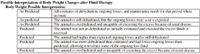
Possible Interpretation of Body Weight Changes after Fluid Therapy Body Weight Possible Interpretation
Reference List
Hansen B, DeFrancesco T. Relationship between hydration estimate and body weight change after fluid therapy in critically ill dogs and cats. J Vet Emerg Crit Care 2002;12(4):235-43.


