Fluid therapy: Finding the best options for perfusion, oxygen supply
Could you review fluid therapy in dogs and cats?
Q: Could you review fluid therapy in dogs and cats?
A: Dr. William W. Muir III at the 2007 American College of Veterinary Internal Medicine (ACVIM) Forum in Seattle gave an excellent lecture titled: "Advances in Fluid Therapy: New Uses for Old and New Crystalloids." Here are some relevant points:
It is believed that the administration of fluids almost always is the single most important therapy during the perioperative period and for the treatment of dogs and cats suffering from any cause of shock. Though few would argue the necessity of fluids, there are many diverging opinions regarding the type of fluid, volumes and rates of fluid administration, and when and how to determine that enough fluid has been administered (Table 1).

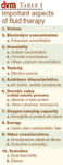

Table 1: Important aspects of fluid therapy
The basis for these dilemmas stems from obvious differences in the types of fluids available (water-containing, crystalloids, colloids, oxygen-carrying), the effects of fluid volume losses and their various causes (cardiogenic, hemorrhagic, traumatic, septic) on the body-fluid compartments.
During septic shock, for example, severe hypotension may necessitate the administration of large volumes of fluid, leading to hemodilution and hypoproteinemia. Plasma albumin concentration and oncotic pressure may decrease which, with increased capillary membrane permeability, predisposes to fluid shifts into the interstitial fluid compartment.
The vasodilation associated with sepsis results in sequestration of blood cells and plasma in venules and peripheral tissues. If vascular volume is not restored and if arterial blood pressure is not returned to near normal (greater than 60 mmHg) values, tissue hypoxia and the release of additional vasoactive substances lead to the inability of cellular membrane pumps and transport mechanisms to maintain cellular and vascular integrity.
Vasoactive substances (prostaglandins, leukotrienes, platelet-activating factor, oxygen-free radicals) alter neuro-regulatory control, dilate arterioles (vascular hyporeactivity), constrict venules and increase capillary-membrane permeability. Decreases in venous return decrease cardiac output and, when coupled with hypovolemia, eventually result in a decrease in peripheral perfusion, tissue-oxygen deprivation and metabolic (non-respiratory) acidosis. Therefore, peripheral perfusion is dependent on intact autoregulatory and compensatory responses, and the exogenous administration of appropriate fluids in order to maintain vascular volume.
Catecholamines may be needed to maintain cardiac (contractile) pumping function and appropriate vascular tone.
Fluid therapy is an inexact science at best. This statement is based on the knowledge that the volumes of the various body-fluid compartments (blood volume, extracellular fluid volume, interstitial fluid volume, etc.) can only be estimated and are in a continuous state of flux during pathophysiologic processes (Table 2).


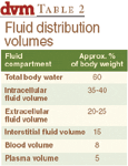
Table 2: Fluid distribution volumes
Are pathophysiologic problems known?
Regardless of these limitations and because the goal of fluid therapy is to optimize vascular volume, restore circulatory function and tissue perfusion and ultimately deliver oxygen to tissues, the more appropriate questions should be:
What type of fluid should be administered? How much should be given? How fast?
These questions, however, are best answered if the pathophysiologic problems are known, the end point(s) of fluid administration have been decided and the methods to assess or evaluate fluid therapy defined.
The most efficacious methods available for the evaluation of fluid therapy in patients are not routinely used in veterinary practice or in veterinary institutions.
Fluid therapy is best evaluated by the ability to restore tissue perfusion, which can be assessed by circulatory function (changes in cardiac output and arterial blood pressure), but more specifically by indices that reflect oxygen transport (delivery) and oxygen consumption.
Numerous studies indicate that oxygen-transport variables (O2 delivery, O2 uptake) are the major predictors of appropriate circulatory function and are excellent predictors of survival or death. Since veterinarians do not routinely make the type of measurements required to determine oxygen-transport variables, does this mean that fluid therapy is a shot in the dark, or only partially effective at best?
The answer to this question is an emphatic, "No."
Appropriate fluid therapy — combined with frequent patient evaluation, periodic cardiovascular and blood-chemical monitoring techniques — can produce astounding and at times miraculous results.
The message of clinical importance is that fixed fluid regimens (i.e., lactated Ringer's), fixed volumes (ml/lb, ml/kg) and rules of thumb are in many instances outdated, inappropriate and oftentimes inadequate. Stated another way, fluid therapy is a dynamic process, which must be reassessed at frequent intervals and adjusted in order to obtain the maximum results (i.e., optimal tissue perfusion).
Understanding options
What, then, are the best fluids, fluid-therapy regimens and monitoring techniques needed to maximize tissue perfusion and oxygen delivery to tissues? (See Table 3.)

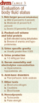
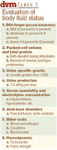
Table 3: Evaluation of body fluid status
There are four types of fluids that can be used for the treatment of shock:
- Those that deliver water (5 percent dextrose),
- Electrolytes (crystalloids: lactated or acetated Ringer's),
- Colloids (fluids that exert a colloid osmotic pressure and which expand the plasma volume)
- Oxygen-carrying solutions (Oxyglobin®; see Table 3).
Although relatively popular in the past, the administration of a large volume of 0.9 percent saline (NaCl) or 5 percent dextrose in water is not recommended for fluid replacement due to dilutional (decreased potassium, total protein, packed cell volume; NaCl, 5 percent dextrose) and acidifying effects (NaCl).
Large amounts of normal saline can potentiate or precipitate hypokalemic, hyperchloremic metabolic (non-respiratory) acidosis.
Balanced electrolyte solutions (Ringer's) containing either acetate or lactate as base replacements are preferred as initial therapy for the treatment of hypovolemia from all forms of shock.
Although these solutions dilute blood components (protein, red-blood cells, etc.), they do not induce or worsen electrolyte or acid-base abnormalities. Providing the packed-cell volume can be maintained above 20 percent and the total protein above 3.5 g/dl, Ringer's solutions help a patient maintain vascular volume and peripheral perfusion.
Small volumes (3 to 5 ml/kg) of hypertonic saline solutions (3-14 percent) administered alone or in combination with colloidal solutions (6 percent dextran 70; hetastarch) are superior to crystalloids for the treatment of shock, including septic shock. The administration of 7 percent NaCl (approximately 2,400 mOsm/L) with colloids draws fluid from the interstitial fluid compartment into the vascular space.
Indeed, the administration of hypertonic solutions and colloids reduces the amount of crystalloid needed. Most plasma or blood-volume deficits are effectively replaced with two to four times less colloid than crystalloid.
Because fluid is drawn from the interstitial compartment into the vascular space, the potential for the development of peripheral or pulmonary edema is reduced. More specifically, cardiac filling pressures, cardiac output, cardiac stroke volume, arterial blood pressure and rates of oxygen delivery and oxygen consumption are improved. This improvement, however, is relatively short-lived (less than 30 minutes in some instances) when hypertonic saline is administered alone, suggesting that they are more effective when administered with colloids.
Since colloids remain within the vascular compartment for comparatively long periods, their combination with hypertonic solutions should produce a sustained hemodynamic effect (Table 4).


Table 4: Volume expansion
The most significant advance in fluid therapy has been the development and clinical approval of an oxygen-carrying blood substitute. Oxyglobin is a solution of approximately 13 g/dl of purified, polymerized bovine hemoglobin diluted in a modified lactated Ringer's solution and has an osmolality of approximately 300 mOsm/kg and a colloid osmotic pressure of approximately 40 mm Hg. The average molecular weight of this solution is 200 kD and its oxygen affinity is regulated by physiologic chloride concentrations, not 2,3-DPG.
This solution increases the oxygen-carrying capability of blood while reducing blood viscosity. What all this means is that Oxyglobin can be administered at any time to dogs or cats in need of a blood transfusion or during acute and critical situations in which an acute increase in blood volume and oxygen-carrying capacity are needed. It is an excellent fluid to use in animals suffering from acute blood loss secondary to trauma or surgical mishap. It should be administered to patients that have become hemodiluted (PCV less than 15 percent to 18 percent) from crystalloids. The product can be administered to patients suffering from chronic anemia with PCV values less than 10 percent. Importantly, Oxyglobin has a two-year shelf life and requires no cross matching because it contains no red-cell membranes.
Fluid-therapy guidelines for patients in shock should include frequent physical examination, hemodynamic monitoring and blood-chemical monitoring (Table 5).


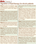
Table 5: Algorithm of fluid therapy for shock patients
Finally, remember that fluid-therapy protocols are guides only and must be modified to meet the changing needs (volume, electrolyte, acid-base, oxygen) of the patient for fluid therapy to be successful without producing side effects.
Dr. Hoskins is owner of Docu-Tech Services. He is a diplomate of the American College of Veterinary Internal Medicine with specialities in small animal pediatrics. He can be reached at (225) 955-3252, fax: (214) 242-2200 or e-mail: jdhoskins@mindspring.com


