Feline oral tumors (Proceedings)
Oral tumors in cats present a challenge. These tumors are usually noticed only at very advanced stages.
Please be aware that these notes are not designed to be a complete reference. It is advisable to consult with an oncologist for current treatment recommendations prior to developing a therapeutic plan for your patient.
Oral tumors in cats present a challenge. These tumors are usually noticed only at very advanced stages. The small size of the feline mouth and tongue limits the extent of surgical resection possible and the most common tumor is squamous cell carcinoma which is exceptionally invasive. In this lecture, we will discuss the diagnosis and treatment of oral tumors in cats.
Histopathologic Types and Incidence
Oral tumors account for about 10% of feline tumors and represent the 4th most common site for tumors in cats. The majority of oral tumors in cats are malignant. Squamous cell carcinoma (SCC) represents 60-80% of oral tumors in cats. About 10-20% are fibrosarcoma. Other less common oral tumors include osteosarcoma, lymphoma, eosinophilic granuloma, epulides (acanthomatous, fibromatous, ossifying; benign, but invasive, tumors arising from the periodontal ligament), malignant melanoma (can be melanotic or amelanotic), odontogenic tumors/ameloblastoma (benign tumors arising from dental laminar epithelium), salivary gland adenocarcinoma, plasma cell tumor, chondrosarcoma, hemangiosarcoma, and mast cell tumor.
Oral malignant melanoma is rare in cats. Pigmented spots can be seen around the mouth/lips (lentigo). Lymphoma and eosinophilic granuloma are more common in cats than dogs. Osteosarcoma was the 3rd most common oral tumor in a study of mandibular tumors in cats. Nasopharyngeal polyps are non-neoplastic masses that occur in the oral cavity of cats.
Etiology
Exposure to environmental tobacco smoke is associated with increased risk of oral SCC in cats. In addition, wearing a flea collar, high canned food intake, and canned tuna fish intake are associated with increased risk of oral SCC. It is thought that cats are exposed to environmental carcinogens through grooming behavior.

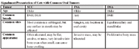
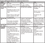
Signalment/Presentation of Cats with Common Oral Tumors
History and Presentation
Most commonly, owners notice a mass. Unfortunately, cats usually present with advanced disease. Ptyalism, halitosis, decreased appetite, dysphagia, and weight loss are also common signs. Exophthalmos may be noted. Ulcers and loose teeth, even with no mass, should make you suspect SCC in cats. Veterinarians should always have an index of suspicion for oral SCC in cats and biopsy any ulcerated areas or sites of loose teeth at dentistry. In addition, it is important for veterinarians to perform a thorough oral examination as part of routine physical examination (when possible) to facilitate early detection of oral tumors. Owners should be educated to brush their cats' teeth and examine their mouths – this will also facilitate early detection.
Diagnosis and Staging
Fine needle aspiration of oral masses in conscious cats is generally impossible. Sedation or anesthesia, biopsy, and histopathologic examination are necessary for diagnosis. Incisional biopsy (diagnostic intent) is recommended for the majority of feline oral tumors. This allows diagnosis and planning of definitive treatment. Excisional biopsy (curative intent) is appropriate only for very small lesions. It is important to clearly document the tumor site and size with a photograph or clear drawing to facilitate further treatment planning.
All cats with oral tumors should have a thorough physical examination with oral exam and lymph node palpation. Remember to retropulse eyes, check nostrils for airflow, and palpate the palate. Tumor measurement and location should be documented in the medical record with a photograph or clear drawing. It is a good idea to repeat the oral exam under anesthesia at the time of biopsy. In addition, the tonsils can be evaluated at that time. The minimum database for cats with oral tumors should include a CBC, chemistry profile, urinalysis, FeLV/FIV test, and T4 level in older cats. Hypercalcemia is occasionally seen as a paraneoplastic syndrome in cats with oral SCC. Retroviruses have not been associated with oral tumors in cats, but retrovirus status important for knowledge of a cat's overall health.
Additional tests are necessary for tumor localization for treatment planning and evaluation for metastasis. If suspected that this is a malignant tumor (older cat, large or invasive oral mass, adherent to bone, enlarged mandibular lymph nodes, etc), some/all of these tests may be done before biopsy.
Evaluation of primary tumor
High detail skull radiographs and dental radiographs can be taken to look for lysis. It is important to get an intraoral view if skull films are taken. CT scan is better for visualization of the extent of the tumor. MRI can provide excellent visualization of tumor but is less available at this time
Evaluation of regional lymph nodes
Most oral tumors that metastasize will first spread to regional lymph nodes so it is important to attempt node cytology even if the nodes are normal in size (however, in cats, if the nodes are normal they can be difficult to aspirate). It may be easier to do when the cat is under anesthesia for biopsy. The role of routine lymph node excision and biopsy is unclear at this time. Lymph nodes other than mandibular nodes may be affected. Enlarged or abnormal tonsils should be biopsied and removal of abnormal mandibular lymph nodes should be considered unless cytology suggests that they are reactive or hyperplastic.
Evaluation for distant metastasis
Most oral tumors that develop distant metastasis will spread to the lungs. For the majority of feline oral tumors, pulmonary metastasis is very rare. Three-view thoracic radiographs are indicated for cats with malignant oral tumors. Clinicians should consider abdominal radiographs and ultrasound in older cats (to look for other diseases) and for cats with round cell tumors.


Behavior of Common Oral Tumors in the Cat
Treatment
Surgery and radiation therapy are used to treat local disease. More recently, chemotherapy has been used as a radiation sensitizer to improve tumor response. Treatment is most successful for small rostral tumors (except for benign tumors and odontogenic tumors). For larger malignant tumors, palliative therapies are often most appropriate. For SCC response has been poor and a multimodality approach is generally recommended.
Surgery
Surgical resection is the treatment of choice for resectable dental tumors (epulides and ameloblastomas) and for malignant oral tumors, depending on size, extent of invasion, and location. Maxillectomy or mandibulectomy are generally necessary for resection of SCC, FSA, or OSA. Surgery is most successful for small tumors of the rostral mandible or maxilla. Mandibulectomy and maxillectomy can negatively impact quality of life and even cause life-threatening morbidity (ex. inability to eat), so careful case selection and surgical planning are essential for success. A retrospective study showed that mandibulectomy was beneficial for some cats with oral tumors and resulted in good quality of life after the post-operative recovery period. However, 98% of cats experienced acute morbidity (dysphagia, inappetance, ptyalism, mandibular drift, tongue protrusion, pain, difficulty grooming, dehiscence, malocclusion with palate injury, and temporomandibular joint crepitus) and 76% experienced long term morbidity (dysphagia, inappetance, ptyalism, mandibular drift, tongue protrusion, difficulty grooming, malocclusion with palate injury, and temporomandibular joint crepitus). Twelve percent of cats had severe anorexia/dysphagia that prevented them from eating to the time of their death. Case selection and treatment planning are important. Removal of >50% of the mandible appears to be too aggressive to allow functional outcome in most cats. An enteral feeding tube should be placed for post-operative support after aggressive oral surgery in cats.
Radiation Therapy
Radiation therapy (RT) is useful for local control of feline odontogenic tumors (> 3 yr control, Moore et al 2000) and oral lymphoma. For SCC, FSA, and OSA RT may improve tumor control after incomplete excision. If a tumor is not amenable to surgery, palliative RT can be used for tumor shrinkage and pain relief. Limited information is available regarding RT for SCC. These tumors are radiation responsive, but response durations are short with survival times of 2-3 months. Nine of 9 cats in a recent study responded to radiation therapy for oral SCC. (Fidel et al 2007) The median survival time was 86 days. Another study questioned the efficacy of RT for feline oral SCC, but only 4/7 cats finished the treatment protocol. (Bregazzi et al 2001) An older study of mandibulectomy + definitive RT reported a median survival time of 14 months for 7 cats, but 6 of the 7 cats developed recurrence. (Hutson et al 1992) Use in feline oral FSA or OSA for improving local control or palliation has not been evaluated, but is logical and has been useful anecdotally. Five cats with oral melanoma treated with coarse fractionated RT were euthanized for progression at a median of 146 days. (Farrelly et al 2004) Acute side effects of radiation therapy typically occur in the 3rd or 4th week of treatment can include mucositis, glossitis, pharyngitis, mild desquamation, and if an eye is in the treatment field keratitis, conjunctivitis, and corneal ulcers. Expected late side effects include leukotrichia, hyperpigmentation, cataract formation, and decreased tear production. Rare late side effects include bone necrosis, oronasal fistula, non-healing corneal ulcers, and retinal degeneration. Most cats with SCC will not live long enough to experience these effects.
Chemotherapy
Chemotherapy has not been well-evaluated in treating feline oral tumors, mainly due to their local nature. Chemotherapy as a single treatment modality has not been effective for oral SCC in cats and is not likely to be recommended for FSA or OSA. Palliative RT + carboplatin and definitive RT + mitoxantrone have been used in small numbers of cats with oral SCC. Median survival times of 5-6 months were reported. Other drugs are being evaluated as radiation sensitizers, but increased toxicity is also possible. Low dose gemcitabine in conjunction with RT resulted in responses in 6/8 cats for a median of 43 days and a median survival of 112 days (range, 11-234 days). (Jones et al 2003) Piroxicam has not been beneficial in treating cats with oral SCC and is associated with some risk of gi bleeding and renal injury. COX-2 expression has been detected in approximately 18% of feline oral SCC, suggesting that NSAIDs may be beneficial in some cats. More recently, increased EGFR expression has been demonstrated in feline oral SCC and may represent a target for novel therapies for this tumor.
Enteral Feeding Tubes
Enteral feeding tubes should be placed for nutritional support for all cats undergoing aggressive oral surgery or cats with inappetance undergoing any treatment for oral tumors. Ideally, the tumor will respond to treatment and the need for a tube will be temporary. Esophagostomy tubes are generally easy to place and maintain.
Symptomatic Therapies
Control of secondary infections (antibiotic therapy) and pain is important. Anti-inflammatory prednisone can reduce inflammation and oral buprenorphine can be palliative for discomfort caused by an in-operable or recurrent oral tumor. Another option would be meloxicam (not with a steroid because of risk of gi ulceration).


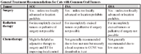
General Treatment Recommendations for Cats with Common Oral Tumors
Prognosis
The majority of cats with oral tumors die of local disease. Metastasis is rare for the most common oral tumors. Tumor size and location are important in determining appropriate treatment and prognosis. Cats with OSA and FSA can enjoy very long survival times if the tumor is operable. There is little information describing the effectiveness of treatment of oral FSA and OSA.
SCC is better described and is more likely to recur and tends to recur quickly. Recently a report described 54 cats with oral SCC treated by general practitioners in the UK. (Hayes et al 2007) Four of these cats underwent surgery and the rest were treated palliatively with antibiotics, NSAIDs, or steroids. Twenty six cats received no treatment. The median survival time of all cats was 44 days with 9.5% cats living 1 year (similar to what has been reported historically).
Ameloblastoma is a benign tumor that can be cured with surgery and cats can still enjoy long-term control with radiation therapy if an ameloblastoma is too large for surgery. Epulides are cured with complete excision. Some cats will develop multiple epulides which seem to be associated with a high incidence of recurrence if wide excision is not performed. Prognosis with oral lymphoma has not been well described. Anecdotally, cats with oral lymphoma respond to chemotherapy and radiation therapy. These cats may have local disease only or local and systemic lymphoma.

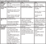
Prognosis and Prognostic Indicators for Common Oral Tumors in Cats
Although few are reported, one half or more of oral melanomas in cats have been reported to be malignant, both locally invasive and metastatic. Five cats treated with palliative radiation therapy for oral melanoma died due to progression of disease at a median of 146 days (range, 66-224 days). (Farrelly et al 2004)