F is for fractured teeth
What happens when your patient breaks a tooth?
Imagine examining a dog or cat’s mouth, and you spot a fractured tooth. The pet seems to be eating fine, with no apparent discomfort. Should you keep your findings silent, share your thoughts, recommend extraction, or refer for vital pulp or root canal therapy? Let’s dive deeper into the world of fractured teeth in our patients.
Dental mishaps happen
Our patients experience dental misfortunes from accidents, fights, chewing on hard toys, antlers, and falls. Sometimes these incidents can lead to fractures in their teeth. The maxillary and mandibular canines are usually the teeth that are most prone to fracture, followed by incisors and the maxillary fourth premolars.
Cats and dogs have different tooth structures. In cats, the pulp chamber extends within millimeters under the crown tip, whereas there’s usually considerable protective dentin beneath the enamel in mature dogs. When trauma exposes the pulp, oral bacteria invade, causing infections that often spread to the surrounding tissues and bone.
Figure 1: Periapical lucency consistent with periapical periodontitis affecting the right mandibular canine. Extraction or root canal therapy is indicated.
Figure 2a: Discolored left first maxillary incisor.
Figure 2b: The radiograph shows an enlarged root canal compared to the adjacent teeth consistent with a nonvital tooth-root canal therapy or extraction is indicated.
To get a better picture, intraoral dental radiographs come in handy; they help identify pulpal and periapical issues and can evaluate the width of the root canal compared with the contralateral tooth (assuming it’s still vital). A nonvital tooth might have a wider root canal, indicating past pulp damage and necrosis (Figures 1, 2a, and 2b).
The many faces of fractured teeth
The presentation regarding teeth with endodontic pathology (issues inside
the tooth) can vary. Concussive trauma can lead to pulpitis in some cases, with no obvious fracture present. Internal bleeding of the pulp is often noticeable as a discolored tooth (Figure 3). Sometimes you’ll see a fractured crown without pulp exposure (uncomplicated crown fracture), whereas other times, the pulp might be exposed (complicated fracture). In advanced scenarios, you might even spot fistulous tracts below the gumline (Figures 4a and 4b).
Figure 3: Discolored left maxillary canine secondary to pulpitis.
Figure 4a: Fistula apical to the mucogingival line secondary apical periodontitis in the fractured left maxillary canine.
Figure 4b: 3-D CBCT confirming marked periapical bone loss.
Most pets with fractured teeth do not display signs of discomfort, even though they’re experiencing pain like humans. Cats, especially, are experts at hiding their pain. However, signs like occasionally chewing on one side, drooling excessively, or avoiding hard food or treats are present. (Table 1)


The treatment dilemma: To save or extract?
What should you do when faced with a fractured tooth that exposes the pulp? (Table 2) Well, you have 2 primary choices:
1. Tooth extraction: Removing the damaged tooth entirely, which stops the pain but may affect oral function.
2. Endodontic therapy: This approach aims to save the tooth and restore its function, and it includes root canal therapy and vital pulp therapy.


Factors that influence your decision


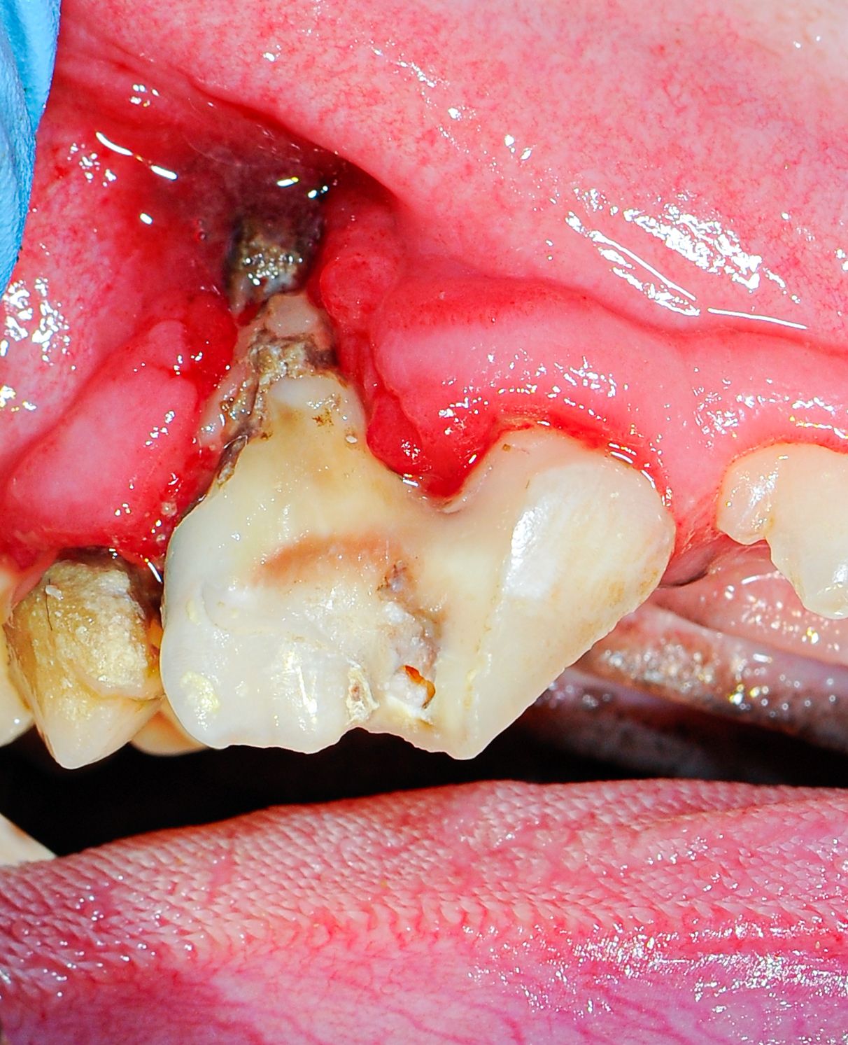
Figure 5: Fractured right maxillary fourth premolar affected with advanced periodontal disease, extraction indicated.
Several factors come into play when deciding between extraction and endodontic therapy.
1. Severity of tooth damage: If the fracture is limited to enamel or involves dentin in a mature dog, without exposing the pulp, no further therapy is often needed if probing and intraoral radiographs do not show evidence of periodontal disease. Action is necessary for an uncomplicated crown fracture if the animal is young (with a large pulp chamber and root canal) and when the pulp is exposed (complicated crown fracture).
2. Periapical and periodontal changes: Tooth support is vital for successful endodontic treatment. The prognosis may be unfavorable if there’s moderate to advanced periodontal disease. In those cases, extraction is the treatment of choice (Figure 5).
3. Tooth function: Although any tooth can be treated endodontically, certain teeth are more critical for oral function, including canines and maxillary fourth premolars. Saving these is a priority (Figures 6a and 6b).
Figure 6a: Complicated left maxillary canine.
Figure 6b: Radiograph of root canal.
4. Owner’s expectations: Recent veterinary literature reports that root canal therapy carries a high (94%) long-term success rate. Ensure your pet caregivers understand the prognosis, costs, and follow-up care involved in endodontic therapy.1
5. Your expertise and comfort: If you’re uncomfortable with advanced endodontic procedures or if the client refuses them (referral), extraction may be the only option for pulp-exposed teeth.
Vital pulp therapy: A lifesaver for some teeth
Vital pulp therapy includes partial coronal pulpectomy, direct pulp capping, and restoration. This approach works well for teeth that have experienced acute (less than 48 hours) pulp exposure; it can save the tooth’s function without the need of root canal therapy. (Figures 7a and 7b).
Figure 7a: Acute complicated crown fracture of the left maxillary canine.
Figure 7b: Medication placed over the exposed pulp before crown restoration.
Age matters, too
The patient’s age and the fracture’s timing can also impact treatment choice. Young patients with open root apices may benefit from procedures like vital pulp therapy, whereas older patients with closed root apices can undergo standard root canal therapy.
Fractured teeth in our pets are common and demand careful attention. Whether it’s tooth extraction or endodontic therapy, your decision should consider the severity of damage, periapical and periodontal changes, the tooth’s function, the owner’s expectations, and your expertise. Intraoral dental radiographs are essential in making informed decisions. Managing fractured teeth relieves pain and ensures your furry patients’ long-term oral health and happiness.
Reference
Kuntsi-Vaattovaara H, Verstraete FJ, Kass PH. Results of root canal treatment in dogs: 127 cases (1995-2000). J Am Vet Med Assoc. 2002;220(6):775-780. doi:10.2460/ javma.2002.220.775
Jan Bellows, DVM, DAVDC, DABVP, FAVD, received his undergraduate training at the University of Florida and his doctorate in veterinary medicine from Auburn University. After completing an internship at the Animal Medical Center in New York, New York, he returned to Florida, where he practices companion animal medicine surgery and dentistry at All Pets Dental in Weston. He has been certified by the American Board of Veterinary Practitioners (canine and feline) since 1986 and the American Veterinary Dental College (AVDC) since 1990. He was president of the AVDC from 2012 to 2014 and currently president of the Foundation for Veterinary Dentistry.