Exploring the perception and physiology of pain in horses
"Anatomically and physiologically, horses have all the structures required for pain processing," says Glenn Pettifer, DVM, DACVA, at the Veterinary Emergency Clinic, Toronto, Ontario, Canada.
"Anatomically and physiologically, horses have all the structures required for pain processing," says Glenn Pettifer, DVM, DACVA, at the Veterinary Emergency Clinic, Toronto, Ontario, Canada.


Telling behavior: A horse with anterior enteritis and colic pain often may manifest the pain by exhibiting a depressed demeanor.
"It's not a matter any more of wondering whether these animals actually do experience pain or feel pain," Pettifer says. "We know they have the capability of doing that."
Horses certainly experience pain from mechanical, thermal and/or chemical insults, but whether they also feel the emotional aspects of pain is difficult to assess.
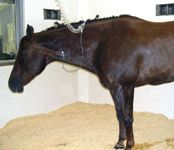
Horses known to be in pain, especially chronic pain, may seem depressed or at least more sedentary, and exhibit reduced self-maintenance behaviors. They certainly are in discomfort and their quality of life is negatively affected.
"Pain increases morbidity," says Lesley Smith, DVM, DACVA, professor of anesthesiology at the University of Wisconsin College of Veterinary Medicine. "It makes the hospital stay longer. Animals don't bounce back as quickly," Smith says.


Signaling distress: A horse with a wound from where a large sarcoid was surgically removed was resistant to wound management after the surgery because of pain.
Smith notes that horses with untreated pain show distress and may be difficult to work with. Other symptoms include decreased water and feed intake, a predominantly catabolic metabolism, abnormal endocrine profile, delayed recovery from surgery, rapid-shallow respiration, muscle spasms, disuse atrophy and decreased mobility.
Acute and chronic pain
Horses experience both acute and chronic pain.
Although felt, a horsefly bite or the smack of a racing whip would be considered mild acute noxious stimuli, with no lasting sensation.
Acute pain usually is of sudden onset and short duration (less than three months), of variable severity, has an identifiable cause, is focal to the injury site and usually is a symptom or warning, an endogenous protective mechanism that signals real or potential serious trauma and/or tissue damage.
For horses in acute pain, there is a reluctance to be handled; restlessness, which may evolve into depression; often a rigid stance and an anxious appearance. They may show dilated pupils, glassy eyes, flared nostrils, muscle tremors, profuse sweating and an increased respiratory and pulse rate.
Acute pain also usually occurs with colic, fracture or surgery and often can be treated with NSAIDs or opioids.
Chronic or debilitating pain is considered pathologic, or having the characteristics of a diseased state. It often is unrelenting, may have no identifiable cause, may spread beyond the original site of an injury or insult and serves no biological function.
Due to increased production of excitatory neurotransmitters, peripheral and spinal-chord sensitization, chronic pain often is associated with musculoskeletal injury or disease (e.g., laminitis, osteoarthritis or degenerative joint disease). It requires complex treatment strategies and typically has a poor prognosis.
Horses suffering chronic pain show reduced food and water intake, often accompanied with weight loss; changes in sleeping habits and increased periods of recumbency; and changes in social behavior, such as irritability or aggressiveness toward people and other horses.
Visceral and somatic pain
Visceral and somatic pain occur within both acute and chronic pain.
The form of pain a horse feels when it steps on a nail or develops laminitis is referred to as somatic. It is processed differently than the visceral pain felt with an illness such as colic.
Somatic pain is associated with bones, ligaments, and tendons. It tends to be more localized and tends to be sharper than visceral pain.
Visceral pain arises from the distension of the viscera, and is associated with conditions of the GI tract and other internal organs. Compared to the skin, the viscera have a lower density of nociceptors, so that the pain tends to be not as well localized. Therefore it is more generalized, as opposed to a sharp or stabbing pain. It may be a gnawing or aching associated with gut torsion, distension or obstruction.
Allodynia and hyperalgesia
Though both acute and chronic pain vary in intensity, allodynia and hyperalgesia are unusual, exaggerated pain sensations.
Allodynia is an exaggerated response to otherwise non-noxious stimuli and may occur in areas other than the one stimulated. It can be either static or mechanical. For example, a person with a severely sunburned arm may perceive light pressure on that area, even from a feather touch or shirtsleeve, as pain radiating up the entire arm.
"I don't know of any specific case reports of allodynia in horses," says Smith. "I suppose one could imagine some types of clinical situation that could be analogous. One example that comes to mind is a horse with chronic back pain that may respond negatively to the saddle pad being placed on its back," says Smith, "but is that allodynia or negative anticipation of pain to come?"
Hyperalgesia is a severe pain response to a mild stimulus, such as extreme sensitivity accompanying inflammation, or a horse's surgical incision becoming tender during healing. Light pressure to the region then becomes painful to an exaggerated degree, with increasingly dramatic response as input increases.
Hyperalgesia is produced by mediator substances in response to tissue damage, including bradykinin, substance P, leukotrienes, tumor necrosis factor alpha (TNF?), interleukins (IL) 1 and 8 and prostaglandins.
Hyperalgesia can be experienced in focal, discrete areas, or in a more diffuse form involving the entire body. The focal form is typically associated with injury, and is divided into two subtypes, primary, which describes pain sensitivity that occurs directly in the damaged tissues, and secondary, which describes pain sensitivity that occurs in surrounding undamaged tissue.
Hyperalgesia is induced by a platelet-aggregating factor, which comes about in an inflammatory or an allergic response. This seems to occur via immune cells interacting with the peripheral nervous system and releasing pain-producing chemicals (i.e., cytokines).
From trauma to pain sensation
"In people, the pain experience is separated into three components: nociception (transduction, transmission, and spinal cord modulation), perception (perceiving the unpleasant experience) and cognition (the behavioral response to pain)," Smith explains.
"We know that there probably are three or four fairly discrete, but interdigitating, processes that take place to provide for the experience of pain," Pettifer explains.
The peripheral nociceptors transduce the stimulus into an electrical signal that is then transmitted from the periphery up to the spinal chord. Some processing occurs there, and the information then is transmitted further up the spinal chord to the (cerebral) cortex.
In the human experience, it's not until there is a perception of a potentially damaging event that we ascribe the notion of pain. There is an experiential component. Prior to its being experienced, it's called nociception. There is a notion of transduction of the stimulus, transmission of it and perception of it.
There also are some events that allow for modulation of the input that is eventually experienced as pain.
From the point of origin of the initial noxious insult or injury to the tissues, nociception, the initial pain stimulus, is encoded as an electrical-action potential, an impulse transmitted to the dorsal horn of the spinal chord, then transmitted via ascending neural pathways to the CNS, including the cerebral cortex, where the awareness of the presence of pain occurs.
The first step in pain processing is the transduction of the stimulus by nociceptors, specialized pain-detecting nerve cells activated by mechanical (touch or pressure), thermal (hot or cold) or chemical (endogenous or exogenous) stimuli.
The nociceptors, found in skin, muscles, joint capsule, visceral organs, pleural membranes and arterial walls, are free nerve endings with cell bodies in dorsal-root ganglia that terminate/synapse in the dorsal horn of the spinal cord.
A-delta and C-fibers carry pain information as an electrical action potential to the dorsal horn of the spinal cord.
"Their function is to transmit pain information from the peripheral tissues at the site of injury up to the spinal cord to synapse with the second-order neuron," states Smith.
They release substance P, glutamate, calcitonin and gene-related peptide (CGRP) at the synapse and that signals the second-order neuron about the pain in the periphery. A-delta sensory neurons are myelinated, fast-conducting, "first-pain" responders to intense noxious mechanical stimulation. C-fiber primary afferent neurons are non-myelinated, slower-conducting, dull, burning, aching "second-pain" responders, but polymodal, i.e., responding to noxious mechanical, chemical and thermal stimuli. The stimuli are noxious in that they are strong and tissue-damaging, whereas nonnociceptive receptors are below the tissue-damaging threshold.
Once the pain signals reach the brain, the various CNS components (i.e., reticular formation, thalamus, hypothalamus) are involved in processing pain.
"We can't say that descending modulation is the same in horses as in people," explains Smith, "but the information after processing in the thalamus, hypothalamus, cortex, etc., converges again in the brainstem and is sent via the dorsolateral funiculus to the spinal cord segment of concern, where inhibitory (e.g., endorphins, serotonin, gaba) and/or excitatory neurotransmitters (e.g., glycine) are released."
The balance of each that is released depends on the degree of windup and the amount of modulation that occurred before the information ascended as well as in the cortex and other higher CNS centers.
Gating is the normal regulatory mechanism that affects the way pain is perceived. "Presumably the same mechanisms (as noted in humans) apply in horses," Smith says.
The first gating mechanism is in the spinal chord, where sensory fibers linked to pain fibers can act to suppress each other. A non-pain sensation, such as massaging or icing-down an injured area, can inhibit the transmission of pain.
A second gating mechanism is found in the brain, where endorphin release may suppress pain signals, reducing the pain perception.
Knowledge improves control
"When it comes to looking at therapeutics for pain control, we look at those different types of processes (of the physiology of pain)," states Pettifer. "We look at applying a local anesthetic peripherally to prevent transmission of the stimulus, or giving non-steroidal drugs like phenylbutazone that decrease inflammation at the periphery, and that decreases the activity of the receptors in the periphery that effect pain stimuli," Pettifer explains.
Drugs like opioids and NMDA-antagonists function at the level of the spinal cord. There are sedatives that, in combination with analgesics, reduce anxiety around painful situations. "We attack the problem from many points – a multimodal approach to pain control, with the notion that you actually achieve much better control if you look at these different facets of the pain system and target your therapy toward interrupting their function," Pettifer explains.
"The more we understand about pain, the more we are able to manage it," he says. "Once you learn more about the physiology of pain and the various neurological processes involved, the development of a pain-management strategy largely becomes an attempt to modify the activity of these systems to the benefit of the animal."
Ed Kane is a Seattle author, researcher and consultant in animal nutrition, physiology and veterinary medicine, with a background in horses, pets and livestock.