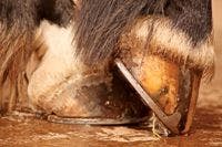
Equine veterinarians make steps toward solving the laminitis mystery
An intensive area of study, laminitis still confounds veterinary researchers looking to advance our knowledge of disease origin, prevention and treatment. But some promising new research projects are looking to make great strides toward better understanding.
This past March, the Daily Racing Form reported that several equine practitioners were making progress in solving the mystery of laminitis by exploring the multitude of factors that lead to this devastating disease. Several researchers at the University of Pennsylvania New Bolton Center, the Hoof Diagnostic and Rehabilitation Clinic (HDRC) in Bryan, Texas, and other institutions are actively working toward that end.
"I believe we've taken some giant steps toward better understanding this multifaceted disease," says James Orsini, DVM, DACVS, associate professor of surgery at the New Bolton Center.


GETTY IMAGES/KATIE PETEF PHOTOGRAPHY
As molecular biology techniques continue to improve and the core of dedicated researchers expands, the goal is to conquer laminitis by the year 2020, says Rustin Moore, DVM, PhD, DACVS, chair of clinical studies at The Ohio State University College of Veterinary Medicine and author of Laminitis Vision: 20/20 by 2020—Conquer Laminitis by 2020.
"Not that laminitis will be totally eliminated," Orsini says, "but that we better understand the complexities of the disease well enough that we prevent at-risk horses from developing the disease more often than we treat at-risk horses. The clinical cases prevented from getting laminitis are clearly on the rise. Rather than treating many of these cases that are chronic and marginally responsive to medical management, we are recognizing them earlier and stopping the disease from happening. This is our ultimate goal."
The research group
Kurt Hankenson, DVM, MS, PhD, Dean W. Richardson Chair for Equine Disease Research and associate professor of musculoskeletal research and orthopedic surgery at the New Bolton Center, is studying mesenchymal stem cells and musculoskeletal regeneration. His research explores laminitis from the molecular level.
A graduate of the University of Illinois School of Veterinary Medicine, Hankenson went into equine practice, but his research interest pushed him from clinician to researcher. After receiving a master's degree from Purdue University in musculoskeletal research, Hankenson ended up at the University of Washington, working on his PhD. His first faculty position was at the University of Michigan's medical school—not at the veterinary school—in orthopedics, with a background in comparative musculoskeletal research.
Always interested in disease pathogenesis and treatment, Hankenson was recruited in 2006 to the University of Pennsylvania and worked in the department of animal biology until 2012, when he took over the Dean W. Richardson Chair in Equine Disease Research. It was a great opportunity for him to transition to doing research that was more impactful in veterinary medicine. As the Richardson Chair, Hankenson will continue to build and to better integrate the university's research program in laminitis.
After the tragic injury and death of 2006 Kentucky Derby winner Barbaro, an influx of goodwill and philanthropy funded the research chair and helped establish the Laminitis Institute, which Orsini directs. The institute offers outreach as well as clinical and research components. Hankenson will oversee the research component.
Hannah Galantino-Homer, VMD, PhD, DACT, senior investigator in laminitis research at the New Bolton Center, established the Laminitis Laboratory at the center in 2008 and has a three-phase attack on the disease:
1. Creating and maintaining the Laminitis Discovery Database, an archive of tissue samples from natural and experimental laminitis cases
2. Investigating global changes in gene and protein expression during laminitis pathogenesis
3. Developing an organotypic culture system for lamellar epidermal cells to allow in vitro laminitis studies and reduce reliance on live animal experimental laminitis induction models as well as to lay the groundwork for equine epidermal regenerative therapies for the hoof, skin and eye.
David Hood, DVM, PhD, with the HDRC, is studying the chronically foundered horse as opposed to acute cases. The New Bolton Center's experimental model samples focus on the developmental and acute phases. The natural cases represent all stages of the disease but primarily reflect chronic disease.
The chronically foundered horse
Central to the chronic rehabilitation research underway at the HDRC is a herd of horses with chronic laminitis that has been donated to the Hoof Project Foundation in College Station, Texas, the organization that funds the HDRC. The foundation funds help maintain the horses, and investigators from other institutions are also using these animals for their research.
"The overall goal of the rehabilitation research is optimizing the clinical management of the difficult cases," says Hood. Given the central role that lameness plays in chronic laminitis, a force plate system has been developed and tested that allows an objective assessment of lameness severity in the standing horse. While the system's data analysis package is still being studied, this system is already a key component of the rehabilitation project.
"One of the primary goals of the project is to obtain a clearer picture of what pathologies are present in the foot and to define how the presence (and severity) of a pathology impacts the severity of lameness shown by the horse," Hood explains.
In addition to standard diagnostic tests (e.g., radiographs, venograms) special diagnostic approaches are being developed. One of these is a laminar biopsy—full thickness down into the dermis, below the laminar interface. It's a fairly simple surgical procedure done under a local anesthetic, Hood says. In addition to tissue from necropsied horses, tissues from biopsies are examined and used to index the healing response that is present in the laminar interface. The endpoint that they are working toward is to develop a treatment regimen for the chronically foundered horse. Diagnostic nerve blocking is also being used to define the regional location of lameness.
As more is learned, data are being organized into diagnostic, prognostic paradigms that allow specific rehabilitation approaches for affected horses to be developed. "For example, once we index the lameness and define what pathological lesions are present in the foot, we have a better focus on what an individual horse needs to optimize its rehabilitation," Hood says. "We are developing a diagnostic battery of tests and logic paradigms to establish data about the foundered horse, other than just noting that the horse has sore feet. With our nerve blocking diagnostic tool, we can assess where the pain is coming from—it may be coming from the sole surface, from the laminar interface, from the heel or from above the foot, the fetlock."
Much has already been learned about the hoof of the foundered horse, says Hood. "We have also learned that a significant number of chronically foundered horses in this age group have acquired injuries to the back, hock or stifle that impact the lameness diagnosis. At this time we are attributing this to some arthritic or traumatic-type injury induced by the horse getting up and down without using the front feet."
Another component of the project is assessing treatments to see if they are impacting lameness severity. "We need to know if the type of shoe used actually does what we think it does. Does it make the horse more comfortable?" says Hood. "Consider that in clinical practice, trimming, shoeing and maybe bute [phenylbutazone] are administered nearly simultaneously. Even with the same treatment protocol, some horses will show a decreased lameness where others demonstrate an increased lameness. In this component of the work, we are separating the specific trimming, shoeing and pain management protocols and evaluating the effect of each on lameness severity. We are also looking at the short- and long-term effect of treatments on the pathologies present in the foot."
The 'shock organ'
Every species has a "shock organ"—an organ whose response to a given medical condition determines the nature and, to a large extent, the outcome for a given disease, says Orsini. "We always knew the horse's foot was at risk, especially during any systemic illness, such as enterocolitis, pleural pneumonia and septicemia. And in cases of contralateral limb laminitis we often see failure of the foot as well, but for different reasons. Contralateral laminitis differs because it is not the septicemia or system disease that results in failure of a supporting foot but the prolonged loading of the foot and lamellar tissues associated with a severe musculoskeletal injury in the opposite limb.
"The foot is bathed in a complex network of veins and arteries, allowing it to readily compensate for changes in environmental temperature and body conditions. The temperature of the foot can fluctuate almost moment-to-moment. This is one of several reasons that the foot is so susceptible to systemic illnesses and behaves like a shock organ, as other organs do in other species," continues Orsini.
Another important consideration is the lamellar tissue's need for glucose—a need that is higher for the foot than it is for the brain. "Add the mechanical loading of the foot during laminitis, and we have the 'perfect storm' for failure of the foot's supporting structures," says Orsini.
During an inflammatory condition, such as laminitis, the needs for glucose are even greater. "This is one of the big differences that we see with the horse's foot compared with other animal species. The inflammatory condition overwhelms the foot, and the dermal lamellar tissue cannot get enough life support (glucose and oxygen) to sustain itself during these severe conditions," Orsini says. "This is one reason that the foot is considered a shock organ during multiple organ dysfunction syndrome.
"We know the kidneys, intestinal tract and coagulation system can fail," Orsini continues. "In the horse, it's interesting we don't see as many cases where the lung is the primary organ of failure. There are other systems that fail first. Comparatively, the horse's lungs may get pulmonary edema but not the pneumonia and severe respiratory problems that determine life and death as in people."
In the horse's foot, however, there are elusive circulating factors, termed laminitis trigger factors, that cause an acute inflammatory condition that could lead to laminitis, says Orsini. The systemic inflammatory response, termed systemic inflammatory response syndrome, results in such a marked inflammatory response that the outcome is loss of the suspensory apparatus supporting the coffin bone (P3) with resulting sinking or rotation.
"The ultimate question is, How do we protect the foot as one of the horse's shock organs?" Orsini says. "There are proven preventive measures like cryotherapy for the very sick horse with diarrhea. With endocrinopathic diseases, such as Cushing's disease and equine metabolic syndrome, we have done a good job recognizing the disease much sooner and prescribing drugs (such as pergolide) specifically formulated for the horse to manage the disease and prevent laminitis as a sequela. With these endocrine diseases, the foot does not behave as the shock organ as described earlier. But using the 'all roads lead to Rome' theory, the supporting tissues of the foot—the lamellar tissue—fail and founder the outcome."
Laminitis research at the New Bolton Center
Hankenson is hoping to take a new approach to research. He has submitted a paper, a collaboration with Julie Engiles, VMD, DACVP, assistant professor of pathology at the New Bolton Center, looking at the changes associated in P3 following laminitis, using samples and case information from the Laminitis Discovery Database run by Galantino-Homer. "What we found is that there are some very pronounced changes that occur rapidly in the morphology of P3," says Hankenson. "We are interested in why those changes occur and what the role of those changes might be in the pathogenesis of laminitis."
This is a novel area in the field laminitis. Hopefully, Hankenson's studies will give us insight into laminitis treatment from looking at the bone perspective and insight into the pathogenesis of the disease, the relationship of the bone changes and that of the hoof. "My research has been focused on bone biology for many years—bone regeneration and healing and how bone changes over time," he says. "I'll be bringing that expertise into the project.
"Also related to bone biology, what I'm quite interested in, which is very specific to the Barbaro situation, is the condition of support limb laminitis (contralateral limb laminitis)," continues Hankenson. "With orthopedic injuries, if we can expedite the healing process it would be a way we could prevent laminitis. While bone heals well, we recognize that if we could accelerate bone regeneration, that might be an opportunity to get at-risk horses to bear weight on the injured limb faster, thereby preventing the support limb laminitis. So we'll also have an effort on how we can heal bone faster and also identify those bones that might be most at risk for injury. Can we identify horses that might be at risk for breakdown injuries, and can we return them to bearing weight in an expedited fashion?"
Hankenson has been studying mesenchymal stem cells for about 15 years. "That will be another area that we'll be focusing on—whether mesenchymal stem cells have any potential for treating laminitis, but also can mesenchymal stem cell therapeutics be better developed as an approach to treating bone regeneration," he says.
"What I find fascinating is that there are multiple factors that contribute to the development of laminitis," says Hankenson. "One of the questions that I think we need to explore is whether there is a point where there is a unifying mechanism that results in the breakdown of that laminar tissue, resulting in the clinical manifestations of founder. Or do we have multiple pathways that reach some common end result that is actually is something unique? We don't really have a good sense of this yet."
Perhaps the common systemic mechanisms have a common factor that leads to the cascade of laminitis. But does that agree with the contralateral limb problem? "I think that is something that still needs to be explored," Hankenson says. "In my opinion, the way to do that is exactly what we have been doing and will continue to focus on—building a large set of clinical data in naturally occurring horses with disease, and doing careful pathological analysis of the tissue as well as doing detailed molecular tissue analysis both locally and systemically. I think this is how we will get at the origin of the disease."
Galantino-Homer says the Laminitis Discovery Database can help. The database includes tissue and serum samples, gross images and histology slides from various sources, including the experimental models of University of Queensland Professor Chris Pollitt, BVSc, PhD; the oligofructose model of pasture laminitis; and the hyperinsulinemia model, which is a recent model for endocrinopathy-associated laminitis in horses with insulin resistance and hyperinsulinemia. It also includes samples from cases of naturally occurring laminitis from the hospital or from horses that are donated to the laboratory. Laminitis cases include supporting limb laminitis cases, sepsis and colitis cases, and horses with insulin resistance, pars pituitary intermedia dysfunction (Cushing's disease) and obesity.
"We're also developing an in vitro system, so that we can study some aspects of the disease without using live animals—in particular, the basal cells of the secondary epidermal lamellae, which are at 'ground zero' for laminitis pathogenesis," Galantino-Homer says. "There is only so much you can study using the naturally occurring laminitis cases, as these cases are typically euthanized late in the disease process, so it's hard to study the early stages of the disease. The disease 'snap shot' that we get from tissues collected at euthanasia or biopsy only provide information about a single stage of the disease, whereas the in vitro system can be used to determine what's happening at the cellular level in real time. That is difficult to do with the live animal."
They've been working with epithelial stem cell biologist Makoto Senoo, PhD, at the University of Pennsylvania's Department of Biology, to adapt the epithelial stem cell selective and "organotypic culture systems" used in human medicine to generate skin and eye cornea tissue grafts for transplantation and research studies. "This will allow us to investigate the effects of environmental factors associated with the development of laminitis, such as inflammatory mediators or supraphysiologic insulin levels, on the ability of lamellar cells to remain viable, maintain cell-matrix and cell-cell adhesions and maintain their normal gene and protein expression patterns," says Galantino-Homer.
"The major part of our work has been looking at the molecular studies, the pathophysiology of laminitis using large-scale quantitative analysis of protein expression and gene expression," Galantino-Homer explains. "We are so indebted to the horse genome project, which has allowed me to do the kind of work that we and other equine disease researchers are doing. We're taking samples from the model systems and using the most current quantitative proteomics methods and analysis relative to the published equine genome sequence to identify and quantify all of the proteins that are present within a certain range of protein sizes.
"If I had tried to do that prior to the release of the annotated horse genome sequence, my results would have been without value," she continues. "Whereas now I'm able to match those protein sequences to the published horse genome sequence and predict protein and gene identities. Compared to previous gene and protein expression studies based on selected targets, the global high-throughput approaches that are now available to us are rapidly accelerating equine disease research progress. The equine genome has also allowed us and other researchers to use new technologies for large-scale 'transcriptomics' to investigate global gene expression patterns. We are now getting a more complete picture of the gene and protein players in laminitis pathogenesis."
An ongoing collaboration with Samantha Brooks, PhD, at Cornell University's Department of Animal Science, is using next-generation high-throughput RNA-sequencing to investigate global changes in gene expression in laminitic horses and to use the information generated to refine genome annotation of hoof-specific gene expression.
The focus of Galantino-Homer's work is to improve our understanding of laminitis pathophysiology with the overall goal of making laminitis easier to diagnose, prevent and treat. "One of our major goals is to discover diagnostic serum biomarkers," she says. "The Grayson-Jockey Club Research Foundation recently funded our serum biomarker studies with Dr. Julie Engiles from the University of Pennsylvania and Dr. Bettina Wagner from Cornell. Target diagnostic proteins have been selected based on our previous GJCRF-funded proteomics studies and will be investigated in experimental and naturally occurring laminitis cases from the Laminitis Discovery Database.
"However, it's unlikely that there will be a single biomarker that is specific for laminitis due to the overlap with other inflammatory diseases and musculoskeletal injury," she continues. "Fortunately, the technology is now in place for multiplex assays that allow the simultaneous evaluation of multiple factors using a single serum sample. We've been working with Dr. Wagner, the director of Automated Serology/Immunology at Cornell's Animal Health Diagnostic Center to develop these assays. We're hoping to find a specific signature that we can use to monitor the at-risk cases, also in regard to aggressive intervention and eventual prognosis."
Looking to the future
As we reflect on the progress during the past 10 years, we find more pieces of the laminitis puzzle that are inextricably linked. The overriding feeling is that of accomplishment in getting closer to solving the mystery. This is because of the dedication of many trailblazers, veterinarians, farriers, researchers, owners and caregivers who continually advance the understanding, prevention and treatment of laminitis. As a profession and industry, we stand on the shoulders of all these achievements. This ground work positions other researchers and collaborators on the launching pad for even greater future accomplishments as we approach 2020 and the goal of conquering laminitis.
Ed Kane, PhD, is a researcher and consultant in animal nutrition. He is an author and editor on nutrition, physiology and veterinary medicine with a background in horses, pets and livestock. Kane is based in Seattle.