Equine protozoal myeloencephalitis: Etiology, diagnosis and treatment
Early identification is key in this widespread neurologic disease.
The somewhat evasive neurologic disease equine protozoal myeloencephalitis (EPM) occurs when horses are exposed to certain protozoal parasites, most commonly Sarcocystis neurona (secondarily, Neospora hughesi), that infect and invade the central nervous system (CNS). The disease is widespread throughout North and South America.


(LEA ROTH/GETTY IMAGES)
EPM is seen any time of year, with an estimated 50 percent of horses exposed to S. neurona, though less than 1 percent develop clinical EPM. Although there's a low incidence of EPM in the general horse population, 14 cases per 10,000 horses per year (23 percent of the horses that died with neurologic signs) showed S. neurona antibodies in their CNS, according to studies done at the University of California-Davis (UC Davis).1
Clinical signs
A large variation of neurologic signs is possible with EPM, depending on the degree of CNS damage, which can make this disease easy to confuse with other neurologic conditions such as wobbler syndrome, trauma, equine herpesvirus myeloencephalopathy, equine degenerative myeloencephalopathy and West Nile encephalomyelitis.


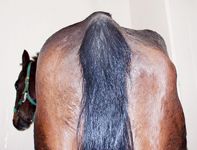
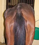
Photos 1 and 2: Two examples of lower motor neuron signs and muscle atrophy associated with EPM. (Photos courtesy Stephen Reed, DVM, Rood & Riddle Equine Hospital, Lexington, Ky.)
Depending on the site and extent of CNS damage, horses may show signs suddenly or progressively. Some signs may be almost imperceptible, such as isolated atrophy of gluteal and masseter muscles. More commonly, neurologic signs occur acutely but also may progress gradually to a noticeable gait abnormality, functional impairment of one or more limbs, weakness, asymmetric muscle atrophy (Photos 1 and 2,), incoordination (leaning to one side), head tilt, stiffness, or severe ataxia, as a horse might suddenly fall over during a race or workout, without any previous apparent signs.


History of research
EPM has been studied since the 1970s. An early pioneer was J.P. Dubey, PhD, a parasitologist and now adjunct professor at the Virginia-Maryland Regional College of Veterinary Medicine, Blacksburg Va., and Animal Parasitic Diseases Laboratory, Agricultural Research Service, United States Department of Agriculture, Beltsville, Md. Dubey, who did this work while at The Ohio State School of Medicine, was one of the early scientists to recognize EPM. It was initially described by James Rooney, DVM, and colleagues at the University of Pennsylvania as an unusual neurologic syndrome, then called segmental myelitis.
Since then, it's been reported in horses from 2 months to 24 years old, typically affecting horses 1 to 6 years of age.2 EPM is common in Thoroughbreds, Standardbreds and Quarter horses. It's also been found in ponies, but not in mules, donkeys or other nonhorse equids.
Etiology
A horse is considered a dead-end host since it cannot transmit the disease to other horses. The definitive host is the opossum, shedding the infected egglike sporocysts into the environment via feces, which are consumed by horses grazing in parasite-infected pasture or in feed or water. Intermediate hosts, which complete the two-host life cycle of S. neurona, include domestic cats, raccoons, skunks, armadillos and passerine birds.
The CNS, including the brain and spinal cord, is infected when the sporocysts migrate from the gastrointestinal tract into the bloodstream to cross the blood-brain barrier. Once infected, horses may show neurologic signs fairly soon after exposure, though most horses similarly exposed do not show signs since their immune response will ward off the disease. Other infected horses may harbor the organism and develop signs at a later time.
Nicola Pusterela, DVM, PhD, Dipl. ACVIM, associate professor in the Department of Medicine and Epidemiology at the School of Veterinary Medicine at UC-Davis, notes, "It's only speculation whether the horse will develop immunity to the protozoal organism without developing disease, develop disease soon after exposure to the parasite or develop it at a later time. We don't know. Horses will develop neurologic disease shortly after being experimentally infected, but what may happen under natural conditions, nobody knows. We assume most horses become exposed and mount an immune response, and the organism never gets into the CNS.
"And then there's a small percentage of horses, less than 1 percent, in which the organism will—probably not freely, but bound to white blood cells—enter the CNS and then undergo different cycles of replication within the neuronal cells, leading to inflammation and clinical disease," Pusterela continues.
Robert MacKay, BVSc, PhD, Dipl. ACVIM, professor of large-animal medicine at the University of Florida College of Veterinary Medicine, notes, "As far as we know, for every horse that develops neurologic signs we can see, there are at least 50 that are infected without [visible] neurologic signs. It's typical for the horse to become infected, develop an immune response and never develop neurologic signs. The exposed horse that's infected and develops neurologic signs is an unusual situation. The horse is relatively resistant to that kind of disease. That represents the tip of the iceberg of infections."
The factors that make one horse in 50 develop disease where others don't are unknown, says MacKay. Retrospective studies have looked at populations of horses and identified some risk factors, such as age, previous adverse or stressful events and exposure to environments rich in opossums. "Presumably, there's also a genetic component, which hasn't been thoroughly investigated as to why some horses appear to be susceptible and most horses aren't," he says.
The fact that almost half of horses are exposed to the parasite but less than 1 percent are affected and show clinical signs may be due to variations in the parasite's potency or a horse's ability to effectively mount an immune response. Stephen Reed, DVM, from Rood & Riddle Equine Hospital, Lexington, Ky., thinks the answer is probably both. "But in reality, I would say the horse's immune system is probably more important than the pathogenicity of organism, though we don't have the knowledge to definitively say it's one or the other.
"In all the areas where we have the definitive host—the opossum—you see a large percentage of antibodies in the [horses'] bloodstream, indicating they've been infected and are making antibodies," Reed continues. "Because only a few of them get neurologic signs, you have to believe they're able to clear the organism on their own. But there are some who argue it's only infection with the very rare, highly pathogenic strain that causes clinical EPM. There's most likely truth to both sides. I feel very comfortable that the role of the immune system is really critical."
No one knows if there are genetic factors between horses that affect their immunity. Jennifer Morrow, PhD, co-owner of Equine Diagnostic Solutions (EDS) laboratory, Lexington, Ky., says, "There are certainly different strains of the parasite across the country, so horses aren't becoming infected with the exact same strain of the parasite. There must be factors on both sides that result in the fact that, while many horses are exposed, very few develop clinical EPM disease. Why that is is currently unknown."
Variations in the clinical neurologic signs of disease leading to deficits depend on where the parasite does its damage anatomically within the CNS (i.e., brain, spinal cord or peripheral nerves) and how severe the damage is. The severity also depends on several other factors, namely, the host, pathogen strain and, possibly, environment. "It's very complicated, and we really don't understand it," says Pusterela.
Diagnostic tests
In addition to a neurologic examination and other tests used to rule in or out other neurologic disease, exposure to EPM may be confirmed by diagnostic tests to detect the presence of IgG antibodies to the parasite. For deceased horses, EPM can be confirmed if classic lesions of the brain and spinal cord are seen during postmortem examination.
A few diagnostic tests are available—the older Western blot test, the indirect fluorescent antibody test (IFAT) and a quantitative ELISA based on S. neurona surface antigens (SnSAGs) SAG1 ELISA as well as the newer SAG 2,3,4 ELISA.
The initial diagnostic test was the Western blot, a complex, semiquantitative method first used in the early 1990s. The test describes the presence of specific immunoreactive bands but relies on subjective interpretation for results, often with variation from laboratory to laboratory. It is easy to inadvertently contaminate cerebrospinal fluid (CSF) samples with blood during the sampling procedure, which can result in false positive results.
"A negative Western blot test from a blood sample is a reliable indicator that a horse does not have EPM," says Pusterela. If the test result is positive, though, then a second Western blot test is performed on a sample of CSF to confirm intrathecal antibody production, which is suggestive of CNS infection.
The IFAT is based on quantitative fluorescence of the entire organism under the microscope, SarcoFluor (specific for S. neurona) and NeoFluor (specific for N. hughesi). It's the only test that offers separate assays for the two organisms. However, the incidence of EPM caused by N. hughesi is thought to be low. IFAT was the first established quantitative antibody testing modality available that calculates the theoretical probability of the disease depending on the titer of the immunologically affected animal. As with Western blot test, this test method also is subjective in its interpretation.
ELISA tests based on the S. neurona SnSAGs are nonsubjective and quantitative, generating titers. However, there are significant differences in diagnostic accuracy depending on the specific SAG used in the assay.
"Using the original surface antigen, SnSAG1, about 30 percent of the isolates in nature did not have that surface antigen, causing false negative results," says Reed. "Because the SnSAG 2,3,4 ELISA uses three specific antigens expressed in all S. neurona isolates to date, there's an inherent enhancement of diagnostic power."
"While individual serum and CSF titers can be determined, it's the ratio of serum to CSF titers that's very predictive of an EPM diagnosis," says Morrow. "There's a normal flow of serum antibodies back and forth across the slightly permeable blood-brain barrier with a normal proportionality. As the intrathecal antibody production increases, the titer ratio decreases. Ratios of more than 100 strongly correlate with EPM. Based on the results from over 100 necropsies, a ratio of more than 100 has a sensitivity of 80 percent and a specificity of 97 percent."
Reed calls CSF sampling helpful, and at Rood & Riddle Equine Hospital they've switched to using the surface antigens SAG 2,3,4 titer ratio test for S. neurona. "In a validation study set, we now have more than 400 paired serum and CSF samples from field cases from several institutions around the country, and more than 100 of them have gone to postmortem. So we have the gold standard of diagnosis on about 25 percent."
Pusterela notes that the IFAT and SnSAG 2,3,4 surface antigen diagnostics are both good quantitative tests, though there's no published comparative study looking at both. "The UC Davis test [IFAT] is quantitative and has been evaluated using confirmed EPM cases," says Pusterela. "Further, the titer appears to be directly related to the likelihood of disease in a neurologic horse.
"The second advantage of the UC Davis-developed test is that it tests for antibodies to both S. neurona and N. hughesi," Pusterela continues. "If you use only the SnSAG 2,3,4 ELISA test, if the disease is caused by N. hughesi, you're not going to pick it up. So, you're going to falsely determine the animal does not have EPM. You're not going to treat him, and this horse will not improve."
MacKay says that, although several tests are available, none are "terribly satisfactory." "They're good at identifying horses that don't have any kind of infection," he says. "They've not been good at discriminating between the types of infection—the one with neurologic signs and the one without."
Whichever blood test is used, the information is still available, says Morrow. The presence of antibodies can support an EPM diagnosis in the presence of clinical signs. "But the diagnostic value is better if you add the CSF component, especially with the SAG 2,3,4 ELISAs," she notes.
Reed adds, "It's certainly easier if a practitioner only had to take a blood sample, but as EPM is a neurologic disease, there's CNS involvement, and it's logical that CSF would be an important diagnostic specimen. I realize how important it is to most veterinarians in the field if we could develop a test where you could dependably determine the disease probability from blood alone. That would sure make life easier for most practitioners. To really increase the accuracy of diagnosis, the ratio of antibody in blood to CSF is the best. And if you become used to tapping CSF from every horse, it doesn't become an enormous challenge."
Treatment
Newer antiprotozoal drugs are available to help diagnose and treat EPM. Without treatment, the progression of EPM is somewhat unpredictable, as the neurologic signs in untreated horses usually get worse—from the more mild signs to ataxia, recumbency or death in hours to years, with periods of severe exacerbations possible after prolonged periods of quiescence.
"Most of the cases of EPM are progressive," says Pusterela. "And we know that the more severely affected horses may improve with treatment, but the chance for full remission in the severely affected animal is significantly worse. If the same animal is treated early on, the signs may be fairly mild to moderate."
Four drugs have been approved by the FDA for treatment of EPM: pyrimethamine, sulfadiazine, ponazuril and diclazuril. Approved by the FDA in 2003, nitazoxanide was recently withdrawn because of potentially serious side effects such as enterocolitis, laminitis, anorexia, fever and lethargy. Several other drugs are considered for treating EPM, but none of them are FDA-approved.
Pyrimethamine and sulfadiazine
According to scientists at UC Davis, pyrimethamine and sulfadiazine, previously thought to cure EPM, only provide temporary relief of the condition. Apparently they're effective only in depressing the viability of the causative parasites S. neurona and N. hughesi. The drugs enable the horse's immune system to kick in—to control the infection temporarily during the treatment period. When treatment is curtailed and the horse returns to work or is stressed, the disease often recurs.
The pyrimethamine-sulfadiazine combination is of limited effectiveness, and its treatment period is three to four months, or longer. It also requires supplemental folate, since these drugs counter folate metabolism, which is especially critical to pregnant mares.
"In the compounded form of pyrimethamine-sulfadiazine, there's very little problem with toxicity," says MacKay.
Ponazuril
A newer antiprotozoal drug, ponazuril, was approved by the FDA in 2001. It's highly effective and can be given for a shorter period compared with pyrimethamine-sulfadiazine. The UC Davis Equine Health Center has been using it since 2001.
The drug has a stated success rate of 70 percent improvement or resolution of EPM clinical signs. It's administered once daily for 28 days or more, depending on clinical signs and disease severity. Although not 100 percent effective, ponazuril shows better outcomes when patients are treated earlier in the disease process. If the CNS is already affected, even though the parasite is destroyed, the horse is left with a damaged nervous system. In short, the earlier the horse is treated, the better the outcome.
Diclazuril
Diclazuril, approved by the FDA in 2007, is about to be released. Studies done with diclazuril show it is as effective as ponazuril. It's impregnated into an alfalfa pellet, so it can be easily top-dressed onto feed.
Both ponazuril and diclazuril don't have the concerns of pyrimethamine-sulfadiazine in regard to folate deficiency. "Those concerns are mostly with pregnant mares," MacKay says. "Most other animals don't have a concern as to folate deficiency problems, though it has that potential."
Treatment comparison and adjunct therapy
"I don't think there's any evidence that any of these treatments are better than the others," says MacKay. "The recommendation for treatment with pyrimethamine-sulfadiazine is three to six months, so it has a longer treatment period. Both ponazuril and diclazuril are given for 28 days. With either drug, at least 60 percent of horses improve. There's no information on how many of the treated EPM horses completely recover, but I think that's a factor of how bad they were to start with."
Along with antiprotozoal drug treatment, anti-inflammatories and immunostimulants have been used.
Assisting horses to strengthen their immunity to help ward off EPM, and to limit those exposed from becoming infected, may be accomplished by adding an antioxidant—specifically, supplementing horses with clinical EPM with natural-source vitamin E at 10,000 IU/day to help build their immune response and help them recover from neurologic disease. This form of the vitamin is able to cross the blood-brain barrier, and it is used as a routine treatment in horses with neurologic disease.
Prognosis
MacKay says it's rare for a horse that's severely affected by EPM to completely recover. "If it's treated very early when the signs are mild, maybe 80 to 90 percent may completely recover," he says. "If it's severe, where the horse has difficulty standing up, then 10 percent or less of those horses may completely recover. It definitely depends on the neurologic condition of the horse when it begins treatment."
And when it comes to relapses, it's basically similar, MacKay continues. "The milder cases have less frequent relapses; the bad ones relapse routinely. But we don't know why that is."
Pusterela cautions that many horse owners wait too long to have their affected animals examined. "There's a potential that some of the antiprotozoal drugs, particularly the new formulas ponazuril and diclazuril, may be used as a prophylactic, especially in stressed or immunocompromised patients," Pusterela says. "Diclazuril will be a very good drug to administer because it's in a pelleted form. It will have great potential to be used more like a preventive drug."
Epilogue
As S. neurona is the critical factor (along with N. hughesi in some cases), it would be advantageous to rid the horse environment of the parasites—similar to trying to eradicate caterpillars to limit mare reproductive loss syndrome. Sarcocystis neurona and N. hughesi, though, are ubiquitous in nature along with their definitive host, the opossum, and numerous intermediate hosts (e.g., cats, raccoons, armadillos, skunks). Management of both hosts and parasites could be used, though it is unlikely to be successful.
"With the parasites being so prevalent in nature, horses may be exposed and reexposed—some horses throughout their lifetimes, or at least for many years," says Morrow. "A horse infected at one time may harbor parasites within the CNS that, under stressors such as transport, at a later time may trigger that individual to develop clinical signs of EPM."
The main veterinary solution is to accurately diagnose the disease and treat it early when possible. The use of the newer diagnostic assays, such as the indirect fluorescent antibody test and the quantitative ELISA based on S. neurona surface antigens (SnSAG 2,3,4), along with the newer coccidiostats, ponazuril and diclazuril, will go a long way until additional methodologies for eradication, such as a possible vaccine, are available.
Ed Kane, PhD, is a researcher and consultant in animal nutrition. He is an author and editor on nutrition, physiology and veterinary medicine with a background in horses, pets and livestock. Kane is based in Seattle.
References
1. Dubey JP, Lindsay DS, Saville WJ, et al. A review of Sarcocystis neurona and equine protozoal myeloencephalitis. Vet Parasitol 2001;95:89-131.
2. Saville WJ, Reed SM, Morley PS, et al. Analysis of risk factors for the development of equine protozoal myeloencephalitis in horses. J Am Vet Med Assoc 2000;217:1174-1180.
Suggested Reading
- Yeargen MR, Howe DK. Improved detection of equine antibodies against Sarcocystis neurona using polyvalent ELISAs based on the parasite SnSAG surface antigens. Vet Parasitol 2011;176(1):16-22.
- Saville W, Reed S, Dubey JP. Prevention of equine protozoal myeloencephalitis. Presented at AAEP Annual Convention, Orlando, Fla., 2002. Updated 2011 and posted at www.aaep.org/health_articles_view.php?id=225.
- MacKay RJ, Davis SW, Dubey JP. Equine protozoal myeloencephalitis. Compend Contin Educ Pract Vet 1992;14(10):1359-1367.
- Saville WJ, Reed SM, Granstrom DE, et al. Response of horses exposed to Sarcocystis neurona when monitored biweekly, in Proceedings. 43rd Annual Am Assoc Equine Pract 1997;43:9.
- Bello TR, Allen TM. An intensive approach in the treatment of clinical equine protozoal myeloencephalitis. J Equine Vet Sci 2008;28(8):479-483.