Ears, the basics all practitioners should know (Proceedings)
Ear disease is a very common problem presented to veterinarians. It may be broken down into two major components, pinnal disease and otitis, though the strict definition of otitis would include any inflammatory pinnal disease.
Ear disease is a very common problem presented to veterinarians. It may be broken down into two major components, pinnal disease and otitis, though the strict definition of otitis would include any inflammatory pinnal disease. Otitis is defined as inflammation of the ear and is subdivided into inflammation of three main anatomic regions, externa, media and interna. Chronic otitis externa is a difficult and frustrating problem often referred to dermatology specialists. It is an important problem that veterinary dermatologists must understand and be able to manage. A good approach and understanding of ear disease requires the understanding of what really happens in a case of ear disease and some basic rules that must be followed to achieve a successful outcome.
Otitis cases are often complex and involve more than one etiologic component. For example most plant awns do not just present for foreign body induced inflammation of the ear canal. By the time the case is presented to a veterinarian there is usually inflammation from the plant awn as well an infection from organisms present in the normal ear or introduced by the plant awn. In addition there may be damage to the ear canal skin resulting in abnormal physiology of the ear or damage to the tympanum and extension of the infections into the middle ear. If the plant awn is removed the case may not get better until the infection is treated. Even if the infection is effectively treated the ear may still develop another infection if treatment is stopped before the skin is totally healed and normal or the middle ear inflammation is resolved. This means the diagnosis and management of otitis externa is often much more complex than just recognizing what " caused" the ear disease.
Anatomy and Physiology
Recognizing the components of otitis requires an understanding of the normal anatomy. For a more complete description and anatomic photos the reader is referred to references 1 and 2.
The basics about the anatomy are similar but it is also important to realize there is wide variation between species and even breeds when it comes to the external and middle ear. The ear starts with the pinna, the most dorsal and lateral aspect of the head. The ear canal then travels to the tympanum and middle ear which are all enclosed by bones of the skull. The tympanum and end of the ear canal are located ventral and caudal to the eyeball but almost on the same plane medial to lateral. The ear is then divided into three major portions the external ear, middle ear and inner ear. The external ear main purpose is to collect sound waves and transmit them to the tympanic membrane. To dermatologists the anatomy most important to learn relates to the external and middle ear as well as the communications to the inner ear.
The external ear is formed from two pieces of cartilage and a boney canal that are covered by skin which ends at a specialized epithelial structure the tympanic membrane. From the external orifice the cartilage is rolled onto itself and goes distally with the central open area forming the lumen of the ear canal. The external ear canal is variable in length (5-10cm) and classically divided into the vertical and horizontal portions. As one proceeds down the vertical canal there is another ridge or fold in the cartilage that is called the auricular projection. It creates the "corner" around which one must proceed to allow access down the canal and when otitis is present the lumen ridge is often inflamed and when pushed against by otoscope cones, especially the edge of the cone, may result in pain and the dog resisting examination. The size of the lumen ridge varies between breeds and between individuals within breeds. During otoscopic examination the lumen ridge is moved and decreased from blocking access to the ear canal lumen by pulling on the pinna and straightening the lumen ridge.
The smaller second cartilage is the annular cartilage surrounding the distal portion the horizontal canal and extends between the auricular cartilage and the external portion of the bone of the external acoustic meatus. The annular cartilage and external acoustic meatus overlap so that dorsally the cartilage lies inside the bone of the orifice but ventrally the cartilage passes ventral to the bone. The external acoustic meatus varies but in mid size dogs is about 1 cm long. The skin lining the acoustic meatus lies on bone and therefore is not subject to movement and massage as the skin lining the cartilaginous canal. The medial ring of the acoustic meatus is the location of the tympanic membrane. Often there are larger primary hairs located in the skin adjacent to the tympanum and this is more often seen on the ventral wall of the lumen, a helpful landmark for locating the ventral tympanum with diseased ears.
The skin lining the ear canal is a relatively smooth surface and similar to most body regions has a thin epidermis and dermis that contains adnexa (hair follicles, sebaceous and apocrine glands). The vertical canal has relatively more adnexa than the horizontal canal. To date breed differences in amount of sebaceous glands have not been shown though apocrine gland and hair follicle density does differ. There may also be an association between the development of otitis externa and apocrine gland density. The skin and adnexa are constantly producing exfoliating corneocytes, intercellular material and glandular secretions. This material forms the earwax and cerumen that is believed to play some protective role. This cerumen is constantly being produced throughout the ear canal. If this material were to build up blockage could result. However there is a normal clearing mechanism. The material produced in the ear canal is cleaned or cleared out by the movement of the epidermis. The existence of epithelial migration has been shown in humans and guinea pigs. [The surface of the skin lining the ear canal is constantly moving from the tympanic membrane laterally to the external orifice of the ear canal. It would seem likely that besides removing the cerumen this would also facilitate the removal of surface microorganisms and even small particulate debris that had become trapped in the sticky cerumen.
The tympanic membrane is an epithelial structure that separates the external ear laterally from the middle ear cavity located medially. The tympanic membrane of the dog is made up of the pars flaccida and pars tensa. The pars flaccida is a small area of the dorsal to anterior-dorsal aspect of the tympanum, which is relatively flaccid and quite vascular. The majority of what is seen of the tympanum when it is examined through the otoscope is the large pars tensa. A normal pars tensa is translucent, with striations seen extending from the manubrium of the malleus outward to the periphery. A whitish appearing discoloration can sometimes be seen through the lower to mid section of the tympanum. This whitish structure is the bony ridge that separates the tympanic cavity from the tympanic bulla. The manubrium of the malleus is "C" shaped with the open end of the "C" pointing toward the nose. It is located over the anterior- medial aspect of the tympanum.
The middle ear consists of the tympanic cavity and the medial wall of the tympanic membrane, the auditory ossicles and associated ligaments, muscles and nerves (chorda tympani and other smaller nerves), and the auditory tube. The tympanic cavity is divided into three parts: dorsal, middle, and ventral. The dorsal is the smallest and contains the head of the malleus, incus and stapes. The stapes attaches to the oval (vestibular) window leading to the inner ear. The middle part, also called tympanic cavity proper, is adjacent to the tympanic membrane that lies anterior and lateral and posterior and medially by the round window. The dorso-medial surface of this is primarily made up of the barrel shaped, cochlear promontory. The promontory is situated opposite to about the mid dorsal aspect of the tympanum. At the caudal end of the promontory is the cochlear (round) window, which communicates with the bony labyrinth of the cochlea. This is the structure one must avoid when doing a myringotomy and flushing the middle ear. The ventral portion is the tympanic bulla and is the largest portion. The tympanic bulla is somewhat egg-shaped, with the dorsal aspect open to communicate with the middle part. It is separated dorsally from the tympanic cavity by the septum bulla which is most prominent over the medial and anterior aspects of the bulla and responsible for making passing tubes into the ventral bullae very difficult. It may have many bony ossicles or projections along it lateral free edge in the lumen of the bulla.
Equipment
Otoscopes must have a strong light and power source combined with at least 10x magnification that allows focusing within the normal length of the ear canal. If any of these components is not present otoscopic examinations may not be totally effective. We have borrowed this equipment from human medicine where there are two main types of otoscope heads the diagnostic or medical and the surgical. They differ in the size of the magnifying lens that one looks through as well as the shape of the cone holders. Many practitioners purchase the diagnostic otoscope head. In general we prefer the surgical otoscope head, which allows more manipulation and angulations as well as easier use with cleaning and therapeutic procedures that require passing instruments or tubes into the ear canal with concurrent visualization. One of the most common mistakes made in practice is the use of hand help battery operated otoscopes that no longer have enough power to adequately light the deep ear canal. In general every clinic should have at least one plug in otoscope, which is not dependent of having fresh fully charged batteries. The battery operated is valuable for being readily moveable to different locations in the practice but for abnormal ears that require work a strong well-lighted otoscope is preferred.
Various sizes of otoscope cones are needed to be able to examine the different size and breeds of dogs and cats seen in practice. This equipment is essential to practice and even if the newer fiberoptic video enhanced otoscopes are available the traditional otoscopes are still necessary. These allow larger instruments and tubes to be passed into the ear canal and allow much faster deep ear cleaning. Smaller 3mm cones are fine for routine examinations but when working on ears it is important to use the largest diameter cone that you can get down the ear canal. This will improve visualization and allow more room for manipulating instruments and tubes. A clean cone should be used in each ear for examination and performing procedures. A study has shown that what many practices do for routine cleaning of otoscope cones is not effective in elimination pathogenic bacteria and at least 10minute soaking in cold sterilization fluids that are properely stored and changed should be done.
The advent of fiberoptics, improved lighting and miniaturization of videocameras combined with a rigid endoscope has led to the development of Fiberoptic Video Enhanced Otoscopy( FOVOE). This equipment can be connected to a video monitor and printer, digital recorder or video camera. The fiber optic tip with camera also magnifies and with a focal length of several centimeters can improve the visualization of the ear canal. Besides improving visualization it allows permanent recordings of what is present as well as allowing clients or other veterinarians to see the pathology of the ear canal. In some cases small tears of the tympanic membrane not readily seen with the normal 10x magnified otoscope will be apparent with FOVEO. In addition filling the ear canal with water or saline is sometimes used with FOVEO as it further enhances magnification and keeps the tip of the camera lens from fogging. This cannot be done with normal otoscopes. With water or saline in the ear canal, perforations not even visible with FOVEO will sometimes be found and are recognized by the air bubbles coming from the middle ear cavity. This equipment is relatively expensive but considering the improved diagnostics and more importantly the client education and benefits on gaining client support for recommended procedures makes this a worthwhile investment in a busy practice.
Other equipment needed for doing diagnostic or therapeutic procedures includes:
Bulb syringes and cleaning solutions for initial cleaning of ear canals.
Ear loops or curettes are also helpful in cleaning material from ear canals. They are effective for breaking up debris that is clumped in the deep ear canal or adhered to the canal wall. This is most often encountered near the level of the tympanum where there often are some hairs present that the debris likes to stick to. My favorite ear loops are the Buck and Billeau ear loops. Some prefer the angled buck curette as it may allow better feel especially at the inner ring of the external acoustic meatus. These are passed through the otoscope head and cone with visualization and should be passed down the canal while touching the wall once the tip is approaching the target debris or tympanum. This aids the ability to know the level and location of the tip as only one eye is used in this technique and depth perception is compromised.
Forceps with a narrow alligator is our preference. A biopsy forceps is also valuable and needs to be small enough to be able to pass through the FOVEO.
Long needles, such as Storz injections needle with Teflon guide, that can be passed through the otoscope cone and reach the deep ear canal for doing intralesional injections or for myringotomy and middle ear flush cytology are required equipment. Twenty-two gauge is the preferred size. Storz also makes brushes that are helpful in cleaning, especially when folds are present in proliferative ears.
Feeding tubes (Soverign feeding tube and Urethral catheter) in various sizes especially 5 and 10 French. These soft rubber tubes are very helpful in ear flushing, acquiring samples and palpating for the tympanic membrane. These are cut short with the proximal end still large enough to fit over the hub of a syringe.
Classification schemes
Over the years a variety of classification schemes have been utilized to help understand otitis externa. A shift in understanding chronic otitis occurred with the proposal for a new scheme that modified a scheme that described predisposing factors and primary and secondary causes of otitis. The new scheme, first proposed by August in the Ear Care Manual, was published as a grant from a pharmaceutical company Solvay in 1986 and was not widely distributed. However over time this classification into 3 different factors (predisposing, primary, perpetuating) became widely accepted. What this scheme added to our understanding was that some problems that occur in ear disease result from changes in the ear and these lead to the perpetuation of disease even when the primary factors have been identified and removed or treated. The more recent scheme classifies otitis into causes and factors that may all require separate treatment and monitoring.
Causes and Factor of Otitis
The etiology of otitis externa, listed in relative importance, include the causes, Primary and Secondary as well as factors, Perpetuating and Predisposing (PSPP).

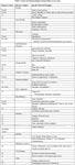

Table 1
PSPP classification scheme for otitis
Causes are diseases or agents that directly produce inflammation in the ear, otitis externa.Table 1 Factors are agents or elements of the disease that contribute to ear disease. Factors combine with causes or facilitate the causes to create more severe inflammation or symptoms. Table 2 Factors can inhibit the response to treatment of the causes of ear disease and can cause recrudescence of disease once treatment of causes is completed.

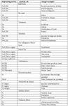
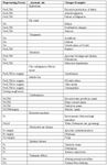
Table 2
Primary causes
Primary causes are usually the actual inciting agent or etiology that directly causes damage to the ear canal skin. These can occur alone and induce otitis externa without any other cause or factor. The primary cause may be very subtle and often go unrecognized by the owner or even veterinarian until a secondary cause occurs. Once a primary etiology alters the aural environment secondary infections often develop. The vast majority of cases will have a primary cause. About the only exception is when predisposing factors combine with secondary causes. The most common causes seen in a dermatology referral practice are atopic disease, food allergy, and epithelialization or metabolic disorders. In general practice foreign bodies and ear mites make up a significant number of cases. Also idiopathic or not diagnosed is common and in one study was reported in 32 of 100 cases.[7] If the case is recurrent or chronic and perpetuating factors are absent then allergy is most likely. It is critical too successful long-term management that a primary cause be found and either eliminated or control be secured.
Secondary causes
The secondary causes do not create disease in a normal ear; they contribute to or cause pathology only in the abnormal ear. As such they occur in combination with primary causes or predisposing factors. Generally secondary causes of otitis externa are easy to eliminate once identified and when they are chronic or recurrent it is usually because primary causes or perpetuating factors have not been adequately addressed. Secondary causes in the past were often considered as primary causes or the "main" diagnosis of an ear case. (ie. Pseudomonas or Malassezia otitis) Even today many clinicians direct all their efforts at diagnosing and treatment of secondary causes. Although their treatment may be important, other causes and factors must be looked for. In some cases such as malassezia, eliminating the concurrent predisposing factor or primary disease may result in the resolution of the secondary problem.
Perpetuating factors
Perpetuating factors are changes in the anatomy and physiology of the ear that occur in response to otitis externa. These factors may be subtle at first but over time can develop into the most severe component of chronic ear disease. These factors are not disease specific and are most commonly seen in chronic cases. Once present, they accentuate or permit the development of secondary causes by providing environments and microscopic niches that favor their persistence. In many cases perpetuating factors prevent the resolution of otitis externa when treatmens are only directed at primary and secondary causes. They cause much frustration to clinicians for several reasons. They often result in animals presenting repetitively with different causes present at each subsequent visit. These factors can become self perpetuating and lead to progressive worsening of disease. They can become severe and end up causing the majority of symptoms exhibited by a pet or be so mild appearing that to many veterinarians as well as owners a pet and its ear canal appear normal. Yet left untreated perpetuating factors, even though primary and secondary causes are controlled or eliminated, result in recrudescence of clinical disease.
In chronic cases often more than one of these factors will be present. Standard treatments of the primary and secondary diseases present often times will not immediately eliminate the perpetuating factors. In early cases, treating the primary cause may be sufficient in controlling a case, but after the establishment of perpetuating factors treatment may need to be directed at them. The treatment for perpetuating factors is often different that what is required to control primary and secondary causes of otitis externa. Their treatment should be continued until they have resolved which may take months of continuous therapy and in some cases they are permanent and will require life long therapy or a surgical solution. Perpetuating factors are the most common reasons otitis externa cases require surgery.
Predisposing factors
Predisposing factors alone do not cause otitis externa but increase the risk of development. These factors work in conjunction with either primary causes or secondary causes to become a significant problem. In rare cases a predisposing factor may combine with a secondary cause to create disease even when no primary cause is present. The best example of this is a dog that gets water in its ear that leads to epidermal maceration or damage and then a secondary bacterial or yeast infection occurs. It is possible this is how environment, increased heat and humidity, also contribute to otitis. However in the authors experience these animals often do have a subtle but mild primary disease still present but controlling that disease does not appear to be necessary.
Interaction Of Causes And Factors
Whenever a case of otitis external presents the best chance for successful treatment and the prognosis is predicated on the recognition of all causes and factors present. Many combinations may be seen and rarely is only one cause present. Most cases will have at least three and often numerous causes and factors present at the same time. It is the combination of the causes and factors that results in how severe a case is and how readily it will respond to treatment. Therefore, it is important that the clinician try to recognize all components present in a case of otitis externa. In addition, the combination of factors often results in more severe symptoms. By combing predisposing or secondary factors, otitis externa may result even without a more classically recognized primary disease being present. On the other hand, primary diseases may not cause clinical signs of otitis until a secondary factor becomes established. This is commonly seen in atopic dogs without apparent clinical otitis until a secondary malassezia or bacterial otitis becomes established or an atopic dog that only develops disease when water also gets into the ear.
Key Points Regarding Perpetuating Factors
In early cases, treating the primary cause may be sufficient in controlling a case, but after the establishment of perpetuating factors treatment may need to be directed at them. Treatment for the perpetuating factors should be continued until they have resolved which may take months of continuous therapy. Perpetuating factors may be the major reason for poor response to therapy, regardless of the predisposing factors and primary causes present.
Progressive pathologic responses occurring from inflammation may affect the epidermis, dermis and subsequently the adnexa and lumen of the ear canal. The epidermis becomes acanthotic and hyperkeratotic which because it is confined to the lumen of the canal and surrounded by a cartilaginous tube results in small epithelial folds. The thickened epidermis and the hyperkeratotic stratum corneum increase the keratin debris that is exfoliated into the canal lumen. The increased secretion and epithelial debris may favor the proliferation of bacteria and yeast. These changes also appear to alter the normal epithelial migration. The abnormal epithelial migration may prevent or impede the removal of the waxes, lipids, and exfoliating corneocytes and associated pathogenic bacteria. The dermis may become edematous, fibrotic and develop nodular pyogranulomas. Fibrosis is more common in end stage otitis in non-cocker breeds.
Tympanic membrane alterations occur in response to the accumulation of inflammatory debris and adjacent infection as well as in response to the buildup of exudate and debris that no longer can migrate out or pass through the occluded lumen. The abnormal tympanic membrane thickens, becomes opaque or slightly colored and loses its transparency. It may appear white, off-white, yellow, brown, or gray. The attachment to the manubrium cannot be seen. Therefore, the abnormal tympanic membrane can appear the same as impacted exudates or keratin plugs. This is a common problem situation where cases referred are diagnosed as having an abnormal tympanic membrane but the referring veterinarian has told the owner the tympanic membrane is intact. Once the tympanum ruptures otitis media likely results.
Assessing The Tympanic Membrane
Diagnosis of otitis media is not as easy to determine as otitis externa as many cases present with only symptoms of otitis externa. Evidence of inflammation to the tissues surrounding the middle ear or the inner ear usually indicates that otitis media has occurred. The presence of nerve damage, temporomandibular disease should be interpreted as indicative of otitis media. Even with otoscopic exam many cases of otitis externa may not be detected and in cases with apparently intact diseased tympanic membranes otitis media may be present. Diagnosis of otitis media can be made when a ruptured tympanic membrane is seen or when radiographic changes are present in the middle ear. Otitis media can only be ruled out when a definitive normal appearing tympanic membrane is present. Palpation of the tympanic membrane with a blunt instrument has been shown to be inaccurate and causes a significant incidence of damage to the tympanic membrane.
The author utilizes a technique of tube palpation and flushing to aid in the diagnosis of otitis media. This technique also may reveal false middle ear cavities. The soft tube can be used to palpate any material located at the approximate level of the tympanic membrane. The feeding tube is passed under visualization with a surgical otoscope head down the ear canal to the level where the tympanic membrane is expected to be located. In a normal ear the tip of the tube will remain visualized. Several features should be observed for:
- Depth and location of tip of tube,
- Loosing the view of the tip
- Movement and retraction of membrane
- Bruising pattern of deep tissue
- Air bubbles that come through a damaged TM
References
Cole, L.K., Anatomy and physiology of the canine ear. Vet Derm, 2009. 20: p. 412-421.
Griffin, C.E., Otitis: Anatomy every practitionar should know. Compend Contin Educ Pract Vet, 2009. November: p. 504-512.
Stout-Graham, M., et al., Morphologic measurements of the external horizontal ear canal of dogs. Am J Vet Res, 1990. 51(7): p. 990-994.
Johnson, A. and M. Hawke, An ink impregnation study of the migratory skin in the external auditory canal of the guinea-pig. Acta Otolaryngol, 1986. 101(3-4): p. 269-277.
Newton, H., et al., Evaluation of otoscope cone cleaning and disinfection procedures commonly used in veterinary medical practices: a pilot study. Vet Dermatol, 2006. 17(2): p. 147-150.
Kirby, A.L., et al., Evaluation of otoscope cone disinfection techniques and contamination level in small animal private practice. Vet Derm, 2010. 21: p. 175-183.
Saridomichelakis, M.N., et al., Aetiology of canine otitis externa: a retrospective study of 100 cases. Vet Dermatol, 2007. 18(5): p. 341-7.
Angus, J.C., et al., Breed Variations in Histopathologic Features of Chronic Severe Otitis Externa in Dogs: 80 Cases (1995-2001). J Am Vet Med Assoc, 2002. 221(7): p. 1000-1006.


