Determining the cause of neurologic signs in neonates
A closer look at genetics is offering new insight into disease conditions.
Q: Please provide an update on neonatal and genetic testing in puppies.
A: Dennis O'Brien, DVM, gave an excellent lecture titled "Neonatal neurology: Assessing the runt" at the 2009 American College of Veterinary Internal Medicine Forum in Montreal, Canada. O'Brien is professor of neurology at the University of Missouri's College of Veterinary Medicine. Some relevant points in this lecture are provided in this column.
Neonatal diseases have received relatively little attention in veterinary medicine. Most ill neonates are dismissed as runts or as "fading," and little effort is put into identifying the underlying cause. With the advent of genomics, the genetics of many hereditary diseases are being elucidated, allowing for disease prevention through DNA testing. Breeders want to decrease the incidence of these diseases both to improve the health of their breed and to improve their bottom line. Precise characterization of neonatal diseases defines syndromes and, in the case of genetic disorders, can provide clues to the underlying genes responsible.
Development and neurologic examination of the neonate
Recognizing and localizing neurologic lesions in neonates is difficult because their nervous systems are undergoing rapid changes during development. For example, crossed extension, which is a sign of upper motor neuron disease in an adult, is normal during the first few weeks of life.
When assessing a neonatal patient, record the animal's precise age, and consider the stage of development when interpreting behavior and neurologic examination findings. Table 1 outlines the different stages of early development and the corresponding behaviors and key elements of neurologic development. Approximate ages for the developmental milestones are given based on experimental observations with limited breeds represented. Note that breed variations are the rule rather than the exception, but these age ranges provide some guidance as to whether an individual is delayed.


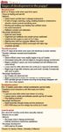
Table 1
As mentioned previously, components of the neurologic examination will change with development.
Diagnostic approach
In general, the diagnostic approach to a neonate will not differ from that in an adult except that the small size of the puppy may preclude some tests, and changes with development may confound the interpretation of others. While I focus on the nervous system in this discussion, it is important to remember that congenital defects can affect multiple organ systems. A thorough evaluation is warranted to detect concurrent deficits affecting other systems such as cardiac, urogenital or musculoskeletal.
When assessing an ill neonate, evaluate a hematocrit and blood glucose concentration and, if sufficient samples are available, perform a complete blood count and serum chemistry profile. Examine a blood smear for evidence of vacuolations or inclusions since some lysosomal storage diseases will affect leukocytes. Consider blood gas analysis if there is a high anion gap or low bicarbonate concentration on the serum chemistry profile. Electrolyte abnormalities are common in other neonates and should be investigated in puppies as well, and thyroid function can be measured to rule out congenital hypothyroidism. Blood ammonia values are valuable when searching for evidence of portosystemic shunts and will detect other congenital diseases such as urea cycle enzyme deficiencies. The test must be run immediately after the sample is taken, however, which limits its usefulness in most practices. Many inborn errors of metabolism produce abnormal byproducts that are excreted in the urine where they can be readily detected through special assays. Ketonuria in a neonate that is not anorectic could indicate an inborn error of metabolism. In contrast to blood, where care must be taken with the volume collected from a neonate, urine can be collected with impunity. Also, thought should be given to collecting whole blood for DNA extraction if a genetic disease is suspected.
There are other diagnostic modalities to consider using in these cases. Imaging of the brain can reveal congenital malformations such as polymicrogyria, Dandy-Walker malformation or hydrocephalus. Ultrasonography may be able to detect the cerebellar atrophy of Dandy-Walker malformation through the foramen magnum and dilated ventricles of hydrocephalus through the fontanels if open. In these patients, magnetic resonance imaging is the most sensitive modality, and because of the small size of the neonate, a high-field magnet will provide the best images. Keep in mind that myelination will be incomplete so the gray-white matter distinction will not be as great as in an adult. If electrodiagnostics are performed to diagnose neuromuscular disease or seizure disorders, the changes with development need to be taken into account.
In many of these cases, postmortem examination offers the best opportunity for a definitive diagnosis, but it is important to collect data such as serum chemistry profile findings or electrodiagnostics antemortem. Ideally, a pathologist should perform the postmortem, but this is sometimes impractical. In addition to routine examination of the liver, kidney, lung, etc., depending on the nature of the condition, brain, spinal cord, peripheral nerve or muscle samples should be obtained for evaluation. If myopathy is suspected, submit fresh muscle overnight to a laboratory that can process it for immunohistochemistry. Liver and kidney are very metabolically active tissues that are often used for biochemical studies. Some studies must be done on brain tissues, but cutting the brain before it is fixed can create artifact. Accessing the brain for laboratory analysis is easier in these patients. The skull of a neonate is often thin and easily removed with rongeurs or even a side-cutting wire cutter. The neonatal brain is very soft, however, so care must be taken not to damage it during removal. If more than one puppy in a litter is affected, one brain could be fixed in toto, while another could be split sagittally and half frozen for potential biochemical analysis. Consideration should also be given to collecting frozen tissues for biochemical assays or DNA studies.
Gene discovery strategies
Many genetic mutations can manifest as neonatal disease. Development of the nervous system is a complex process, and many genes are only expressed for a brief period during development. There is a bias for mutations to interfere with that process.
Inbreeding and founder effects can make genetic diseases more common in purebred animals, but genetic disease can arise even in randomly breeding populations. Littermate and extended family history can help determine whether genetics is playing a role in the disease. Breed tables can be consulted to see what familial diseases have been reported in the breed; the signs can then be compared to the case at hand. Individual breed club Web sites can provide a link to information about genetic diseases and DNA tests available. As with all information on the Internet, a healthy degree of skepticism is needed when evaluating the information.
Modern molecular genetics has provided us with the canine genome map and the tools to identify the genes responsible for hereditary diseases. Breeders are aware of the value of DNA testing, but veterinarians have the background to interpret the results of such tests as well as take the lead in developing new tests. Identifying the specific mutation responsible for a genetic disease can improve therapy and provide breeders the tools to decrease the incidence of the problem. When facing an unknown disease, gene discovery strategies can be used to identify the gene responsible. Current approaches to gene discovery include the candidate gene approach, linkage mapping and single nucleotide polymorphism (SNP) association.
The candidate gene approach involves identifying the comparable disease in humans, rodents or other species for which genes have been identified. The more accurate and molecular the diagnosis in each case, the more refined the list of candidates can be. For example, when two dachshund littermates with a syndrome of blindness, progressive ataxia and terminal myoclonic seizures were necropsied, fluorescent microscopy of brain tissue showed fluorescent material characteristic of neuronal ceroid lipofuscinosis (NCL) within lysosomes. Mutations in eight genes have been identified in humans, mice and sheep with NCL, so these were candidates for causes of the disease in dachshunds. Electron microscopy showed curvilinear bodies in the dog similar to those found in humans with one form of NCL. The gene associated with this form of NCL codes for the lysosomal enzyme TPP1, and the affected dog showed no activity for that enzyme in biochemical assays using frozen brain tissue. Thus, based on electron microscopy and biochemical data, there was one highly likely candidate. Sequencing the comparable canine gene demonstrated a missense mutation that led to a premature stop codon truncating the protein prior to the active site. The candidate gene approach can identify the mutation with a single affected case as was done here, allowing for quick development of a DNA test to identify carriers as well as a definitive diagnosis in affected dogs.
For many diseases, however, there are too many candidates, such as seizure disorders in which hundreds of mutations are known to produce epilepsy in mice. Alternatively, the gene associated with a particular disease may not have been identified yet in any species. In these cases, alternative approaches are necessary.
Linkage mapping uses genetic markers to follow the inheritance of chromosomes within a family. The disease-causing gene will be in the vicinity of some of these markers on a single chromosome. If a set of markers are consistently inherited in affected dogs, then those markers are "linked" to the disease. Neonatal diseases are ideally suited for mapping studies because the entire family, except maybe the sire, is available for DNA sampling at the time the disease is recognized.
Neonatal encephalopathy with seizures (NEWS) in standard poodles is characterized by developmental delay, ataxia, seizures and death before weaning age. There is a linkage between markers on chromosome 36 and NEWS. Recombinations occur when a portion of one chromosome is traded with its pair during meiosis. The resulting shuffle of markers allows more precise localization of the portion of that chromosome where the disease gene resides. This area of a chromosome where the responsible gene is located is called the locus. In NEWS, recombinations within the families studied further narrowed the locus to an area containing 26 genes. Of these, three were clearly involved in central nervous system development and, thus, became the prime candidates to be sequenced. A missense mutation in one of these genes was identified. DNA testing now allows standard poodle breeders to use wise breeding strategies to avoid producing affected dogs while still being able to use carriers in their breeding program.
SNP association is similar to linkage mapping in that marker genes are used. In this case, the markers are single nucleotide polymorphisms, or SNPs. SNPs are much more numerous than the type of markers used in linkage analysis, thus providing more dense coverage of the genome. This increased density allows us to look for a statistical association between having a disease and having a particular set of SNPs. The pattern of SNPs in dogs with the disease is compared with that of healthy dogs of the same breed. Affected dogs will inherit the particular group of SNPs that reside close to the mutant gene more commonly than normal dogs will. This approach can be done without having samples from the extended family. SNP association mapping has identified the mutations responsible for exercise-induced collapse in Labrador retrievers and degenerative myelopathy in a number of breeds.
Conclusion
High-quality breeders understand the role genetic testing will play in their attempts to improve the health of their breed. We, as veterinarians, should offer the diagnostic tests necessary to definitively diagnose a neonate with neurologic signs. In some cases, we may find a treatable condition such as congenital hypothyroidism. We need to be aware of the genetic tests already available and how to interpret them so that we can counsel owners and breeders appropriately. For other cases in which a genetic disorder is suspected, diagnostic tests can provide the clues necessary to help identify the gene responsible for the disease. Then, wise breeding strategies can ensure that affected dogs are not produced in the future and that the incidence of the mutant allele in the population decreases over time.
Dr. Hoskins is owner of Docu-Tech Services. He is a diplomate of the American College of Veterinary Internal Medicine with specialities in small animal pediatrics. He can be reached at (225) 955-3252, fax: (214) 242-2200 or e-mail: jdhoskins@mindspring.com


Urinalysis offers a noninasive, rapid screening for canine cancer detection
February 9th 2024This is the first rapid test using urine developed by the Virginia Tech College of Engineering, College of Agriculture and Life Sciences, and the Virginia-Maryland College of Veterinary Medicine
Read More


