CVC Highlight: 4 veterinary fundamentals of superficial wound healing
Stop infection and speed healing with these wound cleaning and bandaging strategies for veterinarians.

1. Prevent further wound contamination.
First and foremost, you must stabilize the patient and assess for other injuries. Also be sure to protect the wound with an occlusive bandage and provide pain relief.
Once you are ready to evaluate the wound, fill it with a water-soluble lubricant. Clip and clean the surrounding area with generously wide margins. Make sure to wear a cap, mask, and gloves to prevent contaminating a wound further or spreading the wound's infection in your hospital. Next, thoroughly lavage the wound to remove foreign material and minimize infection. Up to 90% of bacteria can be removed with proper lavage.
Perform wound lavage with saline or a balanced electrolyte solution. You want to use large quantities under moderate pressure. I like to use lactated Ringer's solution and microwave 1-L bags for one to three minutes to bring the solution closer to the patient's body temperature instead of room temperature to make it more comfortable for the patient. You want to lavage with 7 to 8 psi, which can be achieved by using a 35- or 60-ml syringe with an 18-ga needle. Wait to culture the wound until you have finished your lavage to ensure your results represent the bacteria still in the wound and not what you have already removed.
You can add antiseptic solutions to your lavage, but proper dilutions and usage are important. Povidone-iodine should be diluted to 1:10 or less and may have a residual activity for four to six hours. However, it forms complexes and becomes inactive with organic material and may be absorbed systemically. Chlorhexidine should be made into a 1:40 solution. It has a long residual activity that increases with reapplication, and side effects are rarer compared with povidone-iodine.
2. Débride dead and dying tissue.
Once the wound is thoroughly lavaged, perform débridement to remove the devitalized tissue and foreign bodies. Débridement can be performed surgically, chemically, or mechanically. Surgical débridement can be layered or en bloc, involving complete excision of the wound as with tumor excision. Chemical or enzymatic débridement is typically used in patients that are poor surgical risks or need minimal débridement of an open wound. Mechanical débridement traps devitalized tissue or foreign material in the primary layer of a bandage. This material is removed with each bandage change.
3. Provide adequate drainage.
Before any wound is bandaged, you must decide whether a drain is indicated. Surgical drains are indicated when dead space cannot be eliminated, fluid accumulation is likely, or infection is present. Disadvantages include an increased risk of infection, and all drains must be bandaged or properly cared for to prevent ascending infections. Drains should not exit through the initial wound or incision, and they should not lie directly under the suture line. Be sure to suture drains to the skin, but best practices discourage tacking buried sutures.
Passive or gravity-dependent drains must be placed ventrally on the wound and have a higher risk of ascending infection. There is no advantage to double-exit drains. Active drains, which use a vacuum, can be purchased or created in-house. They drain deeper tissues and are more efficacious with a lower risk of ascending infection. However, active drains may become obstructed.
Knowing when to remove a drain is as important as recognizing when to use one. Typically a drain is removed when the drainage decreases and becomes a transudate (serous or serosanguinous). Most drains are placed for three to seven days. Drains will always produce some fluid because they are foreign material in the body. A drain will cause 1 to 2 ml/kg/day/drain of fluid from its own irritation to the body.
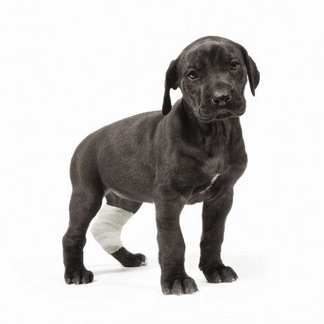
4. Select an appropriate method of closure.
Bandages are used to cover drains and wounds as well as to help with patient comfort, to reduce dead space or edema, to débride wounds, or as vehicles for antiseptics. Bandages consist of three layers-contact or primary, intermediate or secondary, and outer or tertiary.
The contact layer should be sterile and conform to the body contours. This layer must allow drainage to the secondary layer, be nontoxic or nonirritating, and minimize pain. Types of contact layers include dry adherent, wet adherent, and nonadherent. The type of primary layer used on a wound will often change as the wound heals. Numerous options for primary layers with impregnated ointments of medications are available on the market. It is important to remember to use these appropriately as they can result in increased bacterial resistance.
The secondary layer holds drainage, provides support, and decreases dead space. This layer must cover the primary layer completely and be thick enough to absorb all the fluid or drainage from the wound.
The tertiary layer holds the first two layers in place and should be applied with even pressure. Once this layer becomes wet, either from wound fluid or patient contamination, the bandage is useless and must be changed. Always instruct owners to look for slippage and make sure the bandage is clean, dry, and not too tight at least twice a day.


 in general practice")


