Cloning dogs and cats: Where do we stand? (Proceedings)
Assisted reproductive technologies in small animals are not a 21st century invention; the first artificial insemination in dogs was performed in 1780 and the first description of an oocyte at the microscopic level was that of a canine oocyte, in 1827.
Assisted reproductive technologies in small animals are not a 21st century invention; the first artificial insemination in dogs was performed in 1780 and the first description of an oocyte at the microscopic level was that of a canine oocyte, in 1827. However, economic realities have prevented rapid development of reproductive technologies in small animals compared to domestic large animal species. What are the advanced reproductive technologies currently used or proposed in small animals and how likely are they to be commercially available in the near future?
Reproductive physiology and early embryogenesis
The ovaries contain thousands of follicles, each of which contains an egg or ovum. As each estrous cycle begins, a cohort of follicles is selected to begin development. Development is promoted by release of hormones from the hypothalamus (gonadotropin releasing hormone [GnRH]) and pituitary (follicle stimulating hormone [FSH] and luteinizing hormone [LH]). As the follicle develops, it secretes estrogen, which causes the physical and behavioral signs of early heat, or proestrus. In the bitch, estrogen concentrations fall about 9 days after the onset of proestrus; at this time, the bitch will stand to be bred (standing heat or estrus) and a surge of LH is released, causing ovulation. Immature eggs are released from the follicles into the uterine tube, where they undergo two more cell divisions before fertilization can occur. In the queen, estrous behavior occurs when circulating estrogen concentrations are high and copulation stimulates release of GnRH and subsequent ovulation of mature oocytes into the uterine tube.
The egg released into the oviduct is surrounded by the zona pellucida and by a layer of granulosa cells from the follicle, the cumulus oophorus. Spermatozoa introduced into the reproductive tract of the bitch undergo capacitation, a calcium-dependent process involving the acrosome reaction on the head of the spermatozoon and achievement of hypermotility. Capacitated spermatozoa digest the layer of cells surrounding the egg and invade the zona pellucida. As soon as one spermatozoon binds to the inner layer of the zona pellucida, entry of other spermatozoa is blocked by an electrochemical reaction so only one spermatozoon fertilizes each egg. Cell division begins immediately.
Repeated doubling of cells occurs (2 cells – 4 cells – 8 cells – 16 cells) with concomitant changes in cell size and placement. The 16-cell stage is called a morula. The 16 to 64 cell stage is called a blastocyst. The blastocyst is a hollow sphere lined with blastomeres (embryonic cells) and filled with fluid. The blastocyst is divided into the inner cell mass, a group of blastomeres at one pole of the blastocyst will go on to form the embryo itself and two of the fetal membranes (yolk sac and allantois), and the trophoblasts, cells lining the outer surface of the blastocyst that go on to form the other two fetal membranes (chorion and amnion).
Points of technological intervention
No one has demonstrated ability to complete development in an artificial environment – at some point, the embryo must be placed in the uterus of a surrogate dam. The transfer of the embryo usually is accomplished with surgical placement of multiple morulas or blastocysts into the uterus of a recipient dam synchronized to be at the same point in the estrous cycle as the donor dam. It has been demonstrated that the recipient need not be the same species as the embryo but to date, closely related species have been used. This is of value in maintaining threatened or endangered canid and felid species, using domestic bitches and queens as surrogate dams.
Where does the embryo come from? Traditionally, donor dams were allowed to cycle and were bred, and embryos were retrieved from their uterine tubes or uterus surgically. These embryos were then transferred to a recipient dam. This simple form of embryo transfer has been reported successful in dogs and cats since the last 1970s. However, this simple form of embryo transfer does not gain us much in small animals. In large animal species, the donor dam can be superovulated, in which hormone therapy is used to cause release of a larger number of oocytes than normal during one estrous cycle and those many fertilized embryos are transferred into multiple recipient dams. Superovulation has not been demonstrated to work well in bitches and queens, who already release a relatively large number of oocytes during each estrus.
Instead, what people would like to be able to do is to retrieve multiple oocytes from a donor female, fertilize (in vitro fertilization = IVF) and mature them (in vitro maturation = IVM) outside of the donor dam and then implant the embryos produced into multiple recipients. This can be done by surgically retrieving oocytes from ovaries of donor animals and, in cats, by harvesting oocytes from ovaries excised from animals in the event of an untimely death. The oocyte is exposed to capacitated spermatozoa for fertilization. An even more advanced technique, intracytoplasmic sperm injection (ICSI) involves placement of a single capacitated spermatozoon into the oocyte. Maturation through early cleavage is easy in cats but very difficult in dogs. Unique features of the reproductive physiology of the bitch that impair IVM include ovulation of an immature oocyte, ovulation into a low-estrogen / high-progesterone environment, and necessity of continued presence of cumulus cells for ongoing maturation to occur.
Embryos also can be produced by nuclear transfer, commonly termed cloning. In this technique, the nucleus of a single cell from the donor is transferred into an oocyte that has had its DNA removed, that new oocyte fertilized and matured in vitro, and the subsequent embryo transferred into a recipient dam at the morula or blastocyst stage. The beauty of this technique is that any cell of the donor can be used; all reports of cloning in dogs and cats to date used fibroblasts retrieved from skin biopsies from the ear.
The final point to consider is the speed at which this must occur. In human and large animal medicine, embryos can be frozen at various points in development and later thawed and successfully implanted into a recipient dam. This permits creation of many embryos from a given donor and birth of her offspring over an extended period of time.
Success rates
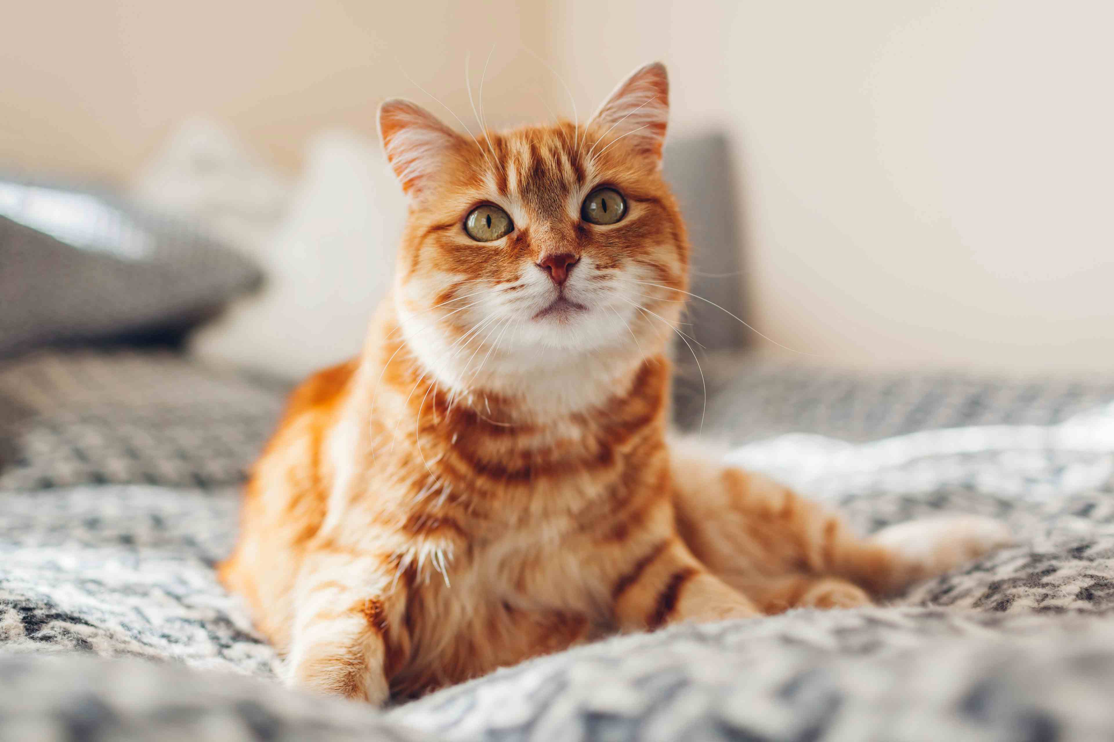
CATS – Assisted reproductive techniques are, in general, more successful in cats than in dogs. IVF and IVM in cats yield up to 50% development to the blastocyst stage; this is comparable to that seen in other species. Frozen-thawed embryos have successfully been transferred in cats. Domestic cats have been cloned; "Copy Cat" was born in 2001. Domestic cats (Felis domesticus) have also been used as surrogate dams carrying embryos constructed from wild cat DNA and domestic cat enucleated oocytes. Species cloned in this way include the African wild cat (Felis silvestris lybica), black-footed cat (Felis nigripes), fishing cats (Prionailurus viverrinus) and caracal (Caracal caracal).
DOGS – Assisted reproduction technologies are not uniformly successful in dogs and research proceeds slowly. IVF and IVM in dogs yield cleavage rates of only 8 to 37% with very few developing to the morula or blastocyst stage. There are no reports of successful embryo transfer to term of embryos matured in vitro or with transfer of frozen-thawed embryos. Domestic dogs have been cloned; "Snuppy" was reported in 2005. Domestic dogs (Canis familiaris) have also been used as surrogate dams carrying embryos constructed from wild canid DNA and domestic dog enucleated oocytes. The single species reported to date is the gray wolf (Canis lupus lupus).
Concerns
"Dolly", the first cloned sheep, died young of chronic disease. It has been estimated that up to 25% of cloned animals of all species (rodents to domestic farm animals) have serious health problems that decrease their quality of life and lifespan. A fairly high proportion of cloned cats in one study were stillborn; of 17 kittens carried to term, 7 (41.2%) were stillborn. Abnormalities of cloned kittens born in other studies included exteriorization of abdominal organs at birth, and respiratory failure and septicemia soon after birth.
Embryo wastage is an ethical concern. In cats, it has been demonstrated that a given recipient queen serving as surrogate to wild cat reconstructed embryos must be implanted with at least 30 embryos to become pregnant. In one study, 12 of 26 queens (46.2%) implanted with 30 embryos became pregnant while none of 24 queens implanted with 25 embryos became pregnant. In that study, 1380 embryos were implanted to produce 17 kittens, of which 2 lived beyond 6 weeks of age. That is a return rate of 1% for kittens born and of 0.1% for kittens living beyond 6 weeks of age. In studies evaluating embryo transfer within domestic cats, use of fresh embryos yielded a return rate of 4.3% for fresh embryos and 2.7% for frozen-thawed embryos for kittens born. In dogs, the picture is even more bleak. "Snuppy", the first cloned dog reported, was the only surviving pup of two carried to term after implantation of 1095 embryos into 123 recipient bitches. That is a return rate of 0.2% for pups born and 0.1% for live pups born.
The Humane Society of the United States (HSUS) does not support cloning of pets. Pet overpopulation is a huge problem in the United States and the $50,000 spent to create one dog or cat by cloning could support many efforts to combat this problem. HSUS sees cloning of pets as having no social value.
Finally, there is a concern about public perception. No animal can be recreated in all respects. Genetic variation alone plays a role; Copy Cat is a white cat with grey tabby stripes in patches and the animal from which she was cloned is a calico cat. Behavior and development are influenced by in utero environment, nutrition, environment in which the animal was raised and trained, and many other factors. Cloning is not a way to "get a beloved pet back." People also have concerns about scientists using animal cloning as a way to perfect human cloning, a hotly debated topic in medicine, ethics, and government.
Commercial availability
One cloned cat has been created and sold. "Little Nicky" was sold to a woman in Texas for $50,000 earlier this decade. The South Korean laboratory that produced the first cloned dog has reportedly agreed to supply cloned dogs as drug-sniffing dogs for the customs service in that country. As of this writing, they have no intention of offering cloning for pet owners and estimate a cost of 50 million to 100 million South Korean won ($55,000 – $100,000 US). Lazaron Biotechnologies of Baton Rouge LA will collect and preserve fibroblasts with a $500 fee for collection and cryopreservation and $10 per month storage fee. This service permits owners to keep their pet's genetic material on hand should cloning become readily available. Genetic Savings and Clone, the company that produced Copy Cat with Texas A&M University and produced Little Nicky, is no longer in business. Their website refers you to ViaGen and contains the statement, "Note that ViaGen has no plans to provide commercial dog or cat cloning services."